 W
WMagnetic resonance imaging (MRI) is a medical imaging technique used in radiology to form pictures of the anatomy and the physiological processes of the body. MRI scanners use strong magnetic fields, magnetic field gradients, and radio waves to generate images of the organs in the body. MRI does not involve X-rays or the use of ionizing radiation, which distinguishes it from CT and PET scans. MRI is a medical application of nuclear magnetic resonance (NMR) which can also be used for imaging in other NMR applications, such as NMR spectroscopy.
 W
WAmyloid-related imaging abnormalities (ARIA) are abnormal differences seen in magnetic resonance imaging of the brain of Alzheimer's disease patients, associated with amyloid-modifying therapies, particularly human monoclonal antibodies such as aducanumab. There are two types of ARIA - ARIA-E and ARIA-H. The phenomenon was first seen in trials of bapineuzumab.
The Brain Imaging Data Structure (BIDS) is a standard for organizing, annotating, and describing data collected during neuroimaging experiments. It is based on a formalized file/folder structure and JSON based metadata files with controlled vocabulary. This standard has been adopted by a multitude of labs around the world as well as databases such as OpenNeuro, SchizConnect, Developing Human Connectome Project, and FCP-INDI, and is seeing uptake in an increasing number of studies.
 W
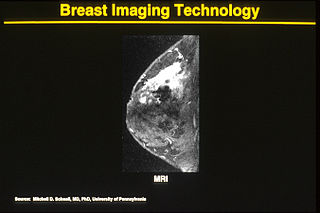
WOne alternative to mammography, Breast MRI or contrast enhanced magnetic resonance imaging (MRI), has shown substantial progress in the detection of breast cancer.
 W
WCardiovascular magnetic resonance imaging is a medical imaging technology for non-invasive assessment of the function and structure of the cardiovascular system. Conventional MRI sequences are adapted for cardiac imaging by using ECG gating and high temporal resolution protocols. The development of CMR is an active field of research and continues to see a rapid expansion of new and emerging techniques.
 W
WCardiac magnetic resonance imaging perfusion, also known as stress CMR perfusion, is a clinical magnetic resonance imaging test performed on patients with known or suspected coronary artery disease to determine if there are perfusion defects in the myocardium of the left ventricle that are caused by narrowing of one or more of the coronary arteries.
 W
WCardiac ventriculography is a medical imaging test used to determine a person's heart function in the right, or left ventricle. Cardiac ventriculography involves injecting contrast media into the heart's ventricle(s) to measure the volume of blood pumped. Cardiac ventriculography can be performed with a radionuclide in radionuclide ventriculography or with an iodine-based contrast in cardiac chamber catheterization.
 W
WDelayed gadolinium-enhanced magnetic resonance imaging of cartilage or dGEMRIC measures the fixed-charge density and relative proteoglycan content of articular cartilage using the spin-lattice relaxation time or T1 relaxation time. Current research is investigating the clinical application of dGEMRIC as a quantitative tool for monitoring cartilage function in diseased or repair cartilage.
 W
WDiffusion-weighted magnetic resonance imaging is the use of specific MRI sequences as well as software that generates images from the resulting data that uses the diffusion of water molecules to generate contrast in MR images. It allows the mapping of the diffusion process of molecules, mainly water, in biological tissues, in vivo and non-invasively. Molecular diffusion in tissues is not random, but reflects interactions with many obstacles, such as macromolecules, fibers, and membranes. Water molecule diffusion patterns can therefore reveal microscopic details about tissue architecture, either normal or in a diseased state. A special kind of DWI, diffusion tensor imaging (DTI), has been used extensively to map white matter tractography in the brain.
 W
WFluid-attenuated inversion recovery (FLAIR) is an MRI sequence with an inversion recovery set to null fluids. For example, it can be used in brain imaging to suppress cerebrospinal fluid (CSF) effects on the image, so as to bring out the periventricular hyperintense lesions, such as multiple sclerosis (MS) plaques. It was invented by Dr. Graeme Bydder. FLAIR can be used with both three-dimensional imaging or two dimensional imaging.
 W
WFunctional magnetic resonance imaging or functional MRI (fMRI) measures brain activity by detecting changes associated with blood flow. This technique relies on the fact that cerebral blood flow and neuronal activation are coupled. When an area of the brain is in use, blood flow to that region also increases.
 W
WFunctional ultrasound imaging (fUS) is a medical ultrasound imaging technique of detecting or measuring changes in neural activities or metabolism, for example, the loci of brain activity, typically through measuring blood flow or hemodynamic changes. The method can be seen as an extension of Doppler imaging.
 W
WHigh-intensity focused ultrasound (HIFU) is a non-invasive therapeutic technique that uses non-ionizing ultrasonic waves to heat or ablate tissue. HIFU can be used to increase the flow of blood or lymph, or to destroy tissue, such as tumors, via thermal and mechanical mechanisms. Given the prevalence and relatively low cost of ultrasound, HIFU has been subject to much research and development. The premise of HIFU is that it is a non-invasive low cost therapy that can at minimum outperform the current standard of care.
High definition fiber tracking (HDFT) is a tractography technique where data from MRI scanners is processed through computer algorithms to reveal the detailed wiring of the brain and to pinpoint fiber tracts. Each tract contains millions of neuronal connections. HDFT is based on data acquired from diffusion spectrum imaging and processed by generalized q-sampling imaging. The technique makes it possible to virtually dissect 40 major fiber tracts in the brain. The HDFT scan is consistent with brain anatomy unlike diffusion tensor imaging (DTI). Thus, the use of HDFT is essential in pinpointing damaged neural connections.
 W
WInterventional magnetic resonance imaging, also Interventional MRI or IMRI, is the use of magnetic resonance imaging (MRI) to do interventional radiology procedures.
 W
WIntravoxel incoherent motion (IVIM) imaging is a concept and a method initially introduced and developed by Le Bihan et al. to quantitatively assess all the microscopic translational motions that could contribute to the signal acquired with diffusion MRI. In this model, biological tissue contains two distinct environments: molecular diffusion of water in the tissue, and microcirculation of blood in the capillary network (perfusion). The concept introduced by D. Le Bihan is that water flowing in capillaries mimics a random walk (Fig.1), as long as the assumption that all directions are represented in the capillaries is satisfied.
 W
Wk-space is a formalism widely used in magnetic resonance imaging introduced in 1979 by Likes and in 1983 by Ljunggren and Twieg.
The Libin Cardiovascular Institute is a partnership between Alberta Health Services and the University of Calgary. Its mandate comprises all cardiovascular research, education and service delivery, with a service area extending from Saskatchewan, Southern Alberta and Eastern British Columbia. The Institute coordinates the activities of over 1,500 individuals in Southern Alberta. Of its more than 175 research and clinician members, over 75 are cardiologists, making it the largest heart or cardiovascular institute in Western Canada by that measure.
 W
WMagnetic resonance angiography (MRA) is a group of techniques based on magnetic resonance imaging (MRI) to image blood vessels. Magnetic resonance angiography is used to generate images of arteries in order to evaluate them for stenosis, occlusions, aneurysms or other abnormalities. MRA is often used to evaluate the arteries of the neck and brain, the thoracic and abdominal aorta, the renal arteries, and the legs.
 W
WMagnetic resonance cholangiopancreatography (MRCP) is a medical imaging technique. It uses magnetic resonance imaging to visualize the biliary and pancreatic ducts non-invasively. This procedure can be used to determine whether gallstones are lodged in any of the ducts surrounding the gallbladder.
 W
WMagnetic resonance elastography (MRE) is a form of elastography that specifically leverages MRI to quantify and subsequently map the mechanical properties of soft tissue. First developed and described at Mayo Clinic by Muthupillai et al. in 1995, MRE has emerged as a powerful, non-invasive diagnostic tool, namely as an alternative to biopsy and serum tests for staging liver fibrosis.
 W
WMagnetic resonance imaging of the brain uses magnetic resonance imaging (MRI) to produce high quality two-dimensional or three-dimensional images of the brain and brainstem without the use of ionizing radiation (X-rays) or radioactive tracers.
 W
WMagnetic resonance microscopy is magnetic resonance imaging (MRI) at a microscopic level down to the scale of microns. The first definition of MRM was MRI having voxel resolutions of better than 100 μm.
 W
WMagnetic resonance myelography is a noninvasive medical imaging technique that can provide anatomic information about the subarachnoid space. It a is type of MRI examination that uses a contrast medium and magnetic resonance imaging scanner to detect pathology of the spinal cord, including the location of a spinal cord injury, cysts, tumors and other abnormalities. The procedure involved the injection of a gadolinium based contrast media into the cervical or lumbar spine, followed by the MRI scan.
 W
WMagnetic resonance velocimetry (MRV) is an experimental method to obtain velocity fields in fluid mechanics. MRV is based on the phenomenon of nuclear magnetic resonance and adapts a medical magnetic resonance imaging system for the analysis of technical flows. The velocities are usually obtained by phase contrast magnetic resonance imaging techniques. This means velocities are calculated from phase differences in the image data that has been produced using special gradient techniques. MRV can be applied using common medical MRI scanners. The term magnetic resonance velocimetry became current due to the increasing use of MR technology for the measurement of technical flows in engineering.
 W
WAn MRI sequence in magnetic resonance imaging (MRI) is a particular setting of pulse sequences and pulsed field gradients, resulting in a particular image appearance.
 W
WMagnetic resonance neurography (MRN) is the direct imaging of nerves in the body by optimizing selectivity for unique MRI water properties of nerves. It is a modification of magnetic resonance imaging. This technique yields a detailed image of a nerve from the resonance signal that arises from in the nerve itself rather than from surrounding tissues or from fat in the nerve lining. Because of the intraneural source of the image signal, the image provides a medically useful set of information about the internal state of the nerve such as the presence of irritation, nerve swelling (edema), compression, pinch or injury. Standard magnetic resonance images can show the outline of some nerves in portions of their courses but do not show the intrinsic signal from nerve water. Magnetic resonance neurography is used to evaluate major nerve compressions such as those affecting the sciatic nerve, the brachial plexus nerves, the pudendal nerve, or virtually any named nerve in the body. A related technique for imaging neural tracts in the brain and spinal cord is called magnetic resonance tractography or diffusion tensor imaging.
 W
WMichael Nobel is a Swedish entrepreneur of Russian origin. He is a member of the Nobel family, a descendant of Ludvig Nobel, a former head of the Nobel Family Society (1995–2006), a co-founder and former Chairman of the Nobel Charitable Trust. At present, Nobel serves on several international boards that focus on scientific, medical and charitable initiatives. He promotes energy efficiency and alternative energy technology.
 W
WPentetic acid or diethylenetriaminepentaacetic acid (DTPA) is an aminopolycarboxylic acid consisting of a diethylenetriamine backbone with five carboxymethyl groups. The molecule can be viewed as an expanded version of EDTA and is used similarly. It is a white solid with limited solubility in water.
 W
WPerfusion MRI or perfusion-weighted imaging (PWI) is perfusion scanning by the use of a particular MRI sequence. The acquired data are then post-processed to obtain perfusion maps with different parameters, such as BV, BF, MTT and TTP.
 W
WPositron emission tomography–magnetic resonance imaging (PET–MRI) is a hybrid imaging technology that incorporates magnetic resonance imaging (MRI) soft tissue morphological imaging and positron emission tomography (PET) functional imaging.
WPhase contrast magnetic resonance imaging (PC-MRI) is a specific type of magnetic resonance imaging used primarily to determine flow velocities. PC-MRI can be considered a method of Magnetic Resonance Velocimetry. It also provides a method of magnetic resonance angiography. Since modern PC-MRI is typically time-resolved, it provides a means of 4D imaging.
 W
WThe physics of magnetic resonance imaging (MRI) concerns fundamental physical considerations of MRI techniques and technological aspects of MRI devices. MRI is a medical imaging technique mostly used in radiology and nuclear medicine in order to investigate the anatomy and physiology of the body, and to detect pathologies including tumors, inflammation, neurological conditions such as stroke, disorders of muscles and joints, and abnormalities in the heart and blood vessels among others. Contrast agents may be injected intravenously or into a joint to enhance the image and facilitate diagnosis. Unlike CT and X-ray, MRI uses no ionizing radiation and is, therefore, a safe procedure suitable for diagnosis in children and repeated runs. Patients with specific non-ferromagnetic metal implants, cochlear implants, and cardiac pacemakers nowadays may also have an MRI in spite of effects of the strong magnetic fields. This does not apply on older devices, details for medical professionals are provided by the device's manufacturer.
 W
WQuantitative Susceptibility Mapping (QSM) provides a novel contrast mechanism in Magnetic Resonance Imaging (MRI) different from traditional Susceptibility Weighted Imaging. The voxel intensity in QSM is linearly proportional to the underlying tissue apparent magnetic susceptibility, which is useful for chemical identification and quantification of specific biomarkers including iron, calcium, gadolinium, and super paramagnetic iron oxide (SPIO) nano-particles. QSM utilizes phase images, solves the magnetic field to susceptibility source inverse problem, and generates a three-dimensional susceptibility distribution. Due to its quantitative nature and sensitivity to certain kinds of material, potential QSM applications include standardized quantitative stratification of cerebral microbleeds and neurodegenerative disease, accurate gadolinium quantification in contrast enhanced MRI, and direct monitoring of targeted theranostic drug biodistribution in nanomedicine.
 W
WReal-time magnetic resonance imaging (MRI) refers to the continuous monitoring ("filming") of moving objects in real time. Because MRI is based on time-consuming scanning of k-space, real-time MRI was possible only with low image quality or low temporal resolution. Using an iterative reconstruction algorithm these limitations have recently been removed: a new method for real-time MRI achieves a temporal resolution of 20 to 30 milliseconds for images with an in-plane resolution of 1.5 to 2.0 mm. Real-time MRI promises to add important information about diseases of the joints and the heart. In many cases MRI examinations may become easier and more comfortable for patients.
 W
WResting state fMRI is a method of functional magnetic resonance imaging (fMRI) that is used in brain mapping to evaluate regional interactions that occur in a resting or task-negative state, when an explicit task is not being performed. A number of resting-state conditions are identified in the brain, one of which is the default mode network. These resting brain state conditions are observed through changes in blood flow in the brain which creates what is referred to as a blood-oxygen-level dependent (BOLD) signal that can be measured using fMRI.
 W
WMagnetic resonance imaging (MRI) is in general a safe technique, although injuries may occur as a result of failed safety procedures or human error. During the last 150 years, thousands of papers focusing on the effects or side effects of magnetic or radiofrequency fields have been published. They can be categorized as incidental and physiological. Contraindications to MRI include most cochlear implants and cardiac pacemakers, shrapnel and metallic foreign bodies in the eyes. The safety of MRI during the first trimester of pregnancy is uncertain, but it may be preferable to other options. Since MRI does not use any ionizing radiation, its use generally is favored in preference to CT when either modality could yield the same information..
The Society for Cardiovascular Magnetic Resonance (SCMR) is an international non-profit medical society based in Mt. Royal, New Jersey, in the United States. It was established as international representative and advocate for physicians, scientists, and technologists working in cardiovascular magnetic resonance (CMR) imaging. It works to improve patient outcomes through education, training, standards, research and development. The society has published more than 30 professional guidelines and expert consensus statements to help standardize and guide education, research and patient management with CMR. The society's members are physicians, scientists, technologists and trainees. In 2018, it is led by Matthias Stuber.
 W
WSodium MRI is a specialised magnetic resonance imaging technique that uses strong magnetic fields, magnetic field gradients, and radio waves to generate images of the distribution of sodium in the body, as opposed to more common forms of MRI that utilise protons present in water (1H-MRI). Like the proton, sodium is naturally abundant in the body, so can be imaged directly without the need for contrast agents or hyperpolarization. Furthermore, sodium ions play a role in important biological processes via their contribution to concentration and electrochemical gradients across cellular membranes, making it of interest as an imaging target in health and disease.
 W
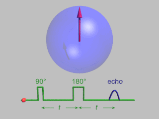
WIn magnetic resonance, a spin echo is the refocusing of spin magnetisation by a pulse of resonant electromagnetic radiation. Modern nuclear magnetic resonance (NMR) and magnetic resonance imaging (MRI) make use of this effect.
 W
WIn physics, the spin–spin relaxation is the mechanism by which Mxy, the transverse component of the magnetization vector, exponentially decays towards its equilibrium value in nuclear magnetic resonance (NMR) and magnetic resonance imaging (MRI). It is characterized by the spin–spin relaxation time, known as T2, a time constant characterizing the signal decay. It is named in contrast to T1, the spin–lattice relaxation time. It is the time it takes for the magnetic resonance signal to irreversibly decay to 37% (1/e) of its initial value after its generation by tipping the longitudinal magnetization towards the magnetic transverse plane. Hence the relation.
 W
WSteady-state free precession (SSFP) imaging is a magnetic resonance imaging (MRI) sequence which uses steady states of magnetizations. In general, SSFP MRI sequences are based on a gradient echo MRI sequence with a short repetition time which in its generic form has been described as the FLASH MRI technique. While spoiled gradient-echo sequences refer to a steady state of the longitudinal magnetization only, SSFP gradient-echo sequences include transverse coherences (magnetizations) from overlapping multi-order spin echoes and stimulated echoes. This is usually accomplished by refocusing the phase-encoding gradient in each repetition interval in order to keep the phase integral constant. Fully balanced SSFP MRI sequences achieve a phase of zero by refocusing all imaging gradients.
 W
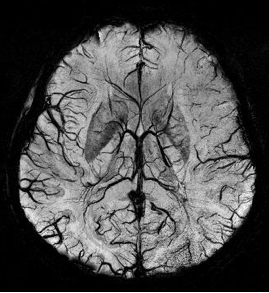
WSusceptibility weighted imaging (SWI), originally called BOLD venographic imaging, is an MRI sequence that is exquisitely sensitive to venous blood, hemorrhage and iron storage. SWI uses a fully flow compensated, long echo, gradient recalled echo (GRE) pulse sequence to acquire images. This method exploits the susceptibility differences between tissues and uses the phase image to detect these differences. The magnitude and phase data are combined to produce an enhanced contrast magnitude image. The imaging of venous blood with SWI is a blood-oxygen-level dependent (BOLD) technique which is why it was referred to as BOLD venography. Due to its sensitivity to venous blood SWI is commonly used in traumatic brain injuries (TBI) and for high resolution brain venographies but has many other clinical applications. SWI is offered as a clinical package by Philips and Siemens but can be run on any manufacturer’s machine at field strengths of 1.0 T, 1.5 T, 3.0 T and higher.
 W
WT2*-weighted imaging is an MRI sequence to quantify observable or effective T2. In this sequence, hemorrhages and hemosiderin deposits become hypointense.
 W
WIn medical imaging, a time-activity curve is a curve of radioactivity plotted on the y-axis against the time plotted on the x-axis. It shows the concentration of a radiotracer within a region of interest in an image, measured over time from a dynamic scan. Generally, when a time-activity curve is obtained within a tissue, it is called as a tissue time-activity curve, which represents the concentration of tracer within a region of interest inside a tissue over time.
 W
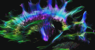
WIn neuroscience, tractography is a 3D modeling technique used to visually represent nerve tracts using data collected by diffusion MRI. It uses special techniques of magnetic resonance imaging (MRI) and computer-based diffusion MRI. The results are presented in two- and three-dimensional images called tractograms.