 W
WIn medicine, breast imaging is a sub-speciality of diagnostic radiology that involves imaging of the breasts for screening or diagnostic purposes. There are various methods of breast imaging using a variety of technologies as described in detail below. Traditional screening and diagnostic mammography uses x-ray technology. Breast tomosynthesis is a new digital mammography technique that produces 3D images of the breast using x-rays. Xeromammography and Galactography also use x-ray technology and are also used infrequently in the detection of breast cancer. Breast ultrasound is another technology employed in diagnosis & screening and specifically can help differentiate between fluid filled and solid lumps that can help determine if cancerous. Breast MRI is, yet, another technology reserved for high-risk patients and can help determine the extent of cancer if diagnosed. Lastly, scintimammography is used in a subgroup of patients who have abnormal mammograms or whose screening is not reliable on the basis of using traditional mammography or ultrasound.
 W
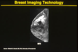
WOne alternative to mammography, Breast MRI or contrast enhanced magnetic resonance imaging (MRI), has shown substantial progress in the detection of breast cancer.
 W
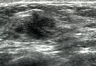
WBreast ultrasound is the use of medical ultrasonography to perform imaging of the breast.
 W
WMammography is the process of using low-energy X-rays to examine the human breast for diagnosis and screening. The goal of mammography is the early detection of breast cancer, typically through detection of characteristic masses or microcalcifications.
 W
WThe Mammography Quality Standards Act (MQSA) was enacted by the United States Congress to regulate the quality of care in mammography. The act was officially effective in 1994, and was extended in 2004 to continue through 2007. The U.S. Food and Drug Administration (FDA) began inspections of mammography facilities to ensure compliance in 1995. In 1997, more comprehensive regulation was added to become effective in 1999.
 W
WNon-contact thermography, thermographic imaging, or medical thermology is the field of thermography that uses infrared images of the human skin to assist in the diagnosis and treatment of medical conditions. Medical thermology is sometimes referred to as medical infrared imaging or tele-thermology and utilizes thermographic cameras. According to the American Academy of Thermology, Medical Thermology practitioners are licensed health care practitioners who utilize IR imaging in consistent with medically established paradigms of care. Non-medically licensed alternative practitioners who are not held to the same standard may offer thermography services but that should not be confused with the field of medical thermology.
 W
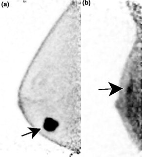
WPositron emission mammography (PEM) is a nuclear medicine imaging modality used to detect or characterise breast cancer. Mammography typically refers to x-ray imaging of the breast, while PEM uses an injected positron emitting isotope and a dedicated scanner to locate breast tumors. Scintimammography is another nuclear medicine breast imaging technique, however it is performed using a gamma camera. Breasts can be imaged on standard whole-body PET scanners, however dedicated PEM scanners offer advantages including improved resolution.
 W
WScintimammography is a type of breast imaging test that is used to detect cancer cells in the breasts of some women who have had abnormal mammograms, or for those who have dense breast tissue, post-operative scar tissue or breast implants.