 W
WAbdominal ultrasonography is a form of medical ultrasonography to visualise abdominal anatomical structures. It uses transmission and reflection of ultrasound waves to visualise internal organs through the abdominal wall. For this reason, the procedure is also called a transabdominal ultrasound, in contrast to endoscopic ultrasound, the latter combining ultrasound with endoscopy through visualize internal structures from within hollow organs.
 W
WCholangiography is the imaging of the bile duct by x-rays and an injection of contrast medium.
 W
WCholescintigraphy or hepatobiliary scintigraphy is scintigraphy of the hepatobiliary tract, including the gallbladder and bile ducts. The image produced by this type of medical imaging, called a cholescintigram, is also known by other names depending on which radiotracer is used, such as HIDA scan, PIPIDA scan, DISIDA scan, or BrIDA scan. Cholescintigraphic scanning is a nuclear medicine procedure to evaluate the health and function of the gallbladder and biliary system. A radioactive tracer is injected through any accessible vein and then allowed to circulate to the liver, where it is excreted into the bile ducts and stored by the gallbladder until released into the duodenum.
 W
WDefecography is a type of medical radiological imaging in which the mechanics of a patient's defecation are visualized in real time using a fluoroscope. The anatomy and function of the anorectum and pelvic floor can be dynamically studied at various stages during defecation.
 W
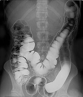
WA double-contrast barium enema is a form of contrast radiography in which x-rays of the colon and rectum are taken using two forms of contrast to make the structures easier to see. A liquid containing barium is put into the rectum. Barium is a silver-white metallic compound that outlines the colon and rectum on an x-ray and helps show abnormalities. Air is also put into the rectum and colon to further enhance the x-ray.
 W
WEndoscopic ultrasound (EUS) or echo-endoscopy is a medical procedure in which endoscopy is combined with ultrasound to obtain images of the internal organs in the chest, abdomen and colon. It can be used to visualize the walls of these organs, or to look at adjacent structures. Combined with Doppler imaging, nearby blood vessels can also be evaluated.
 W
WA lower gastrointestinal series is a medical procedure used to examine and diagnose problems with the human colon. Radiographs are taken while barium sulfate, a radiocontrast agent, fills the colon via an enema through the rectum.
 W
WMagnetic resonance cholangiopancreatography (MRCP) is a medical imaging technique that uses magnetic resonance imaging to visualize the biliary and pancreatic ducts in a non-invasive manner. This procedure can be used to determine if gallstones are lodged in any of the ducts surrounding the gallbladder.
WPercutaneous transhepatic cholangiography, percutaneous hepatic cholangiogram, or percutaneous transhepatic cholangiography and drainage (PTCD) is a radiological technique used to visualize the anatomy of the biliary tract. A contrast medium is injected into a bile duct in the liver, after which X-rays are taken. It allows access to the biliary tree in cases where endoscopic retrograde cholangiopancreatography (ERCP) has been unsuccessful. Initially reported in 1937, the procedure became popular in 1952.
 W
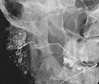
WSialography is the radiographic examination of the salivary glands. It usually involves the injection of a small amount of contrast medium into the salivary duct of a single gland, followed by routine X-ray projections.
 W
WTransrectal ultrasonography, or TRUS in short, is a method of creating an image of organs in the pelvis, most commonly used to perform an ultrasound-guided needle biopsy evaluation of the prostate gland in men with elevated prostate-specific antigen or prostatic nodules on digital rectal exam. TRUS--guided biopsy may reveal prostate cancer, benign prostatic hypertrophy, or prostatitis. TRUS may also detect other diseases of the lower rectum and can be used to stage primary rectal cancer.
 W
WAn upper gastrointestinal series, also called a barium meal, is a series of radiographs used to examine the gastrointestinal tract for abnormalities. A contrast medium, usually a radiocontrast agent such as barium sulfate mixed with water, is ingested or instilled into the gastrointestinal tract, and X-rays are used to create radiographs of the regions of interest. The barium enhances the visibility of the relevant parts of the gastrointestinal tract by coating the inside wall of the tract and appearing white on the film. This in combination with other plain radiographs allows for the imaging of parts of the upper gastrointestinal tract such as the pharynx, larynx, esophagus, stomach, and small intestine such that the inside wall lining, size, shape, contour, and patency are visible to the examiner. With fluoroscopy, it is also possible to visualize the functional movement of examined organs such as swallowing, peristalsis, or sphincter closure. Depending on the organs to be examined, barium radiographs can be classified into "barium swallow", "barium meal", "barium follow-through", and "enteroclysis". To further enhance the quality of images, air or gas is sometimes introduced into the gastrointestinal tract in addition to barium, and this procedure is called double-contrast imaging. In this case the gas is referred to as the negative contrast medium. Traditionally the images produced with barium contrast are made with plain-film radiography, but computed tomography is also used in combination with barium contrast, in which case the procedure is called "CT enterography".
 W
WVirtual colonoscopy is the use of CT scanning or magnetic resonance imaging (MRI) to produce two- and three-dimensional images of the colon, from the lowest part, the rectum, to the lower end of the small intestine, and to display the images on an electronic display device. The procedure is used to screen for colon cancer and polyps, and may detect diverticulosis. A virtual colonoscopy can provide 3D reconstructed endoluminal views of the bowel. VC provides a secondary benefit of revealing diseases or abnormalities outside the colon.