 W
WThe bulboid corpuscles are cutaneous receptors in the human body.
 W
WThe Bulbous corpuscle or Ruffini ending or Ruffini corpuscle is a slowly adapting mechanoreceptor located in the cutaneous tissue between the dermal papillae and the hypodermis. It is named after Angelo Ruffini.
 W
WCampaniform sensilla are a class of mechanoreceptors found in insects, which respond to stress and strain within the animal's cuticle. Campaniform sensilla function as proprioceptors that detect mechanical load as resistance to muscle contraction, similar to mammalian Golgi tendon organs. Sensory feedback from campaniform sensilla is integrated in the control of posture and locomotion.
 W
WCryptochromes are a class of flavoproteins that are sensitive to blue light. They are found in plants and animals. Cryptochromes are involved in the circadian rhythms of plants and animals, and possibly also in the sensing of magnetic fields in a number of species. The name cryptochrome was proposed as a portmanteau combining the cryptic nature of the photoreceptor, and the cryptogamic organisms on which many blue-light studies were carried out.
 W
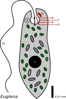
WThe eyespot apparatus is a photoreceptive organelle found in the flagellate or (motile) cells of green algae and other unicellular photosynthetic organisms such as euglenids. It allows the cells to sense light direction and intensity and respond to it, prompting the organism to either swim towards the light, or away from it. A related response occurs when cells are briefly exposed to high light intensity, causing the cell to stop, briefly swim backwards, then change swimming direction. Eyespot-mediated light perception helps the cells in finding an environment with optimal light conditions for photosynthesis. Eyespots are the simplest and most common "eyes" found in nature, composed of photoreceptors and areas of bright orange-red pigment granules. Signals relayed from the eyespot photoreceptors result in alteration of the beating pattern of the flagella, generating a phototactic response.
 W
WHair plates are a type of mechanoreceptor found in insects. Hair plates are tightly packed groups of sensory hairs that sense movements of one body segment relative to an adjoining segment. Hair plates are considered external proprioceptors.
 W
WMalleolus (plural: malleoli) is a fan-shaped chemoreceptor or Racquet Organ, an array of which are carried in pairs on the ventral or undersides of Solpugidae. They are the counterpart of pectines in scorpions, and modified walking limbs in the uropygids and amblypygids. Most species have 5 pairs of malleoli on the ventral surface of the fourth pair of legs of both sexes, while juveniles and other species have 2-3 pairs.In most animals the central pathways of olfactory systems are associated with glomerular neuropil and lack topographic mapping of sensory inputs. Among arthropods, the insect and crustacean olfactory (antennal) pathways are typical examples. Two orders of chelicerate arthropods, the scorpions and solpugids (Cl. Arachnida), present striking exceptions to this generalization. The major chemosensory organs of scorpions are the pectines, two ventral appendages that contact the substrate intermittently as the animal searches for food or mates. In solpugids chemosensory input is from the antennalized pedipalps and first leg pairs, and from ten fan-shaped malleoli extending ventrally to the substrate from the 4th leg pair. The pectinal and malleolar sensory systems have highly ordered arrangement of 105 to 106 primary chemoreceptors, with one (pectines) forming a two-dimensional array and the other (malleoli) assembled in a linear array. The spatial frequencies of these chemoreceptive inputs exceed 100/mm and 1000/mm, respectively, indicating a capacity for resolving structure of chemical deposits on substrates. Using several histological and axonal tracing techniques, the organization of pectinal and malleolar central projections has been resolved. The pectinal projection terminates posteriorly in the cephalothoracic mass and shows a high degree of topographic precision, perhaps to the level of individual receptors in the sensory field. This chemosensory 'map' is imposed on laminar cytoarchitecture posteriorly in the brain but merges anteriorly into glomerular substructures. The sensory projection from the malleoli shows less topographic order with fewer and larger glomeruli reminiscent of the insect olfactory system. These comparisons between arthropod taxa suggest that olfactory projections are, to varying degrees, typically glomerular but may evolve topographic and laminar organization when the stimulus field is of fixed form.
 W
WA nociceptor is a sensory neuron that responds to damaging or potentially damaging stimuli by sending “possible threat” signals to the spinal cord and the brain. If the brain perceives the threat as credible, it creates the sensation of pain to direct attention to the body part, so the threat can hopefully be mitigated; this process is called nociception.
 W
WAn ocelloid is a subcellular structure found in the family Warnowiaceae (warnowiids), which are a members of a group of unicellular organisms known as dinoflagellates. The ocelloid is analogous in structure and function to the eyes of multicellular organisms, which focus, process and detect light. The ocelloid is much more complex than the eyespot, a light-sensitive structure also found in unicellular organisms, and is in fact one of the most complex known subcellular structures. It has been described as a striking example of convergent evolution.
 W
WThe medial orbital gyrus presents a well-marked antero-posterior sulcus, the olfactory sulcus. Its depth is an indicator of congenital anosmia.
 W
WPacinian corpuscles are one of the four major types of mechanoreceptor cell in glabrous (hairless) mammalian skin. They are nerve endings in the skin responsible for sensitivity to vibration and pressure. They respond only to sudden disturbances and are especially sensitive to vibration. The vibrational role may be used to detect surface texture, e.g., rough vs. smooth. Pacinian corpuscles are also found in the pancreas, where they detect vibration and possibly very low frequency sounds. Pacinian corpuscles act as very rapidly adapting mechanoreceptors. Groups of corpuscles respond to pressure changes, e.g. on grasping or releasing an object.
 W
WA pecten is a comb-like structure, widely found in the biological world. Although pectens in various animals look similar, they have a varied range of uses, from grooming and filtering to sensory adaptations.
 W
WPhotopsins are the photoreceptor proteins found in the cone cells of the retina that are the basis of color vision. Iodopsin, the cone pigment system in chicken retina, is a close analog of the visual purple rhodopsin that is used in night vision. Iodopsin consists of the protein component and a bound chromophore, retinal.
 W
WPhytochromes are a class of photoreceptor in plants, bacteria and fungi used to detect light. They are sensitive to light in the red and far-red region of the visible spectrum and can be classed as either Type I, which are activated by far-red light, or Type II that are activated by red light. Recent advances have suggested that phytochromes also act as temperature sensors, as warmer temperatures enhance their de-activation. All of these factors contribute to the plant's ability to germinate.
 W
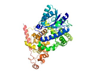
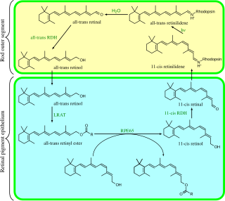
WRhodopsin is a light-sensitive receptor protein involved in visual phototransduction. It is named after ancient Greek ῥόδον for rose, due to its pinkish color, and ὄψις for sight. Rhodopsin is a biological pigment found in the rods of the retina and is a G-protein-coupled receptor (GPCR). It belongs to opsins. Rhodopsin is extremely sensitive to light, and thus enables vision in low-light conditions. When rhodopsin is exposed to light, it immediately photobleaches. In humans, it is regenerated fully in about 30 minutes, after which rods are more sensitive.
 W
WTactile corpuscles or Meissner's corpuscles are a type of mechanoreceptor discovered by anatomist Georg Meissner (1829–1905) and Rudolf Wagner. This corpuscle is a type of nerve ending in the skin that is responsible for sensitivity to light touch. In particular, they have their highest sensitivity when sensing vibrations between 10 and 50 hertz. They are rapidly adaptive receptors. They are most concentrated in thick hairless skin, especially at the finger pads.
 W
WVisual phototransduction is the sensory transduction of the visual system. It is a process by which light is converted into electrical signals in the rod cells, cone cells and photosensitive ganglion cells of the retina of the eye. This cycle was elucidated by George Wald (1906–1997) for which he received the Nobel Prize in 1967. It is so called "Wald's Visual Cycle" after him.