 W
WElectroencephalography (EEG) is an electrophysiological monitoring method to record electrical activity of the brain. It is typically noninvasive, with the electrodes placed along the scalp, although invasive electrodes are sometimes used, as in electrocorticography, sometimes called intracranial EEG.
 W
WAmplitude integrated electroencephalography (aEEG) or cerebral function monitoring (CFM) is a technique for monitoring brain function in intensive care settings over longer periods of time than the traditional electroencephalogram (EEG), typically hours to days. By placing electrodes on the scalp of the patient, a trace of electrical activity is produced which is then displayed on a semilogarithmic graph of peak-to-peak amplitude over time; amplitude is logarithmic and time is linear. In this way, trends in electrical activity in the cerebral cortex can be interpreted to inform on events such as seizures or suppressed brain activity. aEEG is useful especially in neonatology where it can be used to aid in diagnosis of hypoxic ischemic encephalopathy (HIE), and to monitor and diagnose seizure activity.
 W
WThe 10–20 system or International 10–20 system is an internationally recognized method to describe and apply the location of scalp electrodes in the context of an EEG exam, polysomnograph sleep study, or voluntary lab research. This method was developed to maintain standardized testing methods ensuring that a subject's study outcomes could be compiled, reproduced, and effectively analyzed and compared using the scientific method. The system is based on the relationship between the location of an electrode and the underlying area of the brain, specifically the cerebral cortex.
 W
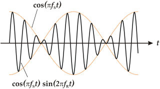
WIn acoustics, a beat is an interference pattern between two sounds of slightly different frequencies, perceived as a periodic variation in volume whose rate is the difference of the two frequencies.
 W
WBurst suppression is an electroencephalography (EEG) pattern that is characterized by periods of high-voltage electrical activity alternating with periods of no activity in the brain. The pattern is found in patients with inactivated brain states, such as from general anesthesia, coma, or hypothermia. This pattern can be physiological, as during early development, or pathological, as in diseases such as Ohtahara syndrome.
 W
WDelta waves are high amplitude neural oscillations with a frequency between 0.5 and 4 hertz. Delta waves, like other brain waves, can be recorded with electroencephalography (EEG) and are usually associated with the deep stage 3 of NREM sleep, also known as slow-wave sleep (SWS), and aid in characterizing the depth of sleep.
 W
WEar-EEG is a method for measuring dynamics of brain activity through the minute voltage changes observable on the skin, typically by placing electrodes on the scalp. In ear-EEG, the electrodes are exclusively placed in or around the outer ear, resulting in both a much greater invisibility and wearer mobility compared to full scalp electroencephalography (EEG), but also significantly reduced signal amplitude, as well as reduction in the number of brain regions in which activity can be measured. It may broadly be partitioned into two groups: those using electrode positions exclusively within the concha and ear canal, and those also placing electrodes close to the ear, usually hidden behind the ear lobe. Generally speaking, the first type will be the most invisible, but also offer the most challenging (noisy) signal. Ear-EEG is a good candidate for inclusion in a hearable device, however, due to the high complexity of ear-EEG sensors, this has not yet been done.
 W
WAn event-related potential (ERP) is the measured brain response that is the direct result of a specific sensory, cognitive, or motor event. More formally, it is any stereotyped electrophysiological response to a stimulus. The study of the brain in this way provides a noninvasive means of evaluating brain functioning.
 W
WFetal electroencephalography, also known as prenatal EEG includes any recording of electrical fluctuations arising from the brain of a fetus. Doctors and scientists use EEGs to detect and characterize brain activity, such as sleep states, potential seizures, or levels of a coma. EEG captures the electrical activity in the vicinity of the recording electrodes. The majority of the neural electrical activity arises from the flow of current from the cell bodies of pyramidal neurons to their apical dendrites, which become depolarized by excitatory inputs from other neurons. To record the most accurate signals, scientists try to minimize the distance between the recording electrode and the neural activity that they want to detect. Given the difficulty of attaching electrodes to a fetus inside a uterus, doctors and scientists use a variety of techniques to record fetal brain activity.
 W
WHypsarrhythmia is very chaotic and disorganized brain electrical activity with no recognizable pattern, whereas a normal brain electrical activity shows clear separation between each signal and visible pattern. It is an abnormal interictal pattern, consisting of high amplitude and irregular waves and spikes in a background of chaotic and disorganized activity seen on electroencephalogram (EEG), and frequently encountered in infants diagnosed with infantile spasms, although it can be found in other conditions.
 W
WA K-complex is a waveform that may be seen on an electroencephalogram (EEG). It occurs during stage 2 of NREM sleep. It is the "largest event in healthy human EEG". They are more frequent in the first sleep cycles.
 W
WA mind-controlled wheelchair is a mind-machine interfacing device that uses thought to command the motorised wheelchair's motion. The first such device to reach production was designed by Diwakar Vaish, Head of Robotics and Research at A-SET Training & Research Institutes. The wheelchair is of great importance to patients suffering from locked-in syndrome (LIS), in which a patient is aware but cannot move or communicate verbally due to complete paralysis of nearly all voluntary muscles in the body except the eyes. Such wheelchairs can also be used in case of muscular dystrophy, a disease that weakens the musculoskeletal system and hampers locomotion.
 W
WThe sensorimotor mu rhythm, also known as mu wave, comb or wicket rhythms or arciform rhythms, are synchronized patterns of electrical activity involving large numbers of neurons, probably of the pyramidal type, in the part of the brain that controls voluntary movement. These patterns as measured by electroencephalography (EEG), magnetoencephalography (MEG), or electrocorticography (ECoG), repeat at a frequency of 7.5–12.5 Hz, and are most prominent when the body is physically at rest. Unlike the alpha wave, which occurs at a similar frequency over the resting visual cortex at the back of the scalp, the mu rhythm is found over the motor cortex, in a band approximately from ear to ear. A person suppresses mu rhythms when he or she performs a motor action or, with practice, when he or she visualizes performing a motor action. This suppression is called desynchronization of the wave because EEG wave forms are caused by large numbers of neurons firing in synchrony. The mu rhythm is even suppressed when one observes another person performing a motor action or an abstract motion with biological characteristics. Researchers such as V. S. Ramachandran and colleagues have suggested that this is a sign that the mirror neuron system is involved in mu rhythm suppression, although others disagree.
 W
WNeurofeedback (NFB), also called neurotherapy or neurobiofeedback, is a type of biofeedback that uses real-time displays of brain activity—most commonly electroencephalography (EEG)—in an attempt to teach self-regulation of brain function. Typically, sensors are placed on the scalp to measure electrical activity, with measurements displayed using video displays or sound. The evidence supporting neurotherapy for generalized treatment of mental disorders numerous.
 W
WOpenBCI is an open-source brain-computer interface platform, created by Joel Murphy and Conor Russomanno, after a successful Kickstarter campaign in late 2013.
 W
WOpenViBE is a software platform dedicated to designing, testing and using brain-computer interfaces. The package includes a Designer tool to create and run custom applications, along with several pre-configured and demo programs which are ready for use.
 W
WThe P3b is a subcomponent of the P300, an event-related potential (ERP) component that can be observed in human scalp recordings of brain electrical activity. The P3b is a positive-going amplitude peaking at around 300 ms, though the peak will vary in latency from 250–500 ms or later depending upon the task and on the individual subject response. Amplitudes are typically highest on the scalp over parietal brain areas.
 W
WThe P300 (P3) wave is an event-related potential (ERP) component elicited in the process of decision making. It is considered to be an endogenous potential, as its occurrence links not to the physical attributes of a stimulus, but to a person's reaction to it. More specifically, the P300 is thought to reflect processes involved in stimulus evaluation or categorization.
 W
WSlow-wave sleep (SWS), often referred to as deep sleep, consists of stage three of non-rapid eye movement sleep. Initially, SWS consisted of both Stage 3, which has 20–50 percent delta wave activity, and Stage 4, which has more than 50 percent delta wave activity.
WSpike-and-wave is a pattern of the electroencephalogram (EEG) typically observed during epileptic seizures. A spike-and-wave discharge is a regular, symmetrical, generalized EEG pattern seen particularly during absence epilepsy, also known as ‘petit mal’ epilepsy. The basic mechanisms underlying these patterns are complex and involve part of the cerebral cortex, the thalamocortical network, and intrinsic neuronal mechanisms. The first spike-and-wave pattern was recorded in the early twentieth century by Hans Berger. Many aspects of the pattern are still being researched and discovered, and still many aspects are uncertain. The spike-and-wave pattern is most commonly researched in absence epilepsy, but is common in several epilepsies such as Lennox-Gastaut syndrome (LGS) and Ohtahara syndrome. Antiepileptic drugs (AEDs) are commonly prescribed to treat epileptic seizures, and new ones are being discovered with fewer adverse effects. Today, most of the research is focused on the origin of the generalized bilateral spike-and-wave discharge. One proposal suggests that a thalamocortical (TC) loop is involved in the initiation spike-and-wave oscillations. Although there are several theories, the use of animal models has provided new insight on spike-and-wave discharge in humans.
WThe visual N1 is a visual evoked potential, a type of event-related electrical potential (ERP), that is produced in the brain and recorded on the scalp. The N1 is so named to reflect the polarity and typical timing of the component. The "N" indicates that the polarity of the component is negative with respect to an average mastoid reference. The "1" originally indicated that it was the first negative-going component, but it now better indexes the typical peak of this component, which is around 150 to 200 milliseconds post-stimulus. The N1 deflection may be detected at most recording sites, including the occipital, parietal, central, and frontal electrode sites. Although, the visual N1 is widely distributed over the entire scalp, it peaks earlier over frontal than posterior regions of the scalp, suggestive of distinct neural and/or cognitive correlates. The N1 is elicited by visual stimuli, and is part of the visual evoked potential – a series of voltage deflections observed in response to visual onsets, offsets, and changes. Both the right and left hemispheres generate an N1, but the laterality of the N1 depends on whether a stimulus is presented centrally, laterally, or bilaterally. When a stimulus is presented centrally, the N1 is bilateral. When presented laterally, the N1 is larger, earlier, and contralateral to the visual field of the stimulus. When two visual stimuli are presented, one in each visual field, the N1 is bilateral. In the latter case, the N1's asymmetrical skewedness is modulated by attention. Additionally, its amplitude is influenced by selective attention, and thus it has been used to study a variety of attentional processes.