 W
WRadiation therapy or radiotherapy, often abbreviated RT, RTx, or XRT, is a therapy using ionizing radiation, generally as part of cancer treatment to control or kill malignant cells and normally delivered by a linear accelerator. Radiation therapy may be curative in a number of types of cancer if they are localized to one area of the body. It may also be used as part of adjuvant therapy, to prevent tumor recurrence after surgery to remove a primary malignant tumor. Radiation therapy is synergistic with chemotherapy, and has been used before, during, and after chemotherapy in susceptible cancers. The subspecialty of oncology concerned with radiotherapy is called radiation oncologist.
 W
WFour-dimensional computed tomography (4DCT) is a type of CT scanning which records multiple images over time. It allows playback of the scan as a video, so that physiological processes can be observed and internal movement can be tracked. The name is derived from the addition of time to traditional 3D computed tomography. Alternatively, the phase of a particular process, such as respiration, may be considered the fourth dimension.
 W
WThe abscopal effect is a hypothesis in the treatment of metastatic cancer whereby shrinkage of untreated tumors occurs concurrently with shrinkage of tumors within the scope of the localized treatment. R.H. Mole proposed the term “abscopal” in 1953 to refer to effects of ionizing radiation “at a distance from the irradiated volume but within the same organism.”
 W
WNeutron capture therapy (NCT) is a nonsurgical therapeutic modality for treating locally invasive malignant tumors such as primary brain tumors, recurrent head and neck cancer, and cutaneous and extracutaneous melanomas. It is a two-step procedure: first, the patient is injected with a tumor-localizing drug containing the non-radioactive isotope boron-10 (10B), which has a high propensity to capture thermal neutrons. The cross section of the 10B is many times greater than that of the other elements present in tissues such as hydrogen, oxygen, and nitrogen. In the second step, the patient is radiated with epithermal neutrons, the source of which is either a nuclear reactor or an accelerator. After losing energy as they penetrate tissue, the neutrons are captured by the 10B, which subsequently emits high-energy alpha particles that kill adjacent cells that have taken up sufficient quantities of 10B. All of the clinical experience to date with NCT is with the non-radioactive isotope boron-10, and this is known as boron neutron capture therapy (BNCT). The use of other non-radioactive isotopes, such as gadolinium, has been limited to experimental studies and has not been used clinically. BNCT has been evaluated clinically as an alternative to conventional radiation therapy for the treatment of high-grade gliomas, meningiomas, and recurrent, locally advanced cancers of the head and neck region and superficial cutaneous and extracutaneous melanomas.
 W
WThe Bragg peak is a pronounced peak on the Bragg curve which plots the energy loss of ionizing radiation during its travel through matter. For protons, α-rays, and other ion rays, the peak occurs immediately before the particles come to rest. This is called Bragg peak, after William Henry Bragg who discovered it in 1903.
 W
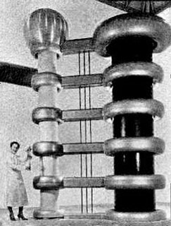
WCobalt therapy is the medical use of gamma rays from the radioisotope cobalt-60 to treat conditions such as cancer. Beginning in the 1950s, cobalt-60 was widely used in external beam radiotherapy (teletherapy) machines, which produced a beam of gamma rays which was directed into the patient's body to kill tumor tissue. Because these "cobalt machines" were expensive and required specialist support, they were often housed in cobalt units. Cobalt therapy was a revolutionary advance in radiotherapy in the post-World War II period but is now being replaced by other technologies such as linear accelerators.
 W
WDeep inspiration breath-hold (DIBH) is a method of delivering radiotherapy while limiting radiation exposure to the heart and lungs. It is used primarily for treating left-sided breast cancer.
 W
WIn External Beam Radiotherapy, transverse and longitudinal dose measurements are taken by a radiation detector in order to characterise the radiation beams from medical linear accelerators. Typically, an ionisation chamber and water phantom are used to create these radiation dose profiles. Water is used due to its tissue equivalence.
 W
WJennifer Clare Jones is an American radiation oncologist and biologist. She is an investigator and head of the translational nanobiology section at the National Cancer Institute.
 W
WMegavoltage X-rays are produced by linear accelerators ("linacs") operating at voltages in excess of 1000 kV (1 MV) range, and therefore have an energy in the MeV range. The voltage in this case refers to the voltage used to accelerate electrons in the linear accelerator and indicates the maximum possible energy of the photons which are subsequently produced. They are used in medicine in external beam radiotherapy to treat neoplasms, cancer and tumors. Beams with the voltage range of 4-25 MV are used to treat deeply buried cancers because radiation oncologists find that they penetrate well to deep sites within the body. Lower energy x-rays, called orthovoltage X-rays, are used to treat cancers closer to the surface.
 W
WA multileaf collimator (MLC) is a collimator or beam-limiting device that is made of individual "leaves" of a high atomic numbered material, usually tungsten, that can move independently in and out of the path of a radiotherapy beam in order to shape it and vary its intensity.
 W
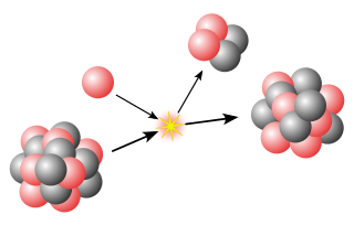
WNeutron generators are neutron source devices which contain compact linear particle accelerators and that produce neutrons by fusing isotopes of hydrogen together. The fusion reactions take place in these devices by accelerating either deuterium, tritium, or a mixture of these two isotopes into a metal hydride target which also contains deuterium, tritium or a mixture of these isotopes. Fusion of deuterium atoms results in the formation of a He-3 ion and a neutron with a kinetic energy of approximately 2.5 MeV. Fusion of a deuterium and a tritium atom results in the formation of a He-4 ion and a neutron with a kinetic energy of approximately 14.1 MeV. Neutron generators have applications in medicine, security, and materials analysis.
 W
WOrthovoltage x-rays are produced by x-ray tubes operating at voltages in the 100–500 kV range, and therefore the x-rays have a peak energy in the 100–500 keV range. Orthovoltage X-rays are sometimes termed "deep" x-rays (DXR). They cover the upper limit of energies used for diagnostic radiography, and are used in external beam radiotherapy to treat cancer and tumors. They penetrate tissue to a useful depth of about 4–6 cm. This makes them useful for treating skin, superficial tissues, and ribs, but not for deeper structures such as lungs or pelvic organs.
 W
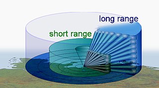
WIn optics, a pencil or pencil of rays is a geometric construct used to describe a beam or portion of a beam of electromagnetic radiation or charged particles, typically in the form of a narrow cone or cylinder.
 W
WPositron emission tomography (PET) is a functional imaging technique that uses radioactive substances known as radiotracers to visualize and measure changes in metabolic processes, and in other physiological activities including blood flow, regional chemical composition, and absorption. Different tracers are used for various imaging purposes, depending on the target process within the body. For example, 18F-FDG is commonly used to detect cancer, NaF-F18 is widely used for detecting bone formation, and oxygen-15 is sometimes used to measure blood flow.
 W
WA radiation burn is a damage to the skin or other biological tissue and organs as an effect of radiation. The radiation types of greatest concern are thermal radiation, radio frequency energy, ultraviolet light and ionizing radiation.
 W
WRadiation proctitis is inflammation and damage to the lower parts of the colon after exposure to x-rays or other ionizing radiation as a part of radiation therapy. Radiation proctitis most commonly occurs after pelvic radiation treatment for cancers such as cervical cancer, prostate cancer, bladder cancer, and rectal cancer. Radiation proctitis involves the lower intestine, primarily the sigmoid colon and the rectum, and is part of the conditions known as pelvic radiation disease and radiation enteropathy.
 W
WIn radiotherapy, radiation treatment planning (RTP) is the process in which a team consisting of radiation oncologists, radiation therapist, medical physicists and medical dosimetrists plan the appropriate external beam radiotherapy or internal brachytherapy treatment technique for a patient with cancer.
 W
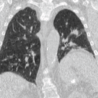
WRadiation-induced lung injury (RILI) is a general term for damage to the lungs as a result of exposure to ionizing radiation. In general terms, such damage is divided into early inflammatory damage and later complications of chronic scarring. Pulmonary radiation injury most commonly occurs as a result of radiation therapy administered to treat cancer.
 W
WRadioimmunotherapy (RIT) uses an antibody labeled with a radionuclide to deliver cytotoxic radiation to a target cell. It is a form of unsealed source radiotherapy. In cancer therapy, an antibody with specificity for a tumor-associated antigen is used to deliver a lethal dose of radiation to the tumor cells. The ability for the antibody to specifically bind to a tumor-associated antigen increases the dose delivered to the tumor cells while decreasing the dose to normal tissues. By its nature, RIT requires a tumor cell to express an antigen that is unique to the neoplasm or is not accessible in normal cells.
 W
WRadiopharmacology is radiochemistry applied to medicine and thus the pharmacology of radiopharmaceuticals. Radiopharmaceuticals are used in the field of nuclear medicine as radioactive tracers in medical imaging and in therapy for many diseases. Many radiopharmaceuticals use technetium-99m (Tc-99m) which has many useful properties as a gamma-emitting tracer nuclide. In the book Technetium a total of 31 different radiopharmaceuticals based on Tc-99m are listed for imaging and functional studies of the brain, myocardium, thyroid, lungs, liver, gallbladder, kidneys, skeleton, blood and tumors.
Sirtex Medical Limited is a medical device company, providing a radioactive treatment for inoperable liver cancer called SIR-Spheres microspheres. Sirtex was established in 1997 in Australia and currently maintains offices and manufacturing facilities in the U.S., Australia, Germany and Singapore. Following an acquisition by China Grand Pharmaceutical and CDH Genetech, Sirtex de-listed from the Australian Securities Exchange (ASX:SRX) on Monday, September 24th 2018.