 W
WProjectional radiography, also known as conventional radiography, is a form of radiography and medical imaging that produces two-dimensional images by x-ray radiation. The image acquisition is generally performed by radiographers, and the images are often examined by radiologists. Both the procedure and any resultant images are often simply called "X-ray". Plain radiography generally refers to projectional radiography. Plain radiography can also refer to radiography without a radiocontrast agent or radiography that generates single static images, as contrasted to fluoroscopy, which are technically also projectional.
 W
WAn abdominal x-ray is an x-ray of the abdomen. It is sometimes abbreviated to AXR, or KUB.
 W
WA coronary catheterization is a minimally invasive procedure to access the coronary circulation and blood filled chambers of the heart using a catheter. It is performed for both diagnostic and interventional (treatment) purposes.
 W
WAngiography or arteriography is a medical imaging technique used to visualize the inside, or lumen, of blood vessels and organs of the body, with particular interest in the arteries, veins, and the heart chambers. This is traditionally done by injecting a radio-opaque contrast agent into the blood vessel and imaging using X-ray based techniques such as fluoroscopy.
 W
WAortography involves placement of a catheter in the aorta and injection of contrast material while taking X-rays of the aorta. The procedure is known as an aortogram. The diagnosis of aortic dissection can be made by visualization of the intimal flap and flow of contrast material in both the true lumen and the false lumen. The catheter has to be inserted through the right femoral artery, because in about two thirds of cases the aortic dissection spreads into the left common iliac artery.
 W
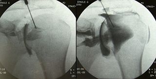
WAn arthrogram is a series of images of a joint after injection of a contrast medium, usually done by fluoroscopy or MRI. The injection is normally done under a local anesthetic such as Novocain or lidocaine. The radiologist or radiographer performs the study using fluoroscopy or x-ray to guide the placement of the needle into the joint and then injects around 10 ml of contrast based on age. There is some burning pain from the anesthetic and a painful bubbling feeling in the joint after the contrast is injected. This only lasts 20 – 30 hours until the Contrast is absorbed. During this time, while it is allowed, it is painful to use the limb for around 10 hours. After that the radiologist can more clearly see what is going on under your skin and can get results out within 24 to 48 hours.
 W
WAlan Archibald Campbell-Swinton FRS was a Scottish consulting electrical engineer, who provided the theoretical basis for the electronic television, two decades before the technology existed to implement it. He began experimenting around 1903 with the use of cathode ray tubes for the electronic transmission and reception of images. Campbell described the theoretical basis for an all electronic method of producing television in a 1908 letter to Nature. Campbell-Swinton's concept was central to the cathode ray television because of his proposed modification of the cathode ray tube that allowed its use as both a transmitter and receiver of light. The cathode-ray tube was the system of electronic television that was subsequently developed in later years, as technology caught up with Campbell-Swinton's initial ideas. Other inventors would use Campbell-Swinton's ideas, as a starting-point to realise the cathode ray tube television as the standard, workable form of all electronic television that it became for decades after his death. It is generally considered that the original credit for the successful theoretical conception of using a cathode ray tube device for imaging should belong to Campbell-Swinton.
WCerebral angiography is a form of angiography which provides images of blood vessels in and around the brain, thereby allowing detection of abnormalities such as arteriovenous malformations and aneurysms. It was pioneered in 1927 by the Portuguese neurologist Egas Moniz at the University of Lisbon, who also helped develop thorotrast for use in the procedure.
 W
WA chest radiograph, called a chest X-ray (CXR), or chest film, is a projection radiograph of the chest used to diagnose conditions affecting the chest, its contents, and nearby structures. Chest radiographs are the most common film taken in medicine.
 W
WPulmonary bay is a medical term which describes a finding on the chest radiograph. In pulmonary bay, there is a concavity where you would normally find the pulmonary artery. Pulmonary bay is most commonly associated with tetralogy of Fallot, however it may also be seen in other conditions where there is a reduced outflow from the pulmonary artery.
 W
WCholangiography is the imaging of the bile duct by x-rays and an injection of contrast medium.
 W
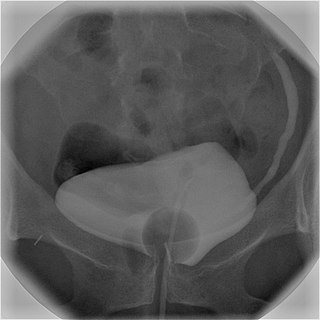
WIn radiology and urology, a cystography is a procedure used to visualise the urinary bladder.
WCystourethrography is a radiographic, fluoroscopic medical procedure that is used to visualize and evaluate the female urethra. Voiding and positive pressure cystourethrograms help to assess lower urinary tract trauma, reflux, suspected fistulas, and to diagnose urinary retention. Magnetic imaging (MRI) has been replacing this diagnostic tool due to its increased sensitivity. This imaging technique is used to diagnose hydronephrosis, voiding anomalies, and urinary tract infections in children. abnormalities.
 W
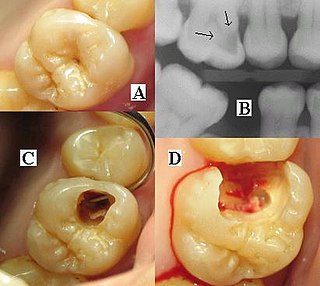
WDental radiographs are commonly called X-rays. Dentists use radiographs for many reasons: to find hidden dental structures, malignant or benign masses, bone loss, and cavities.
 W
WDigital subtraction angiography (DSA) is a fluoroscopy technique used in interventional radiology to clearly visualize blood vessels in a bony or dense soft tissue environment. Images are produced using contrast medium by subtracting a "pre-contrast image" or mask from subsequent images, once the contrast medium has been introduced into a structure. Hence the term "digital subtraction angiography". Subtraction angiography was first described in 1935 and in English sources in 1962 as a manual technique. Digital technology made DSA practical from the 1970s.
 W
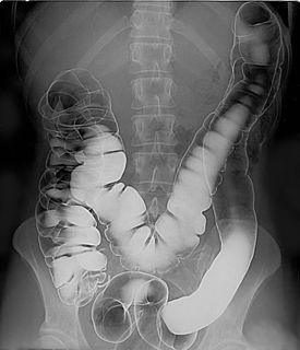
WA double-contrast barium enema is a form of contrast radiography in which x-rays of the colon and rectum are taken using two forms of contrast to make the structures easier to see. A liquid containing barium is put into the rectum. Barium is a silver-white metallic compound that outlines the colon and rectum on an x-ray and helps show abnormalities. Air is also put into the rectum and colon to further enhance the x-ray.
 W
WJohn Francis Hall-Edwards FRSE was a British doctor and pioneer in the medical use of X-rays in the United Kingdom.
 W
WHysterosalpingography (HSG), also known as uterosalpingography, is a radiologic procedure to investigate the shape of the uterine cavity and the shape and patency of the fallopian tubes. This means it is a special x-ray using dye to look at the womb (uterus) and Fallopian tubes. It injects a radio-opaque material into the cervical canal and usually fluoroscopy with image intensification. A normal result shows the filling of the uterine cavity and the bilateral filling of the fallopian tube with the injection material. To demonstrate tubal rupture, spillage of the material into the peritoneal cavity needs to be observed. It has vital role in treatment of infertility especially in case of fallopian tube blockade.
 W
WAn intravenous pyelogram (IVP), also called an intravenous urogram (IVU), is a radiological procedure used to visualize abnormalities of the urinary system, including the kidneys, ureters, and bladder. Unlike a kidneys, ureters, and bladder x-ray (KUB), which is a plain radiograph, an IVP uses contrast to highlight the urinary tract.
 W
WMammography is the process of using low-energy X-rays to examine the human breast for diagnosis and screening. The goal of mammography is the early detection of breast cancer, typically through detection of characteristic masses or microcalcifications.
 W
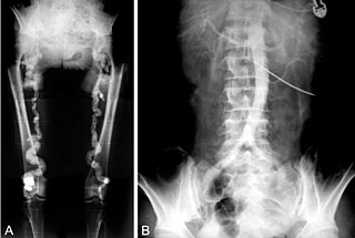
WMyelography is a type of radiographic examination that uses a contrast medium to detect pathology of the spinal cord, including the location of a spinal cord injury, cysts, and tumors. Historically the procedure involved the injection of a radiocontrast agent into the cervical or lumbar spine, followed by several X-ray projections. Today, myelography has largely been replaced by the use of MRI scans, although the technique is still sometimes used under certain circumstances – though now usually in conjunction with CT rather than X-ray projections.
 W
WA panoramic radiograph is a panoramic scanning dental X-ray of the upper and lower jaw. It shows a two-dimensional view of a half-circle from ear to ear. Panoramic radiography is a form of focal plane tomography; thus, images of multiple planes are taken to make up the composite panoramic image, where the maxilla and mandible are in the focal trough and the structures that are superficial and deep to the trough are blurred.
WPercutaneous transhepatic cholangiography, percutaneous hepatic cholangiogram, or percutaneous transhepatic cholangiography and drainage (PTCD) is a radiological technique used to visualize the anatomy of the biliary tract. A contrast medium is injected into a bile duct in the liver, after which X-rays are taken. It allows access to the biliary tree in cases where endoscopic retrograde cholangiopancreatography (ERCP) has been unsuccessful. Initially reported in 1937, the procedure became popular in 1952.
 W
WPneumoencephalography was a common medical procedure in which most of the cerebrospinal fluid (CSF) was drained from around the brain by means of a lumbar puncture and replaced with air, oxygen, or helium to allow the structure of the brain to show up more clearly on an X-ray image. It was derived from ventriculography, an earlier and more primitive method where the air is injected through holes drilled in the skull.
 W
WPulmonary angiography is medical fluoroscopic procedure used to visualize the pulmonary arteries and much less frequently, the pulmonary veins.
 W
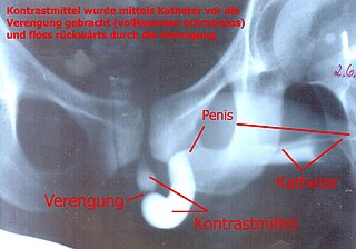
WA retrograde urethrogram is a routine radiologic procedure used to image the integrity of the urethra. Hence a retrograde urethrogram is essential for diagnosis of urethral injury, or urethral stricture.
 W
WThe Röntgen Memorial Site in Würzburg, Germany is dedicated to the work of the German physicist Wilhelm Conrad Röntgen (1845–1923) and his discovery of X-rays, for which he was granted the Nobel Prize in physics. It contains an exhibition of historical instruments, machines and documents.
 W
WWilhelm Conrad Röntgen was a German mechanical engineer and physicist, who, on 8 November 1895, produced and detected electromagnetic radiation in a wavelength range known as X-rays or Röntgen rays, an achievement that earned him the first Nobel Prize in Physics in 1901. In honour of his accomplishments, in 2004 the International Union of Pure and Applied Chemistry (IUPAC) named element 111, roentgenium, a radioactive element with multiple unstable isotopes, after him.
 W
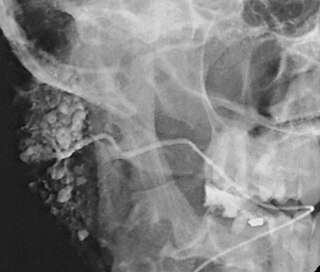
WSialography is the radiographic examination of the salivary glands. It usually involves the injection of a small amount of contrast medium into the salivary duct of a single gland, followed by routine X-ray projections.
 W
WAn upper gastrointestinal series, also called a barium meal, is a series of radiographs used to examine the gastrointestinal tract for abnormalities. A contrast medium, usually a radiocontrast agent such as barium sulfate mixed with water, is ingested or instilled into the gastrointestinal tract, and X-rays are used to create radiographs of the regions of interest. The barium enhances the visibility of the relevant parts of the gastrointestinal tract by coating the inside wall of the tract and appearing white on the film. This in combination with other plain radiographs allows for the imaging of parts of the upper gastrointestinal tract such as the pharynx, larynx, esophagus, stomach, and small intestine such that the inside wall lining, size, shape, contour, and patency are visible to the examiner. With fluoroscopy, it is also possible to visualize the functional movement of examined organs such as swallowing, peristalsis, or sphincter closure. Depending on the organs to be examined, barium radiographs can be classified into "barium swallow", "barium meal", "barium follow-through", and "enteroclysis". To further enhance the quality of images, air or gas is sometimes introduced into the gastrointestinal tract in addition to barium, and this procedure is called double-contrast imaging. In this case the gas is referred to as the negative contrast medium. Traditionally the images produced with barium contrast are made with plain-film radiography, but computed tomography is also used in combination with barium contrast, in which case the procedure is called "CT enterography".
 W
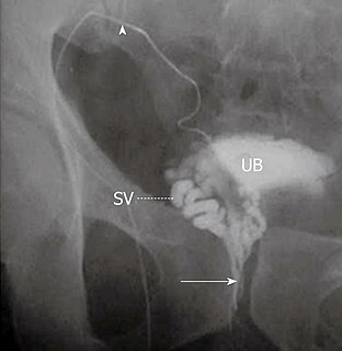
WVasography is an X-ray study of the vas deferens to see if there is blockage, oftentimes in the context of male infertility. An incision is made in the scrotum, contrast is injected in the vas deferens, and X-rays are taken from different angles. Thus, it is an invasive procedure and carries risk of iatrogenic scarring and obstruction of the vas. Vasography has traditionally been considered the gold standard imaging modality for evaluating the seminal tract patency.
 W
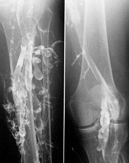
WVenography is a procedure in which an x-ray of the veins, a venogram, is taken after a special dye is injected into the bone marrow or veins. The dye has to be injected constantly via a catheter, making it an invasive procedure. Normally the catheter is inserted by the groin and moved to the appropriate site by navigating through the vascular system.