 W
WA microscope is an instrument used to see objects that are too small to be seen by the naked eye. Microscopy is the science of investigating small objects and structures using such an instrument. Microscopic means invisible to the eye unless aided by a microscope.
 W
WThe Abbe sine condition is a condition that must be fulfilled by a lens or other optical system in order for it to produce sharp images of off-axis as well as on-axis objects. It was formulated by Ernst Abbe in the context of microscopes.
 W
WAdvanced Microscopy Group or AMG is a manufacturer of microscopes based in Bothell, Washington.
 W
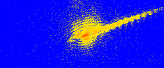
WThe atom probe was introduced at the 14th Field Emission Symposium in 1967 by Erwin Wilhelm Müller and J. A. Panitz. It combined a field ion microscope with a mass spectrometer having a single particle detection capability and, for the first time, an instrument could “... determine the nature of one single atom seen on a metal surface and selected from neighboring atoms at the discretion of the observer”.
 W
WThe atomic de Broglie microscope is an imaging system which is expected to provide resolution at the nanometer scale. Sometimes people call it the nano-scope.
 W
WThe scanning helium microscope (SHeM) is a novel form of microscopy that uses low energy neutral helium atoms to image the surface of a sample without any damage to the sample caused by the imaging process. Since helium is inert and neutral, it can be used to study delicate and insulating surfaces. Images are formed by rastering a sample underneath an atom beam and monitoring the flux of atoms that are scattered into a detector at each point.
 W
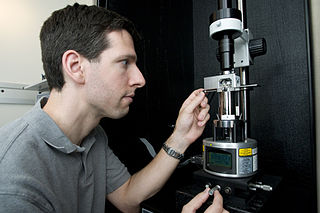
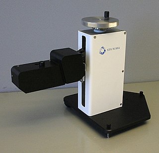
WA Brewster angle microscope (BAM) is a microscope for studying thin films on liquid surfaces, most typically Langmuir films. In a Brewster angle microscope, both the microscope and a polarized light source are aimed towards a liquid surface at that liquid's Brewster angle, in such a way for the microscope to catch an image of any light reflected from the light source via the liquid surface. Because there is no p-polarized reflection from the pure liquid when both are angled towards it at the Brewster angle, light is only reflected when some other phenomenon such as a surface film affects the liquid surface. The technique was first introduced in 1991.
 W
WA cathodoluminescence (CL) microscope combines methods from electron and regular microscopes. It is designed to study the luminescence characteristics of polished thin sections of solids irradiated by an electron beam.
 W
WCoherent diffractive imaging (CDI) is a "lensless" technique for 2D or 3D reconstruction of the image of nanoscale structures such as nanotubes, nanocrystals, porous nanocrystalline layers, defects, potentially proteins, and more. In CDI, a highly coherent beam of x-rays, electrons or other wavelike particle or photon is incident on an object.
 W
WThe contrast transfer function (CTF) mathematically describes how aberrations in a transmission electron microscope (TEM) modify the image of a sample. This contrast transfer function (CTF) sets the resolution of high-resolution transmission electron microscopy (HRTEM), also known as phase contrast TEM.
 W
WThe resolution of an optical imaging system – a microscope, telescope, or camera – can be limited by factors such as imperfections in the lenses or misalignment. However, there is a principal limit to the resolution of any optical system, due to the physics of diffraction. An optical system with resolution performance at the instrument's theoretical limit is said to be diffraction-limited.
 W
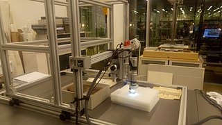
WA digital microscope is a variation of a traditional optical microscope that uses optics and a digital camera to output an image to a monitor, sometimes by means of software running on a computer. A digital microscope often has its own in-built LED light source, and differs from an optical microscope in that there is no provision to observe the sample directly through an eyepiece. Since the image is focused on the digital circuit, the entire system is designed for the monitor image. The optics for the human eye are omitted.
 W
WAn electron microprobe (EMP), also known as an electron probe microanalyzer (EPMA) or electron micro probe analyzer (EMPA), is an analytical tool used to non-destructively determine the chemical composition of small volumes of solid materials. It works similarly to a scanning electron microscope: the sample is bombarded with an electron beam, emitting x-rays at wavelengths characteristic to the elements being analyzed. This enables the abundances of elements present within small sample volumes to be determined, when a conventional accelerating voltage of 15-20 kV is used. The concentrations of elements from lithium to plutonium may be measured at levels as low as 100 parts per million (ppm), material dependent, although with care, levels below 10 ppm are possible The ability to quantify lithium by EPMA became a reality in 2008.
 W
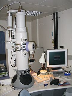
WAn electron microscope is a microscope that uses a beam of accelerated electrons as a source of illumination. As the wavelength of an electron can be up to 100,000 times shorter than that of visible light photons, electron microscopes have a higher resolving power than light microscopes and can reveal the structure of smaller objects. A scanning transmission electron microscope has achieved better than 50 pm resolution in annular dark-field imaging mode and magnifications of up to about 10,000,000× whereas most light microscopes are limited by diffraction to about 200 nm resolution and useful magnifications below 2000×.
 W
Wru:ООС ru:OOСЗМ
 W
WThe Field ion microscope (FIM) was invented by Müller in 1951. It is a type of microscope that can be used to image the arrangement of atoms at the surface of a sharp metal tip.
 W
WA fluorescence microscope is an optical microscope that uses fluorescence instead of, or in addition to, scattering, reflection, and attenuation or absorption, to study the properties of organic or inorganic substances. "Fluorescence microscope" refers to any microscope that uses fluorescence to generate an image, whether it is a more simple set up like an epifluorescence microscope or a more complicated design such as a confocal microscope, which uses optical sectioning to get better resolution of the fluorescence image.
 W
WA Foldscope is an optical microscope that can be assembled from simple components, including a sheet of paper and a lens. It was developed by Manu Prakash and designed to cost less than US$1 to build. It is part of the "frugal science" movement which aims to make cheap and easy tools available for scientific use in the developing world.
 W
WHirox (ハイロックス) is a lens company in Tokyo, Japan that created the first digital microscope in 1985. This company is now known as Hirox Co Ltd. Hirox's main industry is digital microscopes, but still makes the lenses for a variety of items including rangefinders.
 W
WAn inverted microscope is a microscope with its light source and condenser on the top, above the stage pointing down, while the objectives and turret are below the stage pointing up. It was invented in 1850 by J. Lawrence Smith, a faculty member of Tulane University.
 W
WLeica Microsystems GmbH is a manufacturer of optical microscopes, equipment for the preparation of microscopic specimens and related products. There are ten plants in eight countries with distribution partners in over 100 countries. Leica Microsystems emerged in 1997 out of a 1990 merger between Wild-Leitz, headquartered in Heerbrugg Switzerland and Cambridge Instruments of Cambridge England. The merger of those two umbrella companies created an alliance of the following 8 individual manufacturers of scientific instruments. American Optical Scientific Products, Carl Reichert Optische Werke AG, R.Jung, Bausch and Lomb Optical Scientific Products Division, Cambridge Instruments, E.Leitz Wetzlar, Kern & Co. and Wild Heerbrugg AG, bringing much needed modernization and a broader degree of expertise to the newly created entity called Leica Holding B.V. group. In 1997 the name was changed to Leica Microsystems and is a wholly owned entity of Danaher Corporation since July 2005. Danaher is a US venture capital company.
 W
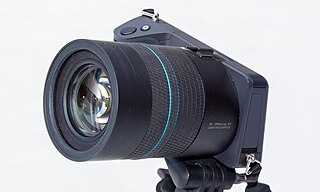
WA light field camera, also known as plenoptic camera, captures information about the light field emanating from a scene; that is, the intensity of light in a scene, and also the direction that the light rays are traveling in space. This contrasts with a conventional camera, which records only light intensity.
 W
WLive cell imaging is the study of living cells using time-lapse microscopy. It is used by scientists to obtain a better understanding of biological function through the study of cellular dynamics. Live cell imaging was pioneered in first decade of the 20th century. One of the first time-lapse microcinematographic films of cells ever made was made by Julius Ries, showing the fertilization and development of the sea urchin egg. Since then, several microscopy methods have been developed which allow researchers to study living cells in greater detail with less effort. A newer type of imaging utilizing quantum dots have been used as they are shown to be more stable. The development of holotomographic microscopy has disregarded phototoxicity and other staining-derived disadvantages by implementing digital staining based on cells’ refractive index.
 W
WMicrographia: or Some Physiological Descriptions of Minute Bodies Made by Magnifying Glasses. With Observations and Inquiries Thereupon. is a historically significant book by Robert Hooke about his observations through various lenses. It is particularly notable for being the first book to illustrate insects, plants etc. as seen through microscopes. Published in January 1665, the first major publication of the Royal Society, it became the first scientific best-seller, inspiring a wide public interest in the new science of microscopy. It is also notable for coining the biological term cell.
 W
WA comparison microscope is a device used to analyze side-by-side specimens. It consists of two microscopes connected by an optical bridge, which results in a split view window enabling two separate objects to be viewed simultaneously. This avoids the observer having to rely on memory when comparing two objects under a conventional microscope.
 W
WIndustrial computed tomography (CT) scanning is any computer-aided tomographic process, usually X-ray computed tomography, that uses irradiation to produce three-dimensional internal and external representations of a scanned object. Industrial CT scanning has been used in many areas of industry for internal inspection of components. Some of the key uses for industrial CT scanning have been flaw detection, failure analysis, metrology, assembly analysis and reverse engineering applications. Just as in medical imaging, industrial imaging includes both nontomographic radiography and computed tomographic radiography.
 W
WX-ray microtomography, like tomography and X-ray computed tomography, uses X-rays to create cross-sections of a physical object that can be used to recreate a virtual model without destroying the original object. The prefix micro- is used to indicate that the pixel sizes of the cross-sections are in the micrometre range. These pixel sizes have also resulted in the terms high-resolution X-ray tomography, micro–computed tomography, and similar terms. Sometimes the terms high-resolution CT (HRCT) and micro-CT are differentiated, but in other cases the term high-resolution micro-CT is used. Virtually all tomography today is computed tomography.
 W
WThe optical microscope, also referred to as a light microscope, is a type of microscope that commonly uses visible light and a system of lenses to generate magnified images of small objects. Optical microscopes are the oldest design of microscope and were possibly invented in their present compound form in the 17th century. Basic optical microscopes can be very simple, although many complex designs aim to improve resolution and sample contrast.
 W
WA petrographic microscope is a type of optical microscope used in petrology and optical mineralogy to identify rocks and minerals in thin sections. The microscope is used in optical mineralogy and petrography, a branch of petrology which focuses on detailed descriptions of rocks. The method is called "polarized light microscopy" (PLM).
 W
WPhenom is a small, table-top sized scanning electron microscope (SEM) originally developed by Philips and FEI and further developed by Phenom-World. The microscope features a combination of optical and electron-optical images; the optical image enables a "Neverlost" function so operators may navigate to any point on the sample. Sample loading takes place in 4 seconds and only 30 seconds into the vacuum space via rapid transfer technology. The system user interface is controlled with a touch screen No SEM experience is required for users to achieve magnifications of up to 100,000 times with a resolution of down to 15 nm. An optional fully integrated X-ray analysis (EDS) system shows the user in just seconds what the sample is made of.
 W
WThe Raman microscope is a laser-based microscopic device used to perform Raman spectroscopy. The term MOLE is used to refer to the Raman-based microprobe. The technique used is named after C. V. Raman who discovered the scattering properties in liquids.
Reynolds & Branson Leeds was a business based at 13 Briggate and 14 Commercial Street in Leeds, England. The business lasted from 1816 to 1972. Edward Matterson managed the company in 1822, and William West F.R.S. took over in 1833. The National Archives Records about the company include a day book, sales ledger, and prescription books. The records were created by Reynolds & Branson Ltd. Reynolds & Branson was registered in July 1898 as a limited corporation with a capital of £34,000 in shares of £10 each by Messrs. R. Reynolds, F. W. Branson. No remuneration was given to Mr. R. Reynolds, but a £700 per annum was given to each of the others. In 1890, Richard Reynold's son, Richard Freshfield (Fred) Reynolds was made a partner. The firm was in the business of wholesale and retail for chemists and surgical instrument makers.
 W
WA scanning acoustic microscope (SAM) is a device which uses focused sound to investigate, measure, or image an object. It is commonly used in failure analysis and non-destructive evaluation. It also has applications in biological and medical research. The semiconductor industry has found the SAM useful in detecting voids, cracks, and delaminations within microelectronic packages.
 W
WA scanning helium ion microscope is an imaging technology based on a scanning helium ion beam. Similar to other focused ion beam techniques, it allows to combine milling and cutting of samples with their observation at sub-nanometer resolution.
 W
WScanning SQUID microscopy is a technique where a superconducting quantum interference device (SQUID) is used to image surface magnetic field strength with micrometre scale resolution. A tiny SQUID is mounted onto a tip which is then rastered near the surface of the sample to be measured. As the SQUID is the most sensitive detector of magnetic fields available and can be constructed at submicrometre widths via lithography, the scanning SQUID microscope allows magnetic fields to be measured with unparalleled resolution and sensitivity. The first scanning SQUID microscope was built in 1992 by Black et al. Since then the technique has been used to confirm unconventional superconductity in several high-temperature superconductors including YBCO and BSCCO compounds.
 W
WA scanning tunneling microscope (STM) is an instrument for imaging surfaces at the atomic level. Its development in 1981 earned its inventors, Gerd Binnig and Heinrich Rohrer, then at IBM Zürich, the Nobel Prize in Physics in 1986. STM senses the surface by using an extremely sharp conducting tip that can distinguish features smaller than 0.1 nm with a 0.01 nm depth resolution. This means that individual atoms can routinely be imaged and manipulated. Most microscopes are built for use in ultra-high vacuum at temperatures approaching zero kelvin, but variants exist for studies in air, water and other environments, and for temperatures over 1000 °C.
 W
WThe scioptic ball is a universal joint allowing an optical instrument mounted on a ball to be swiveled to point anywhere in a wide arc. It was inspired by studies of the human eye. It has a number of applications. The scioptic ball may provide a firm anchor for a microscope, camera or telescope allowing it to be swiveled in all directions, for example to follow the course of an eclipse or for drawing panoramic views. Scioptic balls have been used as camera obscuras, projecting images from the outside on walls in darkened rooms. Scioptic balls have been used simply as light sources. It was an early example of a type of wide-angle lens.
 W
WSingle particle analysis is a group of related computerized image processing techniques used to analyze images from transmission electron microscopy (TEM). These methods were developed to improve and extend the information obtainable from TEM images of particulate samples, typically proteins or other large biological entities such as viruses. Individual images of stained or unstained particles are very noisy, and so hard to interpret. Combining several digitized images of similar particles together gives an image with stronger and more easily interpretable features. An extension of this technique uses single particle methods to build up a three-dimensional reconstruction of the particle. Using cryo-electron microscopy it has become possible to generate reconstructions with sub-nanometer resolution and near-atomic resolution first in the case of highly symmetric viruses, and now in smaller, asymmetric proteins as well.
 W
WA Stanhope lens is a simple, one-piece microscope invented by Charles, the third Earl of Stanhope. It is a cylinder of glass with each end curved outwards, one being more convex than the other. The focal length of the apparatus is at or within the device so that objects to be studied are placed close to or in contact with the less curved end. Because its construction is simple and economical, it was popular in the 19th century. It was useful in medical practice for examining transparent materials such as crystals and fluids.
 W
WThe stereo, stereoscopic or dissecting microscope is an optical microscope variant designed for low magnification observation of a sample, typically using light reflected from the surface of an object rather than transmitted through it. The instrument uses two separate optical paths with two objectives and eyepieces to provide slightly different viewing angles to the left and right eyes. This arrangement produces a three-dimensional visualization of the sample being examined. Stereomicroscopy overlaps macrophotography for recording and examining solid samples with complex surface topography, where a three-dimensional view is needed for analyzing the detail.
 W
WA total internal reflection fluorescence microscope (TIRFM) is a type of microscope with which a thin region of a specimen, usually less than 200 nanometers can be observed.
 W
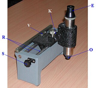
WA travelling microscope is an instrument for measuring length with a resolution typically in the order of 0.01mm. The precision is such that better-quality instruments have measuring scales made from Invar to avoid misreadings due to thermal effects. The instrument comprises a microscope mounted on two rails fixed to, or part of a very rigid bed. The position of the microscope can be varied coarsely by sliding along the rails, or finely by turning a screw. The eyepiece is fitted with fine cross-hairs to fix a precise position, which is then read off the vernier scale. Some instruments, such as that produced in the 1960s by the Precision Tool and Instrument Company of Thornton Heath, Surrey, England, also measure vertically. The purpose of the microscope is to aim at reference marks with much higher accuracy than is possible using the naked eye. It is used in laboratories to measure the refractive index of liquids using the geometrical concepts of ray optics. It is also used to measure very short distances precisely, for example the diameter of a capillary tube. This mechanical instrument has now largely been superseded by electronic- and optically-based measuring devices that are both very much more accurate and considerably cheaper to produce.
 W
WA USB microscope is a low-powered digital microscope which connects to a computer, normally via a USB port. They are widely available at low cost for use at home or in commerce. Their cost varies in the range of tens to thousands of dollars. In essence, USB microscopes are a webcam with a high-powered macro lens and generally do not use transmitted light, but rely on incident light from in-built LEDs lights situated next to the lens. The light reflected from the sample then enters the camera lens. However, the camera is usually sensitive enough not to need additional lighting. As the camera attaches directly to the USB port of a computer, eyepieces are not required and the images are shown directly on the monitor.
 W
WVertico spatially modulated illumination (Vertico-SMI) is the fastest light microscope for the 3D analysis of complete cells in the nanometer range. It is based on two technologies developed in 1996, SMI and SPDM. The effective optical resolution of this optical nanoscope has reached the vicinity of 5 nm in 2D and 40 nm in 3D, greatly surpassing the λ/2 resolution limit applying to standard microscopy using transmission or reflection of natural light according to the Abbe resolution limit That limit had been determined by Ernst Abbe in 1873 and governs the achievable resolution limit of microscopes using conventional techniques.
W. Watson and Son was an optical instrument maker. In 1837, the William Watson business was established in London for the manufacture of optical instruments. By the 1840s, the company moved into lanterns, slides and associated equipment. In 1868, the name was changed to W. Watson & Son and by this time were located at 313 High Holborn, London. In the 1870s, the company added photographic equipment and became known as a leading manufacturer of the Highest Class Photographic Instruments and Apparatus in England. Into the 1940s, the company remained at 313 High Holborn, London, England.
 W
WAn X-ray microscope uses electromagnetic radiation in the soft X-ray band to produce magnified images of objects. Since X-rays penetrate most objects, there is no need to specially prepare them for X-ray microscopy observations.