 W
WX-ray spectroscopy is a general term for several spectroscopic techniques for characterization of materials by using x-ray excitation.
 W
WAn alpha particle X-ray spectrometer (APXS) is a spectrometer that analyses the chemical element composition of a sample from the scattered alpha particles, and fluorescent X-rays after the sample is irradiated with alpha particles and X-rays from radioactive sources. This method of analysing the elemental composition of a sample is most often used on space missions, which require low weight, small size, and minimal power consumption. Other methods are faster, and do not require the use of radioactive materials, but require larger equipment with less modest power requirements. A variation is the alpha proton X-ray spectrometer, such as on the Pathfinder mission, which also detects protons.
 W
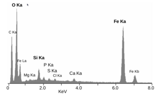
WEnergy-dispersive X-ray spectroscopy, sometimes called energy dispersive X-ray analysis (EDXA) or energy dispersive X-ray microanalysis (EDXMA), is an analytical technique used for the elemental analysis or chemical characterization of a sample. It relies on an interaction of some source of X-ray excitation and a sample. Its characterization capabilities are due in large part to the fundamental principle that each element has a unique atomic structure allowing a unique set of peaks on its electromagnetic emission spectrum. The peak positions are predicted by the Moseley's law with accuracy much better than experimental resolution of a typical EDX instrument.
 W
WMonochromatic wavelength dispersive x-ray fluorescence is an enhanced version of conventional wavelength-dispersive X-ray spectroscopy (WDXRF) elemental analysis. The key difference is that MWD XRF uses a doubly curved crystal X-ray optic between the X-ray source and the sample resulting in monochromatic excitation. This additional optic creates a high-intensity X-ray beam on a small spot size without increasing the power of the X-ray source. An MWD XRF instrument is constructed from a low-power X-ray tube, a point-to-point focusing optic for excitation, a sample cell, a focusing optic that collects the fluorescence from the sample, and an X-ray detector. By using an optic between the X-ray source and the sample, a monochromatic beam free of bremsstrahlung, excites the sample, eliciting the secondary fluorescence X-rays needed for elemental analysis. By restricting the band of wavelengths used for excitation, a much higher signal to background ratio is achieved. This type of excitation allows much lower limits of detection and faster reading times.
 W
WResonant inelastic X-ray scattering (RIXS) is an X-ray spectroscopy technique used to investigate the electronic structure of molecules and materials.
 W
WX-ray emission spectroscopy (XES) is a form of X-ray spectroscopy in which the X-ray line spectra are measured with a spectral resolution sufficient to analyze the impact of the chemical environment on the X-ray line energy and on branching ratios. This is done by exciting electrons out of their shell and then watching the emitted photons of the recombinating electrons.
 W
WX-ray fluorescence (XRF) is the emission of characteristic "secondary" X-rays from a material that has been excited by being bombarded with high-energy X-rays or gamma rays. The phenomenon is widely used for elemental analysis and chemical analysis, particularly in the investigation of metals, glass, ceramics and building materials, and for research in geochemistry, forensic science, archaeology and art objects such as paintings
 W
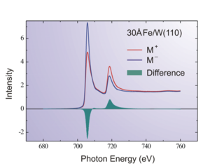
WX-ray magnetic circular dichroism (XMCD) is a difference spectrum of two X-ray absorption spectra (XAS) taken in a magnetic field, one taken with left circularly polarized light, and one with right circularly polarized light. By closely analyzing the difference in the XMCD spectrum, information can be obtained on the magnetic properties of the atom, such as its spin and orbital magnetic moment.