 W
WThe Abelin reaction is a qualitative reaction for demonstrating the presence of arsphenamine and neoarsphenamine in blood and urine.
 W
WThe Ashby technique is a method for determining the volume and life span of red blood cells in humans, first published by Dr. Winifred Ashby in 1919. The technique involves injection of compatible donor red blood cells of a different blood group into a recipient, followed by blood testing periodically afterwards. Differential agglutination of the red cells is then used to determine the number of remaining donor cells, allowing the survival rate to be determined. It does not involve radioisotope technology, and was the first technique to successfully establish the correct red blood cell life span. In particular, Type O blood is first transfused into Type A or B subjects. In subsequent blood samples, the patient's own A and B blood cells are removed by agglutination with either anti-A or anti-B serum. The number of remaining nonagglutinated Type O cells as a function of time defines the survival rate of blood cells. This technique was used extensively during World War II and shortly after but has more recently been replaced by techniques that label one's own blood, due of the dangers of using donor blood.
 W
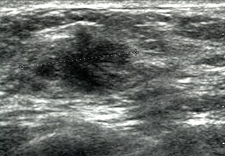
WBreast ultrasound is the use of medical ultrasonography to perform imaging of the breast.
 W
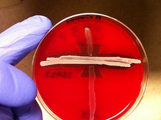
WThe CAMP test (Christie–Atkins–Munch-Peterson) is a test to identify group B β-hemolytic streptococci based on their formation of a substance that enlarges the area of hemolysis formed by the β-hemolysin elaborated from Staphylococcus aureus.
 W
WThe captopril suppression test (CST) is a non-invasive medical test that measures plasma levels of aldosterone. Aldosterone production is suppressed by captopril through the renin–angiotensin–aldosterone system. CST results are used to assist in the diagnososis of primary aldosteronism.
 W
WCuldocentesis is a medical procedure involving the extraction of fluid from the pouch of Douglas through a needle. It can be one diagnostic technique used in identifying pelvic inflammatory disease and ruptured ectopic pregnancies that cause hemoperitoneum.
 W
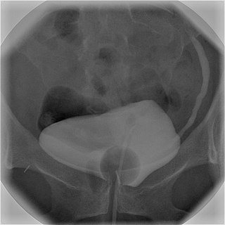
WIn radiology and urology, a cystography is a procedure used to visualise the urinary bladder.
WCystourethrography is a radiographic, fluoroscopic medical procedure that is used to visualize and evaluate the female urethra. Voiding and positive pressure cystourethrograms help to assess lower urinary tract trauma, reflux, suspected fistulas, and to diagnose urinary retention. Magnetic imaging (MRI) has been replacing this diagnostic tool due to its increased sensitivity. This imaging technique is used to diagnose hydronephrosis, voiding anomalies, and urinary tract infections in children. abnormalities.
 W
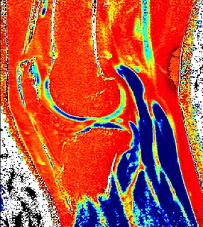
WDelayed gadolinium-enhanced magnetic resonance imaging of cartilage or dGEMRIC measures the fixed-charge density and relative proteoglycan content of articular cartilage using the spin-lattice relaxation time or T1 relaxation time. Current research is investigating the clinical application of dGEMRIC as a quantitative tool for monitoring cartilage function in diseased or repair cartilage.
 W
WDiagnostic medical sonography (DMS), a branch of diagnostic medical imaging, is the use of imaging by medical ultrasound for medical diagnosis. DMS uses non-ionizing ultrasound to produce 2D and 3D images of the body. In Canada, the credentialing for diagnostic medical sonography is the Canadian Association of Registered Ultrasound Professionals. In the United States, the credentialing body is the American Registry for Diagnostic Medical Sonography.
 W
WThe frailty index (FI) is used to measure the health status of older individuals; it serves as a proxy measure of aging and vulnerability to poor outcomes.
 W
WFroin's syndrome – coexistence of xanthochromia, high protein level and marked coagulation of cerebrospinal fluid (CSF). It is caused by meningeal irritation and CSF flow blockage by tumour mass or abscess. Stagnation of the CSF within the thecal sac facilitates exudation from the tumour itself and activation of coagulation factors. A clinical test formerly used for evaluation of spinal stenosis is Queckenstedt's maneuver. Nowadays, a magnetic resonance imaging is used for identification of CSF flow obstruction. It often shows the prolongation of T1 and T2 signal in CSF caudal to a level of block. This phenomenon is named after Georges Froin (1874–1932), a French physician who first described it.
 W
WGandy–Gamna nodules or Gandy-Gamna bodies, sometimes known as Gamna-Gandy bodies or Gamna-Gandy nodules, are small yellow-brown, brown, or rust-colored foci found in the spleen in patients with splenomegaly due to portal hypertension, as well as sickle cell disease. They consist of fibrous tissue with haemosiderin and calcium deposits, and probably form due to scarring at sites of small perivascular haemorrhages. They are visible on MRI scanning due to the presence of haemosiderin.
 W
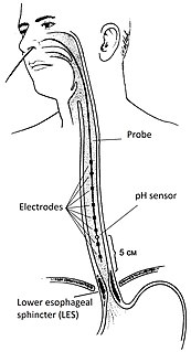
WImpedance–pH monitoring is a technique used in the diagnosis of gastroesophageal reflux disease (GERD), by monitoring both impedance and pH.
 W
WA Lewis Lead is a modified ECG lead used to detect atrial flutter waves when atrial flutter is suspected clinically, based on signs and symptoms, but is not definitely demonstrated on the standard 12 lead ECG. In order to create the Lewis Lead, the right arm electrode is moved to the manubrium adjacent to the sternum. Then the left arm electrode is moved to the right, fifth intercostal space adjacent to the sternum. The left leg electrode is placed on the right lower costal margin. The Lewis Lead is then read as Lead I on the ECG and, since in most patients it will be roughly perpendicular to the wave of ventricular depolarization, atrial flutter waves may be more apparent.
 W
WThe liver span is a measurement performed during physical examination to determine the size of the liver and identify possible hepatomegaly.
 W
WLumbar provocative discography is an invasive diagnostic procedure for evaluation for intervertebral disc pathology. It is usually reserved for persons with persistent, severe low back pain (LBP) who have abnormal spaces between vertebrae on magnetic resonance imaging (MRI), where other diagnostic tests have failed to reveal clear confirmation of a suspected disc as the source of pain, and surgical intervention is being considered.
 W
WThe patellar tap is a technique used in an examination of the knee to test for knee effusion or "water-on-the-knee".
 W
WPercutaneous transhepatic cholangiography, percutaneous hepatic cholangiogram, or percutaneous transhepatic cholangiography and drainage (PTCD) is a radiological technique used to visualize the anatomy of the biliary tract. A contrast medium is injected into a bile duct in the liver, after which X-rays are taken. It allows access to the biliary tree in cases where endoscopic retrograde cholangiopancreatography (ERCP) has been unsuccessful. Initially reported in 1937, the procedure became popular in 1952.
 W
WPrimodos was a hormone-based pregnancy test used in the 1960s and 1970s that consisted of two pills that contained norethisterone and ethinylestradiol. It detected pregnancy by inducing menstruation in women who were not pregnant. The presence or absence of menstrual bleeding was then used to determine whether the user was pregnant. In South Korea it was also used, "perhaps as a double dose" to induce abortions.
 W
WA rapid diagnostic test (RDT) is a medical diagnostic test that is quick and easy to perform. RDTs are suitable for preliminary or emergency medical screening and for use in medical facilities with limited resources. They also allow point-of-care testing in primary care for things that formerly only a laboratory test could measure. They provide same-day results within two hours, typically in approximately 20 minutes.
 W
WScintimammography is a type of breast imaging test that is used to detect cancer cells in the breasts of some women who have had abnormal mammograms, or for those who have dense breast tissue, post-operative scar tissue or breast implants.
 W
WA siderophage is a hemosiderin-containing macrophage. Heart failure cells are siderophages generated in the alveoli of the lungs of people with left heart failure or chronic pulmonary edema, when the high pulmonary blood pressure causes red blood cells to pass through the vascular wall. Siderophages are not specific of heart failure. They are present wherever red blood cells encounter macrophages, such as pulmonary hemorrhage.
 W
WSkull bossing is a descriptive term in medical physical examination indicating a protuberance of the skull, most often in the frontal bones of the forehead. Although prominence of the skull bones may be normal, skull bossing may be associated with certain medical conditions, including nutritional, metabolic, hormonal, and hematologic disorders.
 W
WSloan letters, designed by Louise Sloan in 1959, are a set of optotypes used to test visual acuity generally used in Snellen charts and logMAR charts.
 W
WTechnetium (99mTc) tilmanocept, trade name Lymphoseek, is a radiopharmaceutical diagnostic imaging agent used to locate lymph nodes which may be draining from tumors, and assist doctors in locating lymph nodes for removal during surgery.
 W
WTransrectal ultrasonography, or TRUS in short, is a method of creating an image of organs in the pelvis, most commonly used to perform an ultrasound-guided needle biopsy evaluation of the prostate gland in men with elevated prostate-specific antigen or prostatic nodules on digital rectal exam. TRUS--guided biopsy may reveal prostate cancer, benign prostatic hypertrophy, or prostatitis. TRUS may also detect other diseases of the lower rectum and can be used to stage primary rectal cancer.
 W
WUrine organic acids is a medical diagnostic test that measures organic acid metabolites in the urine. The metabolites can come from host cells or from flora. The test can be used to exclude the possibility that a person has an inborn error of metabolism, usually one of the organic acidemias. It is also used to look for problems with nutrition or evidence of certain infections or bacterial overgrowth. The usual method of analysis is tandem mass spectrometry.
 W
WVenography is a procedure in which an x-ray of the veins, a venogram, is taken after a special dye is injected into the bone marrow or veins. The dye has to be injected constantly via a catheter, making it an invasive procedure. Normally the catheter is inserted by the groin and moved to the appropriate site by navigating through the vascular system.
 W
WThe Wong–Baker Faces Pain Rating Scale is a pain scale that was developed by Donna Wong and Connie Baker. The scale shows a series of faces ranging from a happy face at 0, or "no hurt", to a crying face at 10, which represents "hurts like the worst pain imaginable". Based on the faces and written descriptions, the patient chooses the face that best describes their level of pain.