 W
WAn X-ray, or X-radiation, is a penetrating form of high-energy electromagnetic radiation. Most X-rays have a wavelength ranging from 10 picometers to 10 nanometers, corresponding to frequencies in the range 30 petahertz to 30 exahertz (30×1015Hz to 30×1018 Hz) and energies in the range 124 eV to 124 keV. X-ray wavelengths are shorter than those of UV rays and typically longer than those of gamma rays. In many languages, X-radiation is referred to as Röntgen radiation, after the German scientist Wilhelm Conrad Röntgen, who discovered it on November 8, 1895. He named it X-radiation to signify an unknown type of radiation. Spellings of X-ray(s) in English include the variants x-ray(s), xray(s), and X ray(s).
 W
WX-ray absorption spectroscopy (XAS) is a widely used technique for determining the local geometric and/or electronic structure of matter. The experiment is usually performed at synchrotron radiation facilities, which provide intense and tunable X-ray beams. Samples can be in the gas phase, solutions, or solids.
 W
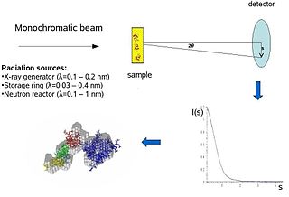
WBiological small-angle scattering is a small-angle scattering method for structure analysis of biological materials. Small-angle scattering is used to study the structure of a variety of objects such as solutions of biological macromolecules, nanocomposites, alloys, and synthetic polymers. Small-angle X-ray scattering (SAXS) and small-angle neutron scattering (SANS) are the two complementary techniques known jointly as small-angle scattering (SAS). SAS is an analogous method to X-ray and neutron diffraction, wide angle X-ray scattering, as well as to static light scattering. In contrast to other X-ray and neutron scattering methods, SAS yields information on the sizes and shapes of both crystalline and non-crystalline particles. When used to study biological materials, which are very often in aqueous solution, the scattering pattern is orientation averaged.
 W
WCargo scanning or non-intrusive inspection (NII) refers to non-destructive methods of inspecting and identifying goods in transportation systems. It is often used for scanning of intermodal freight shipping containers. In the US it is spearheaded by the Department of Homeland Security and its Container Security Initiative (CSI) trying to achieve one hundred percent cargo scanning by 2012 as required by the US Congress and recommended by the 9/11 Commission. In the US the main purpose of scanning is to detect special nuclear materials (SNMs), with the added bonus of detecting other types of suspicious cargo. In other countries the emphasis is on manifest verification, tariff collection and the identification of contraband. In February 2009, approximately 80% of US incoming containers were scanned. To bring that number to 100% researchers are evaluating numerous technologies, described in the following sections.
 W
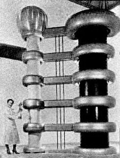
WThe Cockcroft–Walton (CW) generator, or multiplier, is an electric circuit that generates a high DC voltage from a low-voltage AC or pulsing DC input. It was named after the British and Irish physicists John Douglas Cockcroft and Ernest Thomas Sinton Walton, who in 1932 used this circuit design to power their particle accelerator, performing the first artificial nuclear disintegration in history. They used this voltage multiplier cascade for most of their research, which in 1951 won them the Nobel Prize in Physics for "Transmutation of atomic nuclei by artificially accelerated atomic particles". The circuit was discovered in 1919, by Heinrich Greinacher, a Swiss physicist. For this reason, this doubler cascade is sometimes also referred to as the Greinacher multiplier. Cockcroft–Walton circuits are still used in particle accelerators. They also are used in everyday electronic devices that require high voltages, such as X-ray machines, cathode ray tube television sets, microwave ovens and photocopiers.
 W
WIn X-ray crystallography, crystallographic disorder describes the cocrystallization of more than one rotamer, conformer, or isomer where the center of mass of each form is identical or unresolvable. As a consequence of disorder, the crystallographic solution is the sum of the various forms. In many cases, the components of the disorder are equally abundant, and, in other cases, the weighting coefficients for each component differ. Disorder can entail a pair or several components. Disorder usually arises when the forms are nearly equal in energy and the crystal lattice is sufficiently spacious to accommodate the various components.
 W
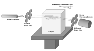
WEnergy-dispersive X-ray diffraction (EDXRD) is an analytical technique for characterizing materials. It differs from conventional X-ray diffraction by using polychromatic photons as the source and is usually operated at a fixed angle. With no need for a goniometer, EDXRD is able to collect full diffraction patterns very quickly. EDXRD is almost exclusively used with synchrotron radiation which allows for measurement within real engineering materials.
 W
WX-ray crystallography (XRC) is the experimental science determining the atomic and molecular structure of a crystal, in which the crystalline structure causes a beam of incident X-rays to diffract into many specific directions. By measuring the angles and intensities of these diffracted beams, a crystallographer can produce a three-dimensional picture of the density of electrons within the crystal. From this electron density, the mean positions of the atoms in the crystal can be determined, as well as their chemical bonds, their crystallographic disorder, and various other information.
 W
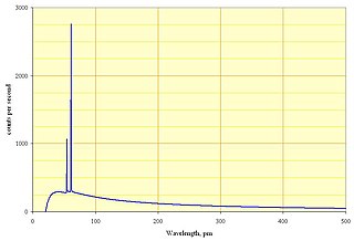
WThe Duane–Hunt law, named after the American physicists William Duane and Franklin L. Hunt, gives the maximum frequency of X-rays that can be emitted by Bremsstrahlung in an X-ray tube by accelerating electrons through an excitation voltage V into a metal target.
 W
WAn electron microprobe (EMP), also known as an electron probe microanalyzer (EPMA) or electron micro probe analyzer (EMPA), is an analytical tool used to non-destructively determine the chemical composition of small volumes of solid materials. It works similarly to a scanning electron microscope: the sample is bombarded with an electron beam, emitting x-rays at wavelengths characteristic to the elements being analyzed. This enables the abundances of elements present within small sample volumes to be determined, when a conventional accelerating voltage of 15-20 kV is used. The concentrations of elements from lithium to plutonium may be measured at levels as low as 100 parts per million (ppm), material dependent, although with care, levels below 10 ppm are possible The ability to quantify lithium by EPMA became a reality in 2008.
 W
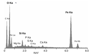
WEnergy-dispersive X-ray spectroscopy, sometimes called energy dispersive X-ray analysis (EDXA) or energy dispersive X-ray microanalysis (EDXMA), is an analytical technique used for the elemental analysis or chemical characterization of a sample. It relies on an interaction of some source of X-ray excitation and a sample. Its characterization capabilities are due in large part to the fundamental principle that each element has a unique atomic structure allowing a unique set of peaks on its electromagnetic emission spectrum. The peak positions are predicted by the Moseley's law with accuracy much better than experimental resolution of a typical EDX instrument.
 W
WX-ray fluorescence (XRF) is the emission of characteristic "secondary" X-rays from a material that has been excited by being bombarded with high-energy X-rays or gamma rays. The phenomenon is widely used for elemental analysis and chemical analysis, particularly in the investigation of metals, glass, ceramics and building materials, and for research in geochemistry, forensic science, archaeology and art objects such as paintings
 W
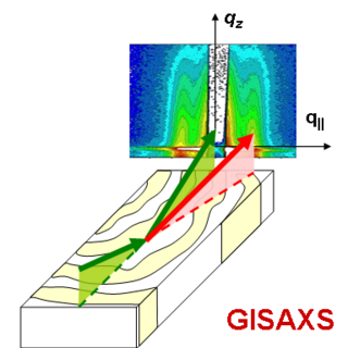
WGrazing-incidence small-angle scattering (GISAS) is a scattering technique used to study nanostructured surfaces and thin films. The scattered probe is either photons or neutrons. GISAS combines the accessible length scales of small-angle scattering and the surface sensitivity of grazing incidence diffraction (GID).
 W
WIn crystallography, a Greninger chart is a chart that allows angular relations between zones and planes in a crystal to be directly read from an x-ray diffraction photograph.
 W
WIn X-ray tubes, the heel effect, or, more precisely, the anode heel effect is a variation of the intensity of X-rays emitted by the anode depending on the direction of emission along the anode-cathode axis. Due to the geometry of the anode, X-rays emitted towards the cathode are in general more intense than those emitted perpendicular to the cathode–anode axis. The effect stems from the absorption of X-ray photons before they leave the anode in which they are produced. The probability of absorption depends on the distance the photons travel within the anode material, which in turn depends on the direction of emission.
 W
W W
WA linear particle accelerator is a type of particle accelerator that accelerates charged subatomic particles or ions to a high speed by subjecting them to a series of oscillating electric potentials along a linear beamline. The principles for such machines were proposed by Gustav Ising in 1924, while the first machine that worked was constructed by Rolf Widerøe in 1928 at the RWTH Aachen University. Linacs have many applications: they generate X-rays and high energy electrons for medicinal purposes in radiation therapy, serve as particle injectors for higher-energy accelerators, and are used directly to achieve the highest kinetic energy for light particles for particle physics.
 W
WMacintyre's X-Ray Film is an 1896 documentary radiography film directed by Scottish medical doctor John Macintyre.
 W
WMedipix is a family of photon counting and particle tracking pixel detectors developed by an international collaboration, hosted by CERN.
 W
WMegavoltage X-rays are produced by linear accelerators ("linacs") operating at voltages in excess of 1000 kV (1 MV) range, and therefore have an energy in the MeV range. The voltage in this case refers to the voltage used to accelerate electrons in the linear accelerator and indicates the maximum possible energy of the photons which are subsequently produced. They are used in medicine in external beam radiotherapy to treat neoplasms, cancer and tumors. Beams with the voltage range of 4-25 MV are used to treat deeply buried cancers because radiation oncologists find that they penetrate well to deep sites within the body. Lower energy x-rays, called orthovoltage X-rays, are used to treat cancers closer to the surface.
 W
WAn X-ray microscope uses electromagnetic radiation in the soft X-ray band to produce images of very small objects.
 W
WAn X-ray microscope uses electromagnetic radiation in the soft X-ray band to produce magnified images of objects. Since X-rays penetrate most objects, there is no need to specially prepare them for X-ray microscopy observations.
 W
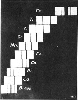
WMoseley's law is an empirical law concerning the characteristic x-rays emitted by atoms. The law had been discovered and published by the English physicist Henry Moseley in 1913-1914. Until Moseley's work, "atomic number" was merely an element's place in the periodic table and was not known to be associated with any measurable physical quantity. In brief, the law states that the square root of the frequency of the emitted x-ray is approximately proportional to the atomic number.
 W
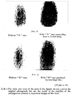
WN rays were a hypothesized form of radiation, described by French physicist Prosper-René Blondlot in 1903, and initially confirmed by others, but subsequently found to be illusory.
 W
WThe hard X-ray nanoprobe at the Center for Nanoscale Materials (CNM), Argonne National Lab advanced the state of the art by providing a hard X-ray microscopy beamline with the highest spatial resolution in the world. It provides for fluorescence, diffraction, and transmission imaging with hard X-rays at a spatial resolution of 30 nm or better. A dedicated source, beamline, and optics form the basis for these capabilities. This unique instrument is not only key to the specific research areas of the CNM; it will also be a general utility, available to the broader nanoscience community in studying nanomaterials and nanostructures, particularly for embedded structures.
 W
WOrthovoltage x-rays are produced by x-ray tubes operating at voltages in the 100–500 kV range, and therefore the x-rays have a peak energy in the 100–500 keV range. Orthovoltage X-rays are sometimes termed "deep" x-rays (DXR). They cover the upper limit of energies used for diagnostic radiography, and are used in external beam radiotherapy to treat cancer and tumors. They penetrate tissue to a useful depth of about 4–6 cm. This makes them useful for treating skin, superficial tissues, and ribs, but not for deeper structures such as lungs or pelvic organs.
 W
WRibs, also known as music on ribs, jazz on bones, bones or bone music (roentgenizdat) are improvised gramophone recordings made from X-ray films. Mostly made through the 1950s and 1960s, ribs were a black market method of smuggling in and distributing music by foreign and emigre musicians that was banned from broadcast in the Soviet Union, such as Pyotr Leshchenko or Alexander Vertinsky, or Western artists such as Elvis, the Beatles, the Rolling Stones, the Beach Boys, Ella Fitzgerald and Chubby Checker.
 W
WThe Röntgen Memorial Site in Würzburg, Germany is dedicated to the work of the German physicist Wilhelm Conrad Röntgen (1845–1923) and his discovery of X-rays, for which he was granted the Nobel Prize in physics. It contains an exhibition of historical instruments, machines and documents.
 W
WX-ray astronomy is an observational branch of astronomy which deals with the study of X-ray observation and detection from astronomical objects. X-radiation is absorbed by the Earth's atmosphere, so instruments to detect X-rays must be taken to high altitude by balloons, sounding rockets, and satellites. X-ray astronomy is the space science related to a type of space telescope that can see farther than standard light-absorption telescopes, such as the Mauna Kea Observatories, via x-ray radiation.
 W
WX‑ray birefringence imaging (XBI) can be considered the X‑ray analogue of the polarizing optical microscope. XBI uses linearly polarized X-rays with an energy tuned to an elemental absorption edge. The tuned X-rays interact solely with the absorbing element, thus allowing the local anisotropy of the bonding environment of the X‑ray absorbing element to be studied. Due to the requirement of linearly polarized tunable X-rays a synchrotron source is necessary. Interaction with the bonding environment of the selected element in the sample changes the incident X-ray polarization plane. A polarization analyzer is used to diffract the rotated component of the polarization plane to an area detector. The greater the vertical component of the polarization plane the greater the intensity observed on the detector. In this way, it is possible to study the distribution of bond environments containing the X-ray absorbing element in a spatially resolved manner.
 W
WX-ray detectors are devices used to measure the flux, spatial distribution, spectrum, and/or other properties of X-rays.
 W
WX-ray emission spectroscopy (XES) is a form of X-ray spectroscopy in which the X-ray line spectra are measured with a spectral resolution sufficient to analyze the impact of the chemical environment on the X-ray line energy and on branching ratios. This is done by exciting electrons out of their shell and then watching the emitted photons of the recombinating electrons.
 W
WThe X-Rays is an 1897 British short silent comedy film, directed by George Albert Smith, featuring a courting couple exposed to X-rays. The trick film, according to Michael Brooke of BFI Screenonline, "contains one of the first British examples of special effects created by means of jump cuts" Smith employs the jump-cut twice; first to transform his courting couple via "X rays," dramatized by means of the actors donning black bodysuits decorated with skeletons and with the woman holding only the metal support work of her umbrella, and then to return them and the umbrella to normal. The couple in question were played by Smith's wife Laura Bayley and Tom Green.
 W
W