 W
WIn genetics, a cytohet is a eukaryotic cell whose non-nuclear genome is heterozygous.
 W
WDiablo homolog (DIABLO) is a mitochondrial protein that in humans is encoded by the DIABLO gene on chromosome 12. DIABLO is also referred to as second mitochondria-derived activator of caspases or SMAC. This protein binds inhibitor of apoptosis proteins (IAPs), thus freeing caspases to activate apoptosis. Due to its proapoptotic function, SMAC is implicated in a broad spectrum of tumors, and small molecule SMAC mimetics have been developed to enhance current cancer treatments.
 W
WA kinetoplast is a network of circular DNA inside a large mitochondrion that contains many copies of the mitochondrial genome. The most common kinetoplast structure is a disk, but they have been observed in other arrangements. Kinetoplasts are only found in Excavata of the class Kinetoplastida. The variation in the structures of kinetoplasts may reflect phylogenic relationships between kinetoplastids. A kinetoplast is usually adjacent to the organism's flagellar basal body, suggesting that it is tightly bound to the cytoskeleton. In Trypanosoma brucei this cytoskeletal connection is called the tripartite attachment complex and includes the protein p166.
 W
WJose V. Lopez is an American-Filipino molecular biologist. He has been faculty and professor of biology at Nova Southeastern University (NSU). in Dania Beach FL since 2007.
 W
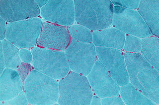
WMitochondrial diseases are a group of disorders caused by dysfunctional mitochondria, the organelles that generate energy for the cell. Mitochondria are found in every cell of the human body except red blood cells, and convert the energy of food molecules into the ATP that powers most cell functions.
 W
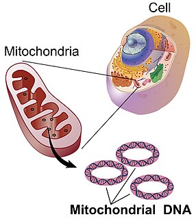
WMitochondrial DNA is the DNA located in mitochondria, cellular organelles within eukaryotic cells that convert chemical energy from food into a form that cells can use, adenosine triphosphate (ATP). Mitochondrial DNA is only a small portion of the DNA in a eukaryotic cell; most of the DNA can be found in the cell nucleus and, in plants and algae, also in plastids such as chloroplasts.
 W
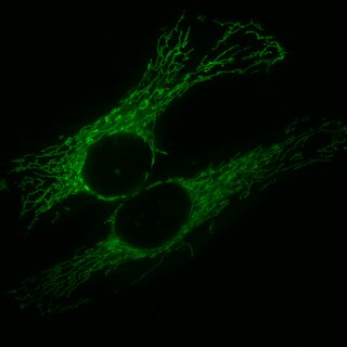
WAlthough mitochondria are commonly depicted as singular oval-shaped structures, it has been known for at least a century that they form a highly dynamic network within most cells where they constantly undergo fission and fusion. Mitochondria can divide by prokaryotic binary fission and since they require mitochondrial DNA for their function, fission is coordinated with DNA replication. Some of the proteins that are involved in mitochondrial fission have been identified and some of them are associated with mitochondrial diseases. Mitochondrial fission has significant implications in stress response and apoptosis.
WMitochondria are dynamic organelles with the ability to fuse and divide (fission), forming constantly changing tubular networks in most eukaryotic cells. These mitochondrial dynamics, first observed over a hundred years ago are important for the health of the cell, and defects in dynamics lead to genetic disorders. Through fusion, mitochondria can overcome the dangerous consequences of genetic malfunction. The process of mitochondrial fusion involves a variety of proteins that assist the cell throughout the series of events that form this process.
 W
WMitochondrial ribosome or mitoribosome is a protein complex that is active in mitochondria and functions as a riboprotein for translating mitochondrial mRNAs encoded in mtDNA. Mitoribosomes, like cytoplasmic ribosomes, consist of two subunits — large (mtLSU) and small (mt-SSU). However, the ratio of rRNA/protein is different from cytoplasmic ribosomes, mitoribosomes consist of several specific proteins and less rRNAs.
 W
WThe mtDNA control region is an area of the mitochondrial genome which is non-coding DNA. This region controls RNA and DNA synthesis. It is the most polymorphic region of the human mtDNA genome, with polymorphism concentrated in hypervariable regions. The average nucleotide diversity in these regions is 1.7%. Despite this variability, an RNA transcript from this region has a conserved secondary structure (pictured) which has been found to be under selective pressure.
 W
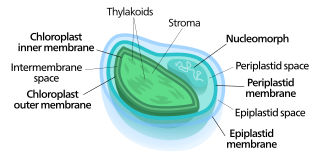
WNucleomorphs are small, vestigial eukaryotic nuclei found between the inner and outer pairs of membranes in certain plastids. They are thought to be vestiges of primitive red and green algal nuclei that were engulfed by a larger eukaryote. Because the nucleomorph lies between two sets of membranes, nucleomorphs support the endosymbiotic theory and are evidence that the plastids containing them are complex plastids. Having two sets of membranes indicate that the plastid, a prokaryote, was engulfed by a eukaryote, an alga, which was then engulfed by another eukaryote, the host cell, making the plastid an example of secondary endosymbiosis.