 W
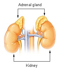
WThe adrenal glands are endocrine glands that produce a variety of hormones including adrenaline and the steroids aldosterone and cortisol. They are found above the kidneys. Each gland has an outer cortex which produces steroid hormones and an inner medulla. The adrenal cortex itself is divided into three zones: the zona glomerulosa, the zona fasciculata and the zona reticularis.
 W
WSituated along the perimeter of the adrenal gland, the adrenal cortex mediates the stress response through the production of mineralocorticoids and glucocorticoids, such as aldosterone and cortisol, respectively. It is also a secondary site of androgen synthesis.
 W
WThe adrenal medulla is part of the adrenal gland. It is located at the center of the gland, being surrounded by the adrenal cortex. It is the innermost part of the adrenal gland, consisting of cells that secrete epinephrine (adrenaline), norepinephrine (noradrenaline), and a small amount of dopamine in response to stimulation by sympathetic preganglionic neurons.
 W
WChromaffin cells, also pheochromocytes, are neuroendocrine cells found mostly in the medulla of the adrenal glands in mammals. These cells serve a variety of functions such as serving as a response to stress, monitoring carbon dioxide and oxygen concentrations in the body, maintenance of respiration and the regulation of blood pressure. They are in close proximity to pre-synaptic sympathetic ganglia of the sympathetic nervous system, with which they communicate, and structurally they are similar to post-synaptic sympathetic neurons. In order to activate chromaffin cells, the splanchnic nerve of the sympathetic nervous system releases acetylcholine, which then binds to nicotinic acetylcholine receptors on the adrenal medulla. This causes the release of catecholamines. The chromaffin cells release catecholamines: ~80% of adrenaline (epinephrine) and ~20% of noradrenaline (norepinephrine) into systemic circulation for systemic effects on multiple organs, and can also send paracrine signals. Hence they are called neuroendocrine cells.
 W
WThe inferior suprarenal arteries usually originate at the trunk of the renal artery before its terminal division, and usually present substantially different diameters corresponding to the age variable. The suprarenal arteries irrigate the suprarenal gland parenchyma, the ureter, and the surrounding cellular tissue and muscles.
 W
WThe middle suprarenal arteries are two small vessels which arise, one from either side of the abdominal aorta, opposite the superior mesenteric artery.
 W
WPrimary pigmented nodular adrenocortical disease (PPNAD) was first coined in 1984 by Carney et al. it often occurs in association with Carney complex (CNC). CNC is a rare syndrome that involves the formation of abnormal tumours that cause endocrine hyperactivity.
 W
WEach superior suprarenal artery is a branch of the inferior phrenic artery on that side of the body. The left and right phrenic arteries supply the diaphragm, and come off the aorta.
 W
WThe suprarenal plexus is formed by branches from the celiac plexus, from the celiac ganglion, and from the phrenic and greater splanchnic nerves, a ganglion being formed at the point of junction with the latter nerve.
 W
WThe Suprarenal veins are two in number:the right ends in the inferior vena cava. the left ends in the left renal or left inferior phrenic vein.
 W
WThe zona fasciculata constitutes the middle and also the widest zone of the adrenal cortex, sitting directly beneath the zona glomerulosa. Constituent cells are organized into bundles or "fascicles."
 W
WThe zona glomerulosa of the adrenal gland is the most superficial layer of the adrenal cortex, lying directly beneath the renal capsule. Its cells are ovoid and arranged in clusters or arches.
 W
WThe zona reticularis is the innermost layer of the adrenal cortex, lying deep to the zona fasciculata and superficial to the adrenal medulla. The cells are arranged cords that project in different directions giving a net-like appearance.