 W
WThe hypothalamus is a portion of the brain that contains a number of small nuclei with a variety of functions. One of the most important functions of the hypothalamus is to link the nervous system to the endocrine system via the pituitary gland. The hypothalamus is located below the thalamus and is part of the limbic system. In the terminology of neuroanatomy, it forms the ventral part of the diencephalon. All vertebrate brains contain a hypothalamus. In humans, it is the size of an almond.
 W
WThe anterior hypothalamic nucleus is a nucleus of the hypothalamus.
WThe arcuate nucleus of the hypothalamus is an aggregation of neurons in the mediobasal hypothalamus, adjacent to the third ventricle and the median eminence. The arcuate nucleus includes several important and diverse populations of neurons that help mediate different neuroendocrine and physiological functions, including neuroendocrine neurons, centrally projecting neurons, and astrocytes. The populations of neurons found in the arcuate nucleus are based on the hormones they secrete or interact with and are responsible for hypothalamic function, such as regulating hormones released from the pituitary gland or secreting their own hormones. Neurons in this region are also responsible for integrating information and providing inputs to other nuclei in the hypothalamus or inputs to areas outside this region of the brain. These neurons, generated from the ventral part of the periventricular epithelium during embryonic development, locate dorsally in the hypothalamus, becoming part of the ventromedial hypothalamic region. The function of the arcuate nucleus relies on its diversity of neurons, but its central role is involved in homeostasis. The arcuate nucleus provides many physiological roles involved in feeding, metabolism, fertility, and cardiovascular regulation.
WThe dorsomedial hypothalamic nucleus is a nucleus of the hypothalamus. It is involved in feeding, drinking, body-weight regulation and circadian activity. More specifically, it is a necessary component for the expression of numerous behavioral and physiological circadian rhythms. The dorsomedial hypothalamic nucleus receives information from neurons and humors involved in feeding regulation, body weight and energy consumption, and then passes this information on to brain regions involved in sleep and wakefulness regulation, body temperature and corticosteroid secretion.
WThe lateral hypothalamus (LH), also called the lateral hypothalamic area (LHA), contains the primary orexinergic nucleus within the hypothalamus that widely projects throughout the nervous system; this system of neurons mediates an array of cognitive and physical processes, such as promoting feeding behavior and arousal, reducing pain perception, and regulating body temperature, digestive functions, and blood pressure, among many others. Clinically significant disorders that involve dysfunctions of the orexinergic projection system include narcolepsy, motility disorders or functional gastrointestinal disorders involving visceral hypersensitivity, and eating disorders.
 W
WThe mammillary bodies are a pair of small round bodies, located on the undersurface of the brain that, as part of the diencephalon, form part of the limbic system. They are located at the ends of the anterior arches of the fornix. They consist of two groups of nuclei, the medial mammillary nuclei and the lateral mammillary nuclei.
WThe median eminence, part of the inferior boundary of the hypothalamus in the brain, is attached to the infundibulum. The median eminence is a small swelling on the tuber cinereum, posterior to and atop the pituitary stalk; it lies in the area roughly bounded on its posterolateral region by the cerebral peduncles, and on its anterolateral region by the optic chiasm.
WThe median preoptic nucleus is located dorsal to the other three nuclei of the preoptic area of the anterior hypothalamus. The hypothalamus is located just beneath the thalamus, the main sensory relay station of the nervous system, and is considered part of the limbic system, which also includes structures such as the hippocampus and the amygdala. The hypothalamus is highly involved in maintaining homeostasis of the body, and the median preoptic nucleus is no exception, contributing to regulation of blood composition, body temperature, and non-REM sleep.
 W
WThe paraventricular nucleus is a nucleus in the hypothalamus. Anatomically, it is adjacent to the third ventricle and many of its neurons project to the posterior pituitary. These projecting neurons secrete oxytocin and a smaller amount of vasopressin, otherwise the nucleus also secretes corticotropin-releasing hormone (CRH) and thyrotropin-releasing hormone (TRH). CRH and TRH are secreted into the hypophyseal portal system and act on different targets neurons in the anterior pituitary. PVN is thought to mediate many diverse functions through these different hormones, including osmoregulation, appetite, and the response of the body to stress.
 W
WThe pituitary stalk is the connection between the hypothalamus and the posterior pituitary. The floor of the third ventricle is prolonged downward as a funnel-shaped recess—the infundibular recess—into the infundibulum, where the apex of the pituitary is attached. It passes through the dura mater of the diaphragma sellae as it carries axons from the magnocellular neurosecretory cells of the hypothalamus down to the posterior pituitary where they release their neurohypophysial hormones, oxytocin and vasopressin, into the blood.
WThe posterior nucleus of the hypothalamus is one of the many nuclei that make up the hypothalamic region of the brain.
WThe preoptic area is a region of the hypothalamus. MeSH classifies it as part of the anterior hypothalamus. TA lists four nuclei in this region,.
 W
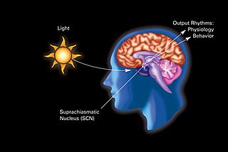
WThe retinohypothalamic tract (RHT) is a photic neural input pathway involved in the circadian rhythms of mammals. The origin of the retinohypothalamic tract is the intrinsically photosensitive retinal ganglion cells (ipRGC), which contain the photopigment melanopsin. The axons of the ipRGCs belonging to the retinohypothalamic tract project directly, monosynaptically, to the suprachiasmatic nuclei (SCN) via the optic nerve and the optic chiasm. The suprachiasmatic nuclei receive and interpret information on environmental light, dark and day length, important in the entrainment of the "body clock". They can coordinate peripheral "clocks" and direct the pineal gland to secrete the hormone melatonin.
WThe suprachiasmatic nucleus or nuclei (SCN) is a tiny region of the brain in the hypothalamus, situated directly above the optic chiasm. It is responsible for controlling circadian rhythms. The neuronal and hormonal activities it generates regulate many different body functions in a 24-hour cycle. The mouse SCN contains approximately 20,000 neurons.
 W
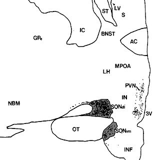
WThe supraoptic nucleus (SON) is a nucleus of magnocellular neurosecretory cells in the hypothalamus of the mammalian brain. The nucleus is situated at the base of the brain, adjacent to the optic chiasm. In humans, the SON contains about 3,000 neurons.
 W
WThe tuber cinereum is a hollow eminence of the middle–ventral hypothalamus, specifically the arcuate nucleus, situated between the mammillary bodies and the optic chiasm. In addition to the ventral hypothalamus, the tuber cinereum includes the median eminence and pituitary gland. Together with the hollow itself, it is sometimes referred to as the pituitary stalk.
 W
WThe tuberoinfundibular pathway refers to a population of dopamine neurons that project from the arcuate nucleus in the tuberal region of the hypothalamus to the median eminence. It is one of the four major dopamine pathways in the brain. Dopamine released at this site inhibits the secretion of prolactin from anterior pituitary gland lactotrophs by binding to D2 receptors.
 W
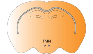
WThe tuberomammillary nucleus (TMN) is a histaminergic nucleus located within the posterior third of the hypothalamus. It consists of, largely, histaminergic neurons and is involved with the control of arousal, learning, memory, sleep and energy balance.
 W
WThe ventrolateral preoptic nucleus (VLPO), also known as the intermediate nucleus of the preoptic area (IPA), is a small cluster of neurons situated in the anterior hypothalamus, sitting just above and to the side of the optic chiasm in the brain of humans and other animals. The brain's sleep-promoting nuclei, together with the ascending arousal system which includes components in the brainstem, hypothalamus and basal forebrain, are the interconnected neural systems which control states of arousal, sleep, and transitions between these two states. The VLPO is active during sleep, particularly during non-rapid eye movement sleep, and releases inhibitory neurotransmitters, mainly GABA and galanin, which inhibit neurons of the ascending arousal system that are involved in wakefulness and arousal. The VLPO is in turn innervated by neurons from several components of the ascending arousal system. The VLPO is activated by the endogenous sleep-promoting substances adenosine and prostaglandin D2. The VLPO is inhibited during wakefulness by the arousal-inducing neurotransmitters norepinephrine and acetylcholine. The role of the VLPO in sleep and wakefulness, and its association with sleep disorders – particularly insomnia and narcolepsy – is a growing area of neuroscience research.
WThe ventromedial nucleus of the hypothalamus is a nucleus of the hypothalamus. "The ventromedial hypothalamus (VMH) is a distinct morphological nucleus involved in terminating hunger, fear, thermoregulation, and sexual activity." This nuclear region is involved with the recognition of the feeling of fullness.