 W
WThe thalamus is a large mass of gray matter located in the dorsal part of the diencephalon. Nerve fibers project out of the thalamus to the cerebral cortex in all directions, allowing hub-like exchanges of information. It has several functions, such as relaying of sensory signals, including motor signals to the cerebral cortex and the regulation of consciousness, sleep, and alertness.
 W
WThe interthalamic adhesion is a flattened band of tissue that connects both parts of the thalamus at their medial surfaces. The medial surfaces form the upper part of the lateral wall to the third ventricle.
 W
WA koniocellular cell is a neuron with a small cell body that is located in the koniocellular layer of the lateral geniculate nucleus (LGN) in primates, including humans.
 W
WLamina affixa is a layer of epithelium growing on the surface of the thalamus and forming the floor of the central part of lateral ventricle, on whose medial margin is attached the choroid plexus of the lateral ventricle; it covers the superior thalamostriate vein and the superior choroid vein. The torn edge of this plexus is called the tela choroidea.
 W
WThe lenticular fasciculus is a tract connecting the globus pallidus (internus) to the thalamus and is a part of the thalamic fasciculus. It is synonymous with field H2 of Forel. The thalamic fasciculus (composed of both the lenticular fasciculus and ansa lenticularis) runs to the thalamus. Basically, it is part of a pathway that connects the globus pallidus and the thalamus.
 W
WThe medial lemniscus, also known as Reil's band or Reil's ribbon, is a large ascending bundle of heavily myelinated axons that decussate in the brainstem, specifically in the medulla oblongata. The medial lemniscus is formed by the crossings of the internal arcuate fibers. The internal arcuate fibers are composed of axons of nucleus gracilis and nucleus cuneatus. The axons of the nucleus gracilis and nucleus cuneatus in the medial lemniscus have cell bodies that lie contralaterally.
 W
WMedullary laminae of thalamus are layers of myelinated fibres that appear on cross sections of the thalamus. They also are commonly referred to as laminae medullares thalami or medullary layers of thalamus. The specific layers are:Lamina medullaris lateralis - separates ventral and lateral thalamus from the subthalamus and thalamic reticular nucleus Lamina medullaris medialis - positioned between the dorsomedial and ventral nuclei of thalamus, encloses the intralaminar nuclei
WThe thalamus is a large mass of gray matter located in the dorsal part of the diencephalon. Nerve fibers project out of the thalamus to the cerebral cortex in all directions, allowing hub-like exchanges of information. It has several functions, such as relaying of sensory signals, including motor signals to the cerebral cortex and the regulation of consciousness, sleep, and alertness.
 W
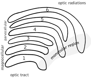
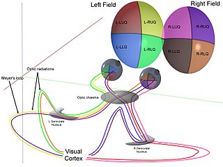
WThe optic radiation are axons from the neurons in the lateral geniculate nucleus to the primary visual cortex. The optic radiation receives blood through deep branches of the middle cerebral artery and posterior cerebral artery.
 W
WThe Papez circuit, or medial limbic circuit, is a neural circuit for the control of emotional expression. In 1937, James Papez proposed that the circuit connecting the hypothalamus to the limbic lobe was the basis for emotional experiences. Paul D. MacLean reconceptualized Papez's proposal and coined the term limbic system. MacLean redefined the circuit as the "visceral brain" which consisted of the limbic lobe and its major connections in the forebrain – hypothalamus, amygdala, and septum. Over time, the concept of a forebrain circuit for the control of emotional expression has been modified to include the prefrontal cortex.
 W
WThe dorsal column–medial lemniscus pathway (DCML) is a sensory pathway of the central nervous system that conveys sensations of fine touch, vibration, two-point discrimination, and proprioception (position) from the skin and joints. It transmits information from the body to the primary somatosensory cortex in the postcentral gyrus of the parietal lobe of the brain. The pathway receives information from sensory receptors throughout the body, and carries this in nerve tracts in the white matter of the dorsal columns of the spinal cord to the medulla, where it is continued in the medial lemniscus, on to the thalamus and relayed from there through the internal capsule and transmitted to the somatosensory cortex. The name dorsal-column medial lemniscus comes from the two structures that carry the sensory information: the dorsal columns of the spinal cord, and the medial lemniscus in the brainstem.
 W
WThe stria medullaris is a part of the epithalamus. It is a fiber bundle containing afferent fibers from the septal nuclei, lateral preoptico-hypothalamic region, and anterior thalamic nuclei to the habenula. It forms a horizontal ridge on the medial surface of the thalamus, and is found on the border between dorsal and medial surfaces of thalamus. Superior and lateral to habenular trigone.
 W
WThe superior thalamostriate vein or terminal vein commences in the groove between the corpus striatum and thalamus, receives numerous veins from both of these parts, and unites behind the crus of the fornix with the superior choroid vein to form each of the internal cerebral veins.
 W
WIn the front, superior surface of the thalamus but separate from the inner, medial surface by a salient margin is the taenia thalami. The bottom epithelial lining of the third ventricle is in between the tela choroidea and the taenia thalami.
 W
WThalamocortical radiations are the fibers between the thalamus and the cerebral cortex.
 W
WThe ventral anterior nucleus (VA) is a nucleus of the thalamus. It acts with the anterior part of the ventral lateral nucleus to modify signals from the basal ganglia.
WThe ventral lateral nucleus (VL) is a nucleus in the ventral nuclear group of the thalamus.
WThe ventral nuclear group is a collection of nuclei on the ventral side of the thalamus. According to MeSH, it consists of the following:ventral anterior nucleus ventral lateral nucleus ventral posterior nucleus – this is made up of two nuclei: the ventral posterolateral nucleus and the ventral posteromedial nucleus
WThe ventral posterior nucleus is the somato-sensory relay nucleus in thalamus of the brain.
WThe ventral posterolateral nucleus (VPL) is a nucleus of the thalamus. Together with the ventral posteromedial nucleus (VPM), ventral posterior inferior nucleus (VPI) and ventromedial posterior nucleus (VMpo), it constitutes the ventral posterior nucleus. There is uncertainty in the location of VMpo, as determined by spinothalamic tract (STT) terminations and staining for calcium-binding proteins, and several authorities do not consider its existence as being proved.
WThe ventral posteromedial nucleus (VPM) is a nucleus of the thalamus.