 W
WThe large intestine, also known as the large bowel, is the last part of the gastrointestinal tract and of the digestive system in vertebrates. Water is absorbed here and the remaining waste material is stored as feces before being removed by defecation.
 W
WIn the anatomy of humans and homologous primates, the ascending colon is the part of the colon located between the cecum and the transverse colon.
 W
WColectomy is bowel resection of the large bowel (colon). It consists of the surgical removal of any extent of the colon, usually segmental resection. In extreme cases where the entire large intestine is removed, it is called total colectomy, and proctocolectomy denotes that the rectum is included.
 W
WThere are two colic flexures, or curvatures in the transverse colon. The one on the right, the right colic flexure is known as the hepatic flexure.
 W
WColon cleansing, also known as colon therapy, or colon hydrotherapy, or a colonic, or colonic irrigation encompasses a number of alternative medical therapies claimed to remove unspecified toxins from the colon and intestinal tract by removing supposed accumulations of feces. Colon cleansing in this context should not be confused with an enema which introduces fluid into the colon, often under mainstream medical supervision, for a limited number of purposes including severe constipation and medical imaging.
 W
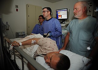
WColonoscopy or coloscopy is the endoscopic examination of the large bowel and the distal part of the small bowel with a CCD camera or a fiber optic camera on a flexible tube passed through the anus. It can provide a visual diagnosis and grants the opportunity for biopsy or removal of suspected colorectal cancer lesions.
 W
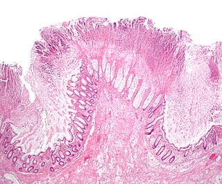
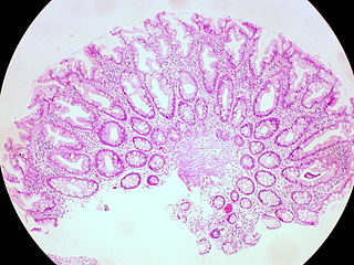
WThe colorectal adenoma is a benign glandular tumor of the colon and the rectum. It is a precursor lesion of the colorectal adenocarcinoma. They often manifest as colorectal polyps.
 W
WA colorectal polyp is a polyp occurring on the lining of the colon or rectum. Untreated colorectal polyps can develop into colorectal cancer.
 W
WIn the anatomy of humans and homologous primates, the descending colon is the part of the large intestine from the splenic flexure to the beginning of the sigmoid colon. The function of the descending colon in the digestive system is to store the remains of digested food that will be emptied into the rectum.
 W
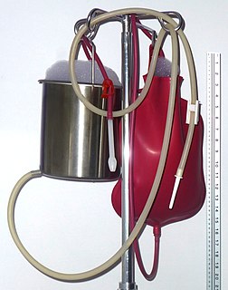
WAn enema, also known as a clyster, is an injection of fluid into the lower bowel by way of the rectum. Also, the word enema can refer to the liquid so injected, as well as to a device for administering such an injection.
 W
WThe haustra of the colon are the small pouches caused by sacculation, which give the colon its segmented appearance. The teniae coli run the length of the colon. Because the taenia coli are shorter than the colon, the colon becomes sacculated between the teniae coli, forming the haustra.
 W
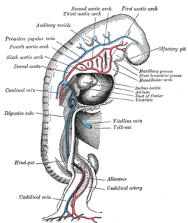
WThe hindgut is the posterior (caudal) part of the alimentary canal. In mammals, it includes the distal third of the transverse colon and the splenic flexure, the descending colon, sigmoid colon and rectum. In zoology, the term hindgut refers also to the cecum and ascending colon.
 W
WA hyperplastic polyp is a type of colorectal polyp.
 W
WThe iliac colon is situated in the left iliac fossa, and is about 12 to 15 cm. long.
 W
WIn human anatomy, the marginal artery of the colon, also known as the marginal artery of Drummond and artery of Drummond is an artery that connects the inferior mesenteric artery with the superior mesenteric artery. It is sometimes absent, as an anatomical variant.
 W
WMegacolon is an abnormal dilation of the colon. The dilation is often accompanied by a paralysis of the peristaltic movements of the bowel. In more extreme cases, the feces consolidate into hard masses inside the colon, called fecalomas, which can require surgery to be removed.
 W
WThe middle colic artery is a branch of the superior mesenteric artery that mostly supplies the transverse colon. It arises just below the pancreas. It passes inferiorly and anteriorly between the layers of the transverse mesocolon, and divides into left and right branches. The right branch anastomoses with the right colic artery, and the left anastomoses with the left colic artery, a branch of the inferior mesenteric artery. This sequence of anastomses are frequently referred to as the marginal artery of the colon.
 W
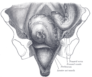
WThe sigmoid colon is the part of the large intestine that is closest to the rectum and anus. It forms a loop that averages about 35–40 cm (13.78-15.75 in) in length. The loop is typically shaped like a Greek letter sigma (ς) or Latin letter S. This part of the colon normally lies within the pelvis, but on account of its freedom of movement it is liable to be displaced into the abdominal cavity.
WThe taeniae coli are three separate longitudinal ribbons of smooth muscle on the outside of the ascending, transverse, descending and sigmoid colons. They are visible and can be seen just below the serosa or fibrosa. There are three teniae coli: mesocolic, free and omental taeniae coli. The teniae coli contract lengthwise to produce the haustra, the bulges in the colon.
WToxic megacolon is an acute form of colonic distension. It is characterized by a very dilated colon (megacolon), accompanied by abdominal distension (bloating), and sometimes fever, abdominal pain, or shock.
 W
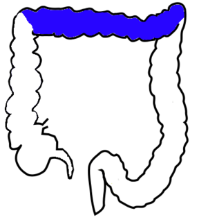
WThe transverse colon is the longest and most movable part of the colon. It crosses the abdomen from the ascending colon at the hepatic or right colic flexure with a downward convexity to the descending colon where it curves sharply on itself beneath the lower end of the spleen forming the splenic or left colic flexure. In its course, it describes an arch, the concavity of which is directed backward and a little upward. Toward its splenic end there is often an abrupt U-shaped curve which may descend lower than the main curve.
 W
WVirtual colonoscopy is the use of CT scanning or magnetic resonance imaging (MRI) to produce two- and three-dimensional images of the colon, from the lowest part, the rectum, to the lower end of the small intestine, and to display the images on an electronic display device. The procedure is used to screen for colon cancer and polyps, and may detect diverticulosis. A virtual colonoscopy can provide 3D reconstructed endoluminal views of the bowel. VC provides a secondary benefit of revealing diseases or abnormalities outside the colon.