 W
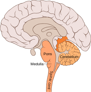
WThe pons is part of the brainstem, and in humans and other bipeds lies inferior to the midbrain, superior to the medulla oblongata and anterior to the cerebellum.
 W
WThe basilar part of pons, also known as basis pontis, is the ventral part of the pons; the dorsal part is known as the pontine tegmentum.
 W
WThe basilar sulcus is a groove in the pons, part of the brainstem.
 W
WThe cochlear nuclear (CN) complex comprises two cranial nerve nuclei in the human brainstem, the ventral cochlear nucleus (VCN) and the dorsal cochlear nucleus (DCN). The ventral cochlear nucleus is unlayered whereas the dorsal cochlear nucleus is layered. Auditory nerve fibers, fibers that travel through the auditory nerve carry information from the inner ear, the cochlea, on the same side of the head, to the nerve root in the ventral cochlear nucleus. At the nerve root the fibers branch to innervate the ventral cochlear nucleus and the deep layer of the dorsal cochlear nucleus. All acoustic information thus enters the brain through the cochlear nuclei, where the processing of acoustic information begins. The outputs from the cochlear nuclei are received in higher regions of the auditory brainstem.
 W
WThe facial colliculus is an elevated area located on the pontine tegmentum in the floor of the fourth ventricle. It is formed by fibers from the facial motor nucleus of the facial nerve as they loop over the abducens nucleus. Thus a lesion to the facial colliculus would result in ipsilateral facial paralysis and ipsilateral unopposed eye medial deviation.
 W
WThe frontopontine fibers are situated in the medial fifth of the base of the cerebral peduncles; they arise from the cells of the frontal lobe and then pass through the anterior limb of internal capsule at last end in the nuclei of the pons.
 W
WThe Keep of Pons is an 830-year-old fortified tower located in Pons, France and is one of the few remnants of the original castle of Pons. The keep is located near the chapel and porch of Saint Gilles and remains of the ramparts. On a hill and visible from a distance, this 33-meter-high (108 ft) edifice is used as the symbol of the city.
WThe locus coeruleus (LC) (\-si-ˈrü-lē-əs\), also spelled locus caeruleus or locus ceruleus, is a nucleus in the pons of the brainstem involved with physiological responses to stress and panic. It is a part of the reticular activating system.
 W
WThe median raphe nucleus, also known as the nucleus raphes medianus (NRM) or superior central nucleus, is a brain region composed of polygonal, fusiform, and piriform neurons, which exists rostral to the nucleus raphes pontis. The MRN is located between the posterior end of the superior cerebellar peduncles and the V. Afferents of the motor nucleus. It is one of two nuclei, the other being the dorsal raphe nucleus (DnR), in the midbrain-pons.
 W
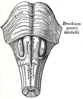
WThe middle cerebellar peduncles are paired structures that connect the cerebellum to the pons and are composed entirely of centripetal fibers, i.e. incoming fibers. The fibers arise from the pontine nucleus to the opposite hemisphere of the cerebellar cortex. The fibers are arranged in three fasciculi: superior, inferior, and deep.The superior fasciculus, the most superficial, is derived from the upper transverse fibers of the pons; it is directed backward and lateralward superficial to the other two fasciculi, and is distributed mainly to the lobules on the inferior surface of the cerebellar hemisphere and to the parts of the superior surface adjoining the posterior and lateral margins. The inferior fasciculus is formed by the lowest transverse fibers of the pons; it passes under cover of the superior fasciculus and is continued downward and backward more or less parallel with it, to be distributed to the folia on the under surface close to the vermis. The deep fasciculus comprises most of the deep transverse fibers of the pons. It is at first covered by the superior and inferior fasciculi, but crosses obliquely and appears on the medial side of the superior, from which it receives a bundle; its fibers spread out and pass to the upper anterior cerebellar folia. The fibers of this fasciculus cover those of the inferior cerebellar peduncle.
WMillard–Gubler syndrome is a lesion of the pons. It is also called ventral pontine syndrome.
WThe paramedian pontine reticular formation, also known as PPRF or paraabducens nucleus, is part of the pontine reticular formation, a brain region without clearly defined borders in the center of the pons. It is involved in the coordination of eye movements, particularly horizontal gaze and saccades.
 W
WThe pontine nuclei are the nuclei of the pons involved in motor activity. The pontine nuclei are located in the ventral pons. Corticopontine fibres carry information from the primary motor cortex to the ipsilateral pontine nucleus in the ventral pons, and the pontocerebellar projection then carries that information to the contralateral cerebellum via the middle cerebellar peduncle. Extension of these nuclei in the medulla oblongata are named arcuate nucleus (medulla) which has the same function.
WThe pontine tegmentum, or dorsal pons, is located within the brainstem, and is one of two parts of the pons, the other being the ventral pons or basilar part of the pons. The pontine tegmentum can be defined in contrast to the basilar pons: basilar pons contains the corticospinal tract running craniocaudally and can be considered the rostral extension of the ventral medulla oblongata; however, basilar pons is distinguished from ventral medulla oblongata in that it contains additional transverse pontine fibres that continue laterally to become the middle cerebellar peduncle. The pontine tegmentum is all the material dorsal from the basilar pons to the fourth ventricle. Along with the dorsal surface of the medulla, it forms part of the rhomboid fossa – the floor of the fourth ventricle.
 W
WThe pontocerebellar fibers are the second order neuron fibers of the corticopontocerebellar tracts that cross to the other side of the pons and run within the middle cerebellar peduncles, from the pons to the contralateral cerebellum.
 W
WThe principal sensory nucleus of trigeminal nerve is a group of second-order neurons which have cell bodies in the caudal pons.
 W
WThe respiratory center is located in the medulla oblongata and pons, in the brainstem. The respiratory center is made up of three major respiratory groups of neurons, two in the medulla and one in the pons. In the medulla they are the dorsal respiratory group, and the ventral respiratory group. In the pons, the pontine respiratory group includes two areas known as the pneumotaxic centre and the apneustic centre.
 W
WThe salivatory nuclei are the superior salivatory nucleus, and the inferior salivatory nucleus that innervate the salivary glands. They are located in the pontine tegmentum in the brainstem. They both are examples of cranial nerve nuclei.
WThe salivatory nuclei are the superior salivatory nucleus, and the inferior salivatory nucleus that innervate the salivary glands. They are located in the pontine tegmentum in the brainstem. They both are examples of cranial nerve nuclei.
 W
WIn the human nervous system the temporopontine fibers, a component of the corticopontine tract are lateral to the cerebrospinal fibers; they originate in the temporal lobe and end in the nuclei pontis.
 W
WThe trapezoid body is part of the auditory pathway where some of the axons coming from the cochlear nucleus decussate to the other side before traveling on to the superior olivary nucleus. This is believed to help with localization of sound.
 W
WThe trigeminal motor nucleus contains motor neurons that innervate muscles of the first branchial arch, namely the muscles of mastication, the tensor tympani, tensor veli palatini, mylohyoid, and anterior belly of the digastric. This nucleus is located in the mid-pons.