 W
WIn human anatomy, the wrist is variously defined as 1) the carpus or carpal bones, the complex of eight bones forming the proximal skeletal segment of the hand; (2) the wrist joint or radiocarpal joint, the joint between the radius and the carpus and; (3) the anatomical region surrounding the carpus including the distal parts of the bones of the forearm and the proximal parts of the metacarpus or five metacarpal bones and the series of joints between these bones, thus referred to as wrist joints. This region also includes the carpal tunnel, the anatomical snuff box, bracelet lines, the flexor retinaculum, and the extensor retinaculum.
 W
WA bracelet is an article of jewellery that is worn around the wrist. Bracelets may serve different uses, such as being worn as an ornament. When worn as ornaments, bracelets may have a supportive function to hold other items of decoration, such as charms. Medical and identity information are marked on some bracelets, such as allergy bracelets, hospital patient-identification tags, and bracelet tags for newborn babies. Bracelets may be worn to signify a certain phenomenon, such as breast cancer awareness, or for religious/cultural purposes.
 W
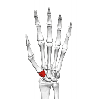
WThe capitate bone is found in the center of the carpal bone region, colloquially known as the wrist, which is at the distal end of the radius and ulna bones. It articulates with the third metacarpal bone and forms the third carpometacarpal joint. The capitate bone is the largest of the carpal bones in the human hand. It presents, above, a rounded portion or head, which is received into the concavity formed by the scaphoid and lunate bones; a constricted portion or neck; and below this, the body. The bone is also found in many other mammals, and is homologous with the "third distal carpal" of reptiles and amphibians.
 W
WThe carpal bones are the eight small bones that make up the wrist that connects the hand to the forearm. The term "carpus" is derived from the Latin carpus and the Greek καρπός (karpós), meaning "wrist". In human anatomy, the main role of the wrist is to facilitate effective positioning of the hand and powerful use of the extensors and flexors of the forearm, and the mobility of individual carpal bones increase the freedom of movements at the wrist.
 W
WCarpal coalition is the abnormal fusion of two or more carpal bones when they fail to segment during intrauterine development. First described by Eduard Sandifort in 1779, carpal coalitions are often an isolated issue which connect two carpal bones in the same row of the wrist. These issues are congenital and occur at various rates throughout the population.
 W
WIn the human body, the carpal tunnel or carpal canal is the passageway on the palmar side of the wrist that connects the forearm to the hand.
 W
WCarpal tunnel surgery, also called carpal tunnel release (CTR) and carpal tunnel decompression surgery, is a surgery in which the transverse carpal ligament is divided. It is a surgical treatment for carpal tunnel syndrome (CTS) and recommended when there is constant numbness, muscle weakness, or atrophy, and when night-splinting no longer controls intermittent symptoms of pain in the carpal tunnel. In general, milder cases can be controlled for months to years, but severe cases are unrelenting symptomatically and are likely to result in surgical treatment. Approximately 500,000 surgical procedures are performed each year, and the economic impact of this condition is estimated to exceed $2 billion annually.
 W
WA cuff is a layer of fabric at the lower edge of the sleeve of a garment at the wrist, or at the ankle end of a trouser leg. The function of turned-back cuffs is to protect the cloth of the garment from fraying, and, when frayed, to allow the cuffs to be readily repaired or replaced, without changing the garment. Cuffs are made by turning back (folding) the material, or a separate band of material can be sewn on, or worn separately, attached either by buttons or studs. A cuff may display an ornamental border or have lace or some other trimming. In US usage, the word trouser cuffs refers to the folded, finished bottoms of the legs of a pair of trousers. In the UK, while this usage is now sometimes followed, the traditional term for the turned up trouser hem is 'turnup'.
 W
WExtensor tendon compartments of the wrist are anatomical tunnels on the back of the wrist that contain tendons of muscles that extend the wrist and the digits.
 W
WThe hamate bone or unciform bone is a bone in the human wrist readily distinguishable by its wedge shape and a hook-like process ("hamulus") projecting from its palmar surface.
 W
WKienböck's disease is a disorder of the wrist. It is named for Dr. Robert Kienböck, a radiologist in Vienna, Austria who described osteomalacia of the lunate in 1910.
 W
WThe lunate bone is a carpal bone in the human hand. It is distinguished by its deep concavity and crescentic outline. It is situated in the center of the proximal row carpal bones, which lie between the ulna and radius and the hand. The lunate carpal bone is situated between the lateral scaphoid bone and medial triquetral bone.
 W
WThe midcarpal joint is formed by the scaphoid, lunate, and triquetral bones in the proximal row, and the trapezium, trapezoid, capitate, and hamate bones in the distal row. The distal pole of the scaphoid articulates with two trapezial bones as a gliding type of joint. The proximal end of the scaphoid combines with the lunate and triquetrum to form a deep concavity that articulates with the convexity of the combined capitate and hamate in a form of diarthrodial, almost condyloid joint.
 W
WPhalen's maneuver is a diagnostic test for carpal tunnel syndrome by an American orthopedist named George S. Phalen.
 W
WThe pisiform bone, also spelled pisiforme, is a small knobbly, sesamoid bone that is found in the wrist. It forms the ulnar border of the carpal tunnel.
 W
WThe scaphoid bone is one of the carpal bones of the wrist. It is situated between the hand and forearm on the thumb side of the wrist. It forms the radial border of the carpal tunnel. The scaphoid bone is the largest bone of the proximal row of wrist bones, its long axis being from above downward, lateralward, and forward. It is approximately the size and shape of a medium cashew.
 W
WThread carpal tunnel release (TCTR) is a minimally-invasive procedure of performing carpal tunnel release using a piece of surgical dissecting thread as a dividing element. This is instead of using a scalpel as in the situation of open carpal tunnel release (OCTR) or endoscopic carpal tunnel release (ECTR).
 W
WThe trapezium bone is a carpal bone in the hand. It forms the radial border of the carpal tunnel.
 W
WThe trapezoid bone is a carpal bone in tetrapods, including humans. It is the smallest bone in the distal row of carpal bones that give structure to the palm of the hand. It may be known by its wedge-shaped form, the broad end of the wedge constituting the dorsal, the narrow end the palmar surface; and by its having four articular facets touching each other, and separated by sharp edges. It is homologous with the "second distal carpal" of reptiles and amphibians.
 W
WThe triquetral bone is located in the wrist on the medial side of the proximal row of the carpus between the lunate and pisiform bones. It is on the ulnar side of the hand, but does not articulate with the ulna. It connects with the pisiform, hamate, and lunate bones. It is the 3rd most commonly fractured carpal bone.
 W
WThe ulnar canal or ulnar tunnel (also known as Guyon's canal or tunnel) is a semi-rigid longitudinal canal in the wrist that allows passage of the ulnar artery and ulnar nerve into the hand. The roof of the canal is made up of the superficial palmar carpal ligament, while the deeper flexor retinaculum and hypothenar muscles comprise the floor. The space is medially bounded by the pisiform and pisohamate ligament more proximally, and laterally bounded by the hook of the hamate more distally. It is approximately 4 cm long, beginning proximally at the transverse carpal ligament and ending at the aponeurotic arch of the hypothenar muscles.
 W
WWrist arthroscopy can be used to look inside the joint of the wrist. It is a minimally invasive technique which can be utilized for diagnostic purposes as well as for therapeutic interventions. Wrist arthroscopy has been used for diagnostic purposes since it was first introduced in 1979. However, it only became accepted as diagnostic tool around the mid-1980s. At that time, arthroscopy of the wrist was an innovative technique to determine whether a problem could be found in the wrist. A few years later, wrist arthroscopy could also be used as a therapeutic tool.