 W
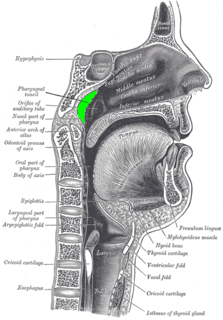
WThe pharynx is the part of the throat behind the mouth and nasal cavity, and above the esophagus and larynx – the tubes going down to the stomach and the lungs. It is found in vertebrates and invertebrates, though its structure varies across species.
 W
WIn anatomy, the adenoid, also known as the pharyngeal tonsil or nasopharyngeal tonsil, is the superior-most of the tonsils. It is a mass of lymphatic tissue located behind the nasal cavity, in the roof of the nasopharynx, where the nose blends into the throat. In children, it normally forms a soft mound in the roof and back wall of the nasopharynx, just above and behind the uvula.
 W
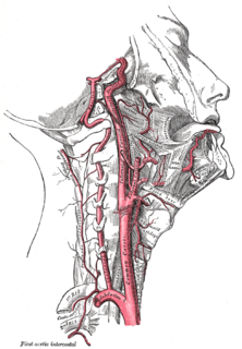
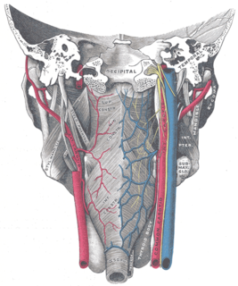
WThe ascending pharyngeal artery is an artery in the neck that supplies the pharynx, developing from the proximal part of the embryonic second aortic arch.
 W
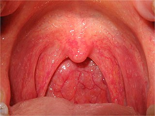
WThe fauces, isthmus of fauces, or the oropharyngeal isthmus, is the opening at the back of the mouth into the throat. It is a narrow passage between the pharynx and the base of the tongue.
 W
WThe glossopharyngeal nerve, known as the ninth cranial nerve, is a mixed nerve that carries afferent sensory and efferent motor information. It exits the brainstem out from the sides of the upper medulla, just anterior to the vagus nerve. The motor division of the glossopharyngeal nerve is derived from the basal plate of the embryonic medulla oblongata, while the sensory division originates from the cranial neural crest.
 W
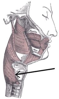
WThe Inferior pharyngeal constrictor, the thickest of the three constrictors, arises from the sides of the cricoid and thyroid cartilage. Similarly to the superior and middle pharyngeal constrictor muscles, it is innervated by the vagus nerve, specifically, by branches from the pharyngeal plexus and by neuronal branches from the recurrent laryngeal nerve.
 W
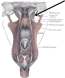
WThe levator veli palatini is the elevator muscle of the soft palate in the human body. During swallowing, it contracts, elevating the soft palate to help prevent food from entering the nasopharynx. It is innervated via the pharyngeal plexus.
 W
WThe lingual artery arises from the external carotid artery between the superior thyroid artery and facial artery. It can be located easily in the tongue.
 W
WThe middle pharyngeal constrictor is a fan-shaped muscle located in the neck. It is one of three pharyngeal constrictors. Similarly to the superior and inferior pharyngeal constrictor muscles, the middle pharyngeal constrictor is innervated by a branch of the vagus nerve through the pharyngeal plexus. The middle pharyngeal constrictor is smaller than the inferior pharyngeal constrictor muscle.
 W
WNasopharyngeal carcinoma (NPC), or nasopharynx cancer, is the most common cancer originating in the nasopharynx, most commonly in the postero-lateral nasopharynx or pharyngeal recess, accounting for 50% of cases. NPC occurs in children and adults. NPC differs significantly from other cancers of the head and neck in its occurrence, causes, clinical behavior, and treatment. It is vastly more common in certain regions of East Asia and Africa than elsewhere, with viral, dietary and genetic factors implicated in its causation. It is most common in males. It is a squamous cell carcinoma of an undifferentiated type. Squamous epithelial cells are a flat type of cell found in the skin and the membranes that line some body cavities. Differentiation means how different the cancer cells are from normal cells. Undifferentiated cells are cells that do not have their mature features or functions
WThe palatopharyngeal arch is larger and projects farther toward the middle line than the palatoglossal arch; it runs downward, lateralward, and backward to the side of the pharynx, and is formed by the projection of the palatopharyngeal muscle, covered by mucous membrane.
 W
WPharyngeal aspiration is the introduction of a substance into the pharynx and its subsequent aspiration into the lungs. It is used to test the respiratory toxicity of a substance in animal testing. It began to be used in the late 1990s. Pharyngeal aspiration is widely used to study the toxicity of a wide variety of substances, including nanomaterials such as carbon nanotubes.
 W
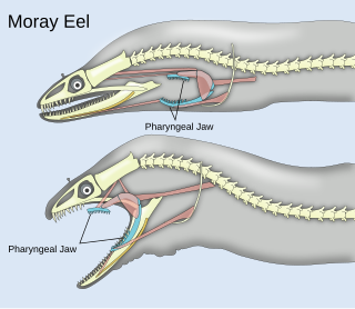
WPharyngeal jaws are a "second set" of jaws contained within an animal's throat, or pharynx, distinct from the primary or oral jaws. They are believed to have originated as modified gill arches, in much the same way as oral jaws. Originally hypothesized to have evolved only once, current morphological and genetic analyses suggest at least two separate points of origin. Based on connections between musculoskeletal morphology and dentition, diet has been proposed as a main driver of the evolution of the pharyngeal jaw. A study conducted on cichlids showed that the pharyngeal jaws can undergo morphological changes in less than two years in response to their diet. Fish that ate hard shelled prey had a robust jaw with molar-like teeth fit for crushing their durable prey. Fish that ate softer prey, on the other hand, exhibited a more slender jaw with thin, curved teeth used for tearing apart fleshy prey. These rapid changes are an example of phenotypic plasticity, wherein environmental factors affect genetic expression responsible for pharyngeal jaw development. Studies of the genetic pathways suggest that receptors in the jaw bone respond to the mechanical strain of biting hard-shelled prey, which prompts the formation of a more robust set of pharyngeal jaws.
 W
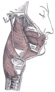
WThe pharyngeal muscles are a group of muscles that form the pharynx, which is posterior to the oral cavity, determining the shape of its lumen, and affecting its sound properties as the primary resonating cavity.
 W
WThe pharyngeal plexus is a network of nerve fibers innervating most of the palate and pharynx.
 W
WPharyngeal slits are filter-feeding organs found among deuterostomes. Pharyngeal slits are repeated openings that appear along the pharynx caudal to the mouth. With this position, they allow for the movement of water in the mouth and out the pharyngeal slits. It is postulated that this is how pharyngeal slits first assisted in filter-feeding, and later with the addition of gills along their walls, aided in respiration of aquatic chordates. These repeated segments are controlled by similar developmental mechanisms. Some hemichordate species can have as many as 200 gill slits. Pharyngeal clefts resembling gill slits are transiently present during the embryonic stages of tetrapod development. The presence of pharyngeal arches and clefts in the neck of the developing human embryo famously led Ernst Haeckel to postulate that "ontogeny recapitulates phylogeny"; this hypothesis, while false, contains elements of truth, as explored by Stephen Jay Gould in Ontogeny and Phylogeny. However, it is now accepted that it is the vertebrate pharyngeal pouches and not the neck slits that are homologous to the pharyngeal slits of invertebrate chordates. Pharyngeal arches, pouches, and clefts are, at some stage of life, found in all chordates. One theory of their origin is the fusion of nephridia which opened both on the outside and the gut, creating openings between the gut and the environment.
 W
WPharyngitis is inflammation of the back of the throat, known as the pharynx. It typically results in a sore throat and fever. Other symptoms may include a runny nose, cough, headache, difficulty swallowing, swollen lymph nodes, and a hoarse voice. Symptoms usually last 3–5 days. Complications can include sinusitis and acute otitis media. Pharyngitis is a type of upper respiratory tract infection.
 W
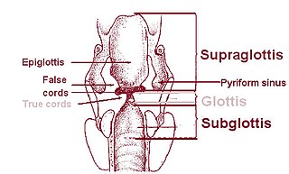
WOn either side of the laryngeal orifice in humans is a recess, termed the pyriform sinus, which is bounded medially by the aryepiglottic fold, laterally by the thyroid cartilage and thyrohyoid membrane. The fossae are involved in speech.
 W
WThe salpingopharyngeus muscle arises from the superior border of the medial cartilage of the pharyngotympanic tube, in the nasal cavity, making the posterior welt of the torus tubarius; it passes downward and blends with the posterior fasciculus of the palatopharyngeus muscle.
 W
WThe superior pharyngeal constrictor muscle is a muscle in the pharynx. It is the highest located muscle of the three pharyngeal constrictors. The muscle is a quadrilateral muscle, thinner and paler than the inferior pharyngeal constrictor muscle and middle pharyngeal constrictor muscle.
 W
WThe superior thyroid artery arises from the external carotid artery just below the level of the greater cornu of the hyoid bone and ends in the thyroid gland.
 W
WThe tensor veli palatini muscle is a broad, thin, ribbon-like muscle in the head that tenses the soft palate.
 W
WWaldeyer's tonsillar ring is a ringed arrangement of lymphoid organs in the pharynx. Waldeyer's ring surrounds the naso- and oropharynx, with some of its tonsillar tissue located above and some below the soft palate.