 W
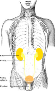
WThe urinary system, also known as the renal system or urinary tract, consists of the kidneys, ureters, bladder, and the urethra. The purpose of the urinary system is to eliminate waste from the body, regulate blood volume and blood pressure, control levels of electrolytes and metabolites, and regulate blood pH. The urinary tract is the body's drainage system for the eventual removal of urine. The kidneys have an extensive blood supply via the renal arteries which leave the kidneys via the renal vein. Each kidney consists of functional units called nephrons. Following filtration of blood and further processing, wastes exit the kidney via the ureters, tubes made of smooth muscle fibres that propel urine towards the urinary bladder, where it is stored and subsequently expelled from the body by urination (voiding). The female and male urinary system are very similar, differing only in the length of the urethra.
 W
WA cystocele, also known as a prolapsed bladder, is a medical condition in which a woman's bladder bulges into her vagina. Some may have no symptoms. Other may have trouble starting urination, urinary incontinence, or frequent urination. Complications may include recurrent urinary tract infections and urinary retention. Cystocele and a prolapsed urethra often occur together and is called a cystourethrocele. Cystocele can negatively affect quality of life.
 W
WThe detrusor muscle, also detrusor urinae muscle, muscularis propria of the urinary bladder and muscularis propria, is smooth muscle found in the wall of the bladder. The detrusor muscle remains relaxed to allow the bladder to store urine, and contracts during urination to release urine. Related are the urethral sphincter muscles which envelop the urethra to control the flow of urine when they contract.
 W
WThe external sphincter muscle of female urethra is a muscle which controls urination in females. The muscle fibers arise on either side from the margin of the inferior ramus of the pubis. They are directed across the pubic arch in front of the urethra, and pass around it to blend with the muscular fibers of the opposite side, between the urethra and vagina.
 W
WThe external sphincter muscle of male urethra, also sphincter urethrae membranaceae, sphincter urethrae externus, surrounds the whole length of the membranous urethra, and is enclosed in the fascia of the urogenital diaphragm.
 W
WA flame cell is a specialized excretory cell found in the simplest freshwater invertebrates, including flatworms, rotifers and nemerteans; these are the simplest animals to have a dedicated excretory system. Flame cells function like a kidney, removing waste materials. Bundles of flame cells are called protonephridia.
 W
WThe foreskin is the double-layered fold of smooth muscle tissue, blood vessels, neurons, skin, and mucous membrane part of the penis that covers and protects the glans penis and the urinary meatus. The foreskin is mobile, fairly stretchable, and acts as a natural lubricant. It is also described as the prepuce, a technically broader term that also includes the clitoral hood in women, to which the foreskin is embryonically homologous.
 W
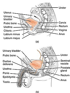
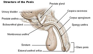
WThe human penis is an external male intromittent organ that additionally serves as the urinal duct. The main parts are the root (radix); the body (corpus); and the epithelium of the penis including the shaft skin and the foreskin (prepuce) covering the glans penis. The body of the penis is made up of three columns of tissue: two corpora cavernosa on the dorsal side and corpus spongiosum between them on the ventral side. The human male urethra passes through the prostate gland, where it is joined by the ejaculatory duct, and then through the penis. The urethra traverses the corpus spongiosum, and its opening, the meatus, lies on the tip of the glans penis. It is a passage both for urination and ejaculation of semen
WThe internal urethral orifice is the opening of the urinary bladder into the urethra. It is placed at the apex of the trigonum vesicae, in the most dependent part of the bladder, and is usually somewhat crescent-shaped; the mucous membrane immediately behind it presents a slight elevation in males, the uvula vesicae, caused by the middle lobe of the prostate.
WThe internal urethral sphincter is a urethral sphincter muscle which constricts the internal urethral orifice. It is located at the junction of the urethra with the urinary bladder and is continuous with the detrusor muscle, but anatomically and functionally fully independent from it. It is composed of smooth muscle, so it is under the control of the autonomic nervous system, specifically the sympathetic nervous system.
 W
WIn male anatomy, the lacuna magna is the largest of several recesses in the roof of the navicular fossa of the urethra. Its embryologic origin is contested, but recent evidence suggests it and the navicular fossa derive from infiltrating endodermal cells of the urethral plate. In young males, the presence of the lacuna magna is associated with painful urination (dysuria), bloody urine (hematuria), and bloody spotting of underwear.
 W
WThe renal medulla is the innermost part of the kidney. The renal medulla is split up into a number of sections, known as the renal pyramids. Blood enters into the kidney via the renal artery, which then splits up to form the interlobar arteries. The interlobar arteries each in turn branch into arcuate arteries, which in turn branch to form interlobular arteries, and these finally reach the glomeruli. At the glomerulus the blood reaches a highly disfavourable pressure gradient and a large exchange surface area, which forces the serum portion of the blood out of the vessel and into the renal tubules. Flow continues through the renal tubules, including the proximal tubule, the Loop of Henle, through the distal tubule and finally leaves the kidney by means of the collecting duct, leading to the renal pelvis, the dilated portion of the ureter.
 W
WMichaelis–Gutmann bodies are concentrically layered basophilic inclusions found in the urinary tract. They are 2 to 10 μm in diameter, and are thought to represent remnants of phagosomes mineralized by iron and calcium deposits.
 W
WPig bladder is the urinary bladder of a domestic pig, similar to the human urinary bladder. Today, this hollow organ has various applications in medicine, and in traditional cuisines and customs. Historically, the pig bladder had several additional uses, all based on its properties as a lightweight, stretchable container that could be filled and tied off.
WThe trigone is a smooth triangular region of the internal urinary bladder formed by the two ureteric orifices and the internal urethral orifice.
 W
WRelated to the urinary bladder, anteriorly there are the following folds:one median umbilical fold on the median umbilical ligament two medial umbilical folds on the occluded umbilical artery two lateral umbilical folds on the inferior epigastric vessels
 W
WThe ureters are tubes made of smooth muscle that propel urine from the kidneys to the urinary bladder. In the human adult, the ureters are usually 20–30 cm (8–12 in) long and around 3–4 mm (0.12–0.16 in) in diameter. The ureter is lined by urothelial cells, a type of transitional epithelium, and has an additional smooth muscle layer in third closest to the bladder that assists with peristalsis.
 W
WThe urethra is a tube that connects the urinary bladder to the urinary meatus for the removal of urine from the body of both females and males. In human females and other primates, the urethra connects to the urinary meatus above the vagina, whereas in marsupials, the female's urethra empties into the urogenital sinus.
 W
WThe urethral crest is an anatomical feature present in the urinary system of both males and females.
WThe urethral sphincters are two muscles used to control the exit of urine in the urinary bladder through the urethra. The two muscles are either the male or female external urethral sphincter and the internal urethral sphincter. When either of these muscles contracts, the urethra is sealed shut.
WThe urinary bladder, or simply bladder, is a hollow muscular organ in humans and other vertebrates that stores urine from the kidneys before disposal by urination. In the human the bladder is a hollow muscular, and distensible organ that sits on the pelvic floor. Urine enters the bladder via the ureters and exits via the urethra. The typical human bladder will hold between 300 and 500 ml before the urge to empty occurs, but can hold considerably more.
 W
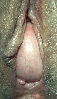
WThe urinary meatus, also known as the external urethral orifice, is the opening of the urethra. It is the point where urine exits the urethra in males and in females and where semen exits the urethra in males. The meatus has varying degrees of sensitivity to touch. The meatus is located on the glans of the penis or in the vulval vestibule.
 W
WUrinothorax means urine in the fluid-filled cavity that surrounds the lungs. It is an extremely rare cause of pleural effusion. It is secondary to obstructive uropathy whereby urine forms a collection in the pleural cavity. The urine arrives in the pleural space either retroperitoneally under the posterior diaphragm, or via the retro peritoneal lymphatics. It remains a rare, possibly under-diagnosed, differential in the case of transudative pleural effusion. Respiratory symptoms are usually mild. Handa et al., described 47 cases between 1967 and 2007, noting that it was more prevalent in males, generally ipsilateral to the obstruction, and in most of the cases it is relieved by clearance of the obstruction.