 W
WAdiposis dolorosa, is an outdated term for many years used synonymously as Dercum's disease, lipedema or Anders disease. While there are numerous references to Adiposis dolorosa, it is recommended that the term no longer be used. Dercum's is now recognized as a separate condition, as is lipedema.
 W
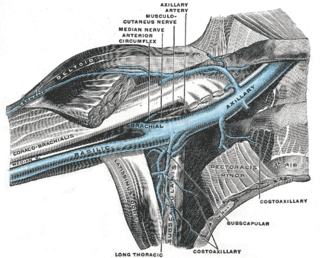
WAxillary lymphadenopathy is lymphadenopathy of the axillary lymph nodes.
 W
WBudd–Chiari syndrome is a very rare condition, affecting one in a million adults. The condition is caused by occlusion of the hepatic veins that drain the liver. It presents with the classical triad of abdominal pain, ascites, and liver enlargement. The formation of a blood clot within the hepatic veins can lead to Budd–Chiari syndrome. The syndrome can be fulminant, acute, chronic, or asymptomatic. Subacute presentation is the most common form.
 W
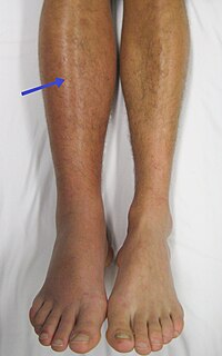
WDeep vein thrombosis (DVT) is the formation of a blood clot in a deep vein, most commonly in the legs or pelvis. Symptoms can include pain, swelling, redness, and enlarged veins in the affected area, but some DVTs have no symptoms. The most common life-threatening concern with DVT is the potential for a clot to detach from the veins (embolize), travel through the right side of the heart, and become stuck in arteries that supply blood to the lungs. This is called pulmonary embolism (PE). Both DVT and PE are considered as part of the same overall disease process, which is called venous thromboembolism (VTE). VTE can occur as DVT only, as PE with DVT, or PE without DVT. The most frequent long-term complication is post-thrombotic syndrome, which can cause pain, swelling, a sensation of heaviness, itching, and in severe cases, ulcers. Also, recurrent VTE occurs in about 30% of those in the ten years following an initial VTE.
 W
WElephantiasis is the enlargement and hardening of limbs or body parts due to tissue swelling. It is characterised by edema, hypertrophy, and fibrosis of skin and subcutaneous tissues, due to obstruction of lymphatic vessels. It may affect the genitalia. The term elephantiasis is often used in reference to parasitic worm infections, but may refer to a variety of diseases where parts of a person's body swell to massive proportions.
 W
WEsophageal varices are extremely dilated sub-mucosal veins in the lower third of the esophagus. They are most often a consequence of portal hypertension, commonly due to cirrhosis; people with esophageal varices have a strong tendency to develop severe bleeding which left untreated can be fatal. Esophageal varices are typically diagnosed through an esophagogastroduodenoscopy.
 W
WGastric varices are dilated submucosal veins in the lining of the stomach, which can be a life-threatening cause of bleeding in the upper gastrointestinal tract. They are most commonly found in patients with portal hypertension, or elevated pressure in the portal vein system, which may be a complication of cirrhosis. Gastric varices may also be found in patients with thrombosis of the splenic vein, into which the short gastric veins which drain the fundus of the stomach flow. The latter may be a complication of acute pancreatitis, pancreatic cancer, or other abdominal tumours, as well as hepatitis C. Gastric varices and associated bleeding are a potential complication of schistosomiasis resulting from portal hypertension.
 W
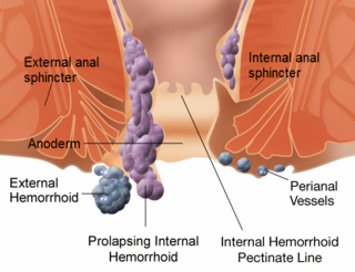
WHemorrhoids, also spelled haemorrhoids, are vascular structures in the anal canal. In their normal state, they are cushions that help with stool control. They become a disease when swollen or inflamed; the unqualified term "hemorrhoid" is often used to refer to the disease. The signs and symptoms of hemorrhoids depend on the type present. Internal hemorrhoids often result in painless, bright red rectal bleeding when defecating. External hemorrhoids often result in pain and swelling in the area of the anus. If bleeding occurs it is usually darker. Symptoms frequently get better after a few days. A skin tag may remain after the healing of an external hemorrhoid.
 W
WInguinal lymphadenopathy causes swollen lymph nodes in the groin area. It can be a symptom of infective or neoplastic processes. Infective aetiologies include Tuberculosis, HIV, non-specific or reactive lymphadenopathy to recent lower limb infection or groin infections. Another notable infectious cause is Lymphogranuloma venereum, which is a sexually transmitted infection of the lymphatic system. Neoplastic aetiologies include lymphoma, leukaemia and metastatic disease from primary tumours in the lower limb, external genitalia or perianal region and melanoma.
 W
WIntranodal palisaded myofibroblastoma (IPM) is a rare primary tumour of lymph nodes, that classically presents as an inguinal mass.
 W
WLipedema is a disorder where there is enlargement of both legs due to deposits of fat under the skin. Typically it gets worse over time, pain may be present, and sufferers bruise easily. In severe cases the trunk and upper body may be involved. Lipedema is commonly misdiagnosed.
 W
WLymphadenopathy or adenopathy is a disease of the lymph nodes, in which they are abnormal in size or consistency. Lymphadenopathy of an inflammatory type is lymphadenitis, producing swollen or enlarged lymph nodes. In clinical practice, the distinction between lymphadenopathy and lymphadenitis is rarely made and the words are usually treated as synonymous. Inflammation of the lymphatic vessels is known as lymphangitis. Infectious lymphadenitis affecting lymph nodes in the neck is often called scrofula.
 W
WLymphangitis is an inflammation or an infection of the lymphatic channels that occurs as a result of infection at a site distal to the channel. The most common cause of lymphangitis in humans is Streptococcus pyogenes, hemolythic streptococci, and in some cases, mononucleosis, cytomegalovirus, tuberculosis, syphilis, and the fungus Sporothrix schenckii. Lymphangitis is sometimes mistakenly called "blood poisoning". In reality, "blood poisoning" is synonymous with sepsis.
 W
WLymphedema, also known as lymphoedema and lymphatic edema, is a condition of localized swelling caused by a compromised lymphatic system. The lymphatic system functions as a critical portion of the body's immune system and returns interstitial fluid to the bloodstream. Lymphedema is most frequently a complication of cancer treatment or parasitic infections, but it can also be seen in a number of genetic disorders. Though incurable and progressive, a number of treatments can improve symptoms. Tissues with lymphedema are at high risk of infection because the lymphatic system has been compromised.
 W
WPaget–Schroetter disease, is a form of upper extremity deep vein thrombosis (DVT), a medical condition in which blood clots form in the deep veins of the arms. These DVTs typically occur in the axillary and/or subclavian veins.
 W
WPortal vein thrombosis (PVT) is a vascular disease of the liver that occurs when a blood clot occurs in the hepatic portal vein, which can lead to increased pressure in the portal vein system and reduced blood supply to the liver. The mortality rate is approximately 1 in 10.
 W
WPost-thrombotic syndrome (PTS), also called postphlebitic syndrome and venous stress disorder is a medical condition that may occur as a long-term complication of deep vein thrombosis (DVT).
 W
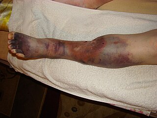
WSuperficial vein thrombosis (SVT) is a type blood clot in a vein, which forms in a superficial vein near the surface of the body. Usually there is thrombophlebitis, which is an inflammatory reaction around a thrombosed vein, presenting as a painful induration with redness. SVT itself has limited significance when compared to a deep vein thrombosis (DVT), which occurs deeper in the body at the deep venous system level. However, SVT can lead to serious complications, and is therefore no longer regarded as a benign condition. If the blood clot is too near the saphenofemoral junction there is a higher risk of pulmonary embolism, a potentially life-threatening complication.
 W
WSuperior vena cava syndrome (SVCS), is a group of symptoms caused by obstruction of the superior vena cava ("SVC"), a short, wide vessel carrying circulating blood into the heart. The majority of cases are caused by malignant tumors within the mediastinum, most commonly lung cancer and non-Hodgkin's lymphoma, directly compressing or invading the SVC wall. Non-malignant causes are increasing in prevalence due to expanding use of intravascular devices, which can result in thrombosis. Other non-malignant causes include benign mediastinal tumors, aortic aneurysm, infections, and fibrosing mediastinitis.
WThrombophlebitis is a phlebitis related to a thrombus. When it occurs repeatedly in different locations, it is known as thrombophlebitis migrans.
 W
WThrombosis prevention or thromboprophylaxis is medical treatment to prevent the development of thrombosis in those considered at risk for developing thrombosis. Some people are at a higher risk for the formation of blood clots than others. Prevention measures or interventions are usually begun after surgery as people are at higher risk due to immobility.
 W
WUltrasonography of suspected or previously confirmed chronic venous insufficiency of leg veins is a risk-free, non-invasive procedure. It gives information about the anatomy, physiology and pathology of mainly superficial veins. As with heart ultrasound (echocardiography) studies, venous ultrasonography requires an understanding of hemodynamics in order to give useful examination reports. In chronic venous insufficiency, sonographic examination is of most benefit; in confirming varicose disease, making an assessment of the hemodynamics, and charting the progression of the disease and its response to treatment. It has become the reference standard for examining the condition and hemodynamics of the lower limb veins. Particular veins of the deep venous system (DVS), and the superficial venous system (SVS) are looked at. The great saphenous vein (GSV), and the small saphenous vein (SSV) are superficial veins which drain into respectively, the common femoral vein and the popliteal vein. These veins are deep veins. Perforator veins drain superficial veins into the deep veins. Three anatomic compartments are described, (N1) containing the deep veins, (N2) containing the perforator veins, and (N3) containing the superficial veins, known as the saphenous compartment. This compartmentalisation makes it easier for the examiner to systematize and map. The GSV can be located in the saphenous compartment where together with the Giacomini vein and the accessory saphenous vein (ASV) an image resembling an eye, known as the 'eye sign' can be seen. The ASV which is often responsible for varicose veins, can be located at the 'alignment sign', where it is seen to align with the femoral vessels.
 W
WVaricose veins are superficial veins that have become enlarged and twisted. Typically they occur just under the skin in the legs. Usually they result in few symptoms but some may experience fullness or pain in the area. Complications may include bleeding or superficial thrombophlebitis. When varices occur in the scrotum it is known as a varicocele while those around the anus are known as hemorrhoids. Varicose veins may negatively affect quality of life due to their physical, social and psychological effects.
 W
WVenostasis, or venous stasis, is a condition of slow blood flow in the veins, usually of the legs.