 W
WA549 cells are adenocarcinomic human alveolar basal epithelial cells, and constitute a cell line that was first developed in 1972 by D. J. Giard, et al. through the removal and culturing of cancerous lung tissue in the explanted tumor of a 58-year-old caucasian male. The cells are used as models for the study of lung cancer and the development of drug therapies against it.
 W
WA brush border is the microvilli-covered surface of simple cuboidal and simple columnar epithelium found in different parts of the body. Microvilli are approximately 100 nanometers in diameter and their length varies from approximately 100 to 2,000 nanometers in length. Because individual microvilli are so small and are tightly packed in the brush border, individual microvilli can only be resolved using electron microscopes; with a light microscope they can usually only be seen collectively as a fuzzy fringe at the surface of the epithelium. This fuzzy appearance gave rise to the term brush border, as early anatomists noted that this structure appeared very much like the bristles of a paintbrush.
 W
WClue cells are epithelial cells of the vagina that get their distinctive stippled appearance by being covered with bacteria. The etymology behind the term "clue" cell derives from the original research article from Gardner and Dukes describing the characteristic cells. The name was chosen for its brevity in describing the sine qua non of bacterial vaginosis.
 W
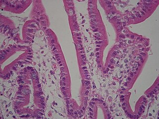
WEnterocytes, or intestinal absorptive cells, are simple columnar epithelial cells which line the inner surface of the small and large intestines. A glycocalyx surface coat contains digestive enzymes. Microvilli on the apical surface increase its surface area. This facilitates transport of numerous small molecules into the enterocyte from the intestinal lumen. These include broken down proteins, fats, and sugars, as well as water, electrolytes, vitamins, and bile salts. Enterocytes also have an endocrine role, secreting hormones such as leptin.
 W
WIn dentistry, the epithelial cell rests of Malassez (ERM) or epithelial rests of Malassez are part of the periodontal ligament cells around a tooth. They are discrete clusters of residual cells from Hertwig's epithelial root sheath (HERS) that didn't completely disappear. It is considered that these cell rests proliferate to form epithelial lining of various odontogenic cysts such as radicular cyst under the influence of various stimuli. They are named after Louis-Charles Malassez (1842–1909) who described them. Some rests become calcified in the periodontal ligament (cementicles).
 W
WGoblet cells are simple columnar goblet shaped like epithelial cells that secrete gel-forming mucins, like mucin MUC5AC. The goblet cells mainly use the merocrine method of secretion, secreting vesicles into a duct, but may use apocrine methods, budding off their secretions, when under stress. The term goblet refers to the cell's goblet-like shape. The apical portion is shaped like a cup, as it is distended by abundant mucus laden granules; its basal portion lacks these granules and is shaped like a stem.
 W
WA koilocyte is a squamous epithelial cell that has undergone a number of structural changes, which occur as a result of infection of the cell by human papillomavirus (HPV).
 W
WLangerhans cells (LC) are tissue-resident dendritic cells of the skin, and contain organelles called Birbeck granules. They are present in all layers of the epidermis and are most prominent in the stratum spinosum. They also occur in the papillary dermis, particularly around blood vessels, as well as in the mucosa of the mouth, foreskin, and vaginal epithelium. They can be found in other tissues, such as lymph nodes, particularly in association with the condition Langerhans cell histiocytosis (LCH).
 W
WMelanocytes are melanin-producing neural crest-derived cells located in the bottom layer of the skin's epidermis, the middle layer of the eye, the inner ear, vaginal epithelium, meninges, bones, and heart. Melanin is a dark pigment primarily responsible for skin color. Once synthesized, melanin is contained in special organelles called melanosomes which can be transported to nearby keratinocytes to induce pigmentation. Thus darker skin tones have more melanosomes present than lighter skin tones. Functionally, melanin serves as protection against UV radiation. Melanocytes also have a role in the immune system.
 W
WMicrovilli are microscopic cellular membrane protrusions that increase the surface area for diffusion and minimize any increase in volume, and are involved in a wide variety of functions, including absorption, secretion, cellular adhesion, and mechanotransduction.
 W
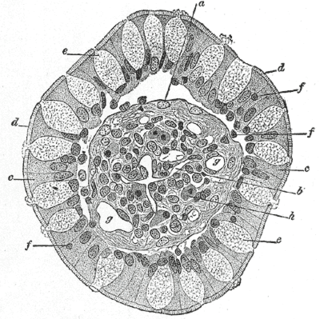
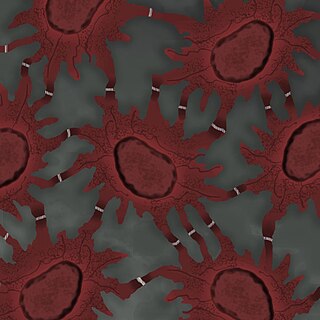
WOlfactory ensheathing cells (OECs), also known as olfactory ensheathing glia or olfactory ensheathing glial cells, are a type of macroglia found in the nervous system. They are also known as olfactory Schwann cells, because they ensheath the non-myelinated axons of olfactory neurons in a similar way to which Schwann cells ensheath non-myelinated peripheral neurons. They also share the property of assisting axonal regeneration.
 W
WAn oncocyte is an epithelial cell characterized by an excessive number of mitochondria, resulting in an abundant acidophilic, granular cytoplasm. Oncocytes can be benign or malignant.
 W
WPaneth cells are cells in the small intestine epithelium, alongside goblet cells, enterocytes, and enteroendocrine cells. Some can also be found in the cecum and appendix. They are below the intestinal stem cells in the intestinal glands and the large eosinophilic refractile granules that occupy most of their cytoplasm.
 W
WParietal cells (also known as oxyntic cells) are epithelial cells in the stomach that secrete hydrochloric acid (HCl) and intrinsic factor. These cells are located in the gastric glands found in the lining of the fundus and cardia regions of the stomach. They contain an extensive secretory network of canaliculi from which the HCl is secreted by active transport into the stomach. The enzyme hydrogen potassium ATPase (H+/K+ ATPase) is unique to the parietal cells and transports the H+ against a concentration gradient of about 3 million to 1, which is the steepest ion gradient formed in the human body. Parietal cells are primarily regulated via histamine, acetylcholine and gastrin signaling from both central and local modulators.
 W
WPodocytes are cells in the Bowman's capsule in the kidneys that wrap around capillaries of the glomerulus. Podocyte cells make up the epithelial lining of Bowman's capsule, the third layer through which filtration of blood takes place. The Bowman's capsule filters the blood, retaining large molecules such as proteins while smaller molecules such as water, salts, and sugars are filtered as the first step in the formation of urine. Although various viscera have epithelial layers, the name visceral epithelial cells usually refers specifically to podocytes, which are specialized epithelial cells that reside in the visceral layer of the capsule.
 W
WA pseudostratified epithelium is a type of epithelium that, though comprising only a single layer of cells, has its cell nuclei positioned in a manner suggestive of stratified epithelia. As it rarely occurs as squamous or cuboidal epithelia, it is usually considered synonymous with the term pseudostratified columnar epithelium.
 W
WA simple columnar epithelium is a single layer of columnar cells attached to the basement membrane, with oval-shaped nuclei located in the basal region. In humans, a simple columnar epithelium lines most organs of the digestive tract including the stomach, small intestine, and large intestine. Simple columnar epithelia line the uterus.
 W
WSimple cuboidal epithelium is a type of epithelium that consists of a single layer of cuboidal (cube-like) cells. These cuboidal cells have large, spherical and central nuclei.
 W
WA simple squamous epithelium is a single layer of flat cells in contact with the basal lamina of the epithelium. This type of epithelium is often permeable and occurs where small molecules need to pass quickly through membranes via filtration or diffusion. Simple squamous epithelia are found in capillaries, alveoli, glomeruli, and other tissues where rapid diffusion is required. Within the cardiovascular system such as lining capillaries or the inside of the heart, simple squamous epithelium is specifically called the endothelium. Cells are flat with flattened and oblong nuclei. It is also called pavement epithelium due to its tile-like appearance. This epithelium is associated with filtration and diffusion. This tissue is extremely thin, and forms a delicate lining. It offers very little protection.
 W
WSpinous cells, or prickle cells, are keratin producing epidermal cells owing their prickly appearance to their numerous intracellular connections. They make up the stratum spinosum of the epidermis and provide a continuous net-like layer of protection for underlying tissue. They are susceptible to mutations caused by sunlight and can become malignant.
 W
WThe stratum basale is the deepest layer of the five layers of the epidermis, the external covering of skin in mammals.
WThe stratum corneum is the outermost layer of the epidermis. There has been a long-standing belief in dermatology that the stratum corneum consisted of dead cells (corneocytes), devoid of biological activity and function. The stratum corneum is now understood to be live tissue that performs protective and adaptive physiological functions including mechanical shear, impact resistance, water flux and hydration regulation, microbial proliferation and invasion regulation, initiation of inflammation through cytokine activation and dendritic cell activity, and selective permeability to exclude toxins, irritants, and allergens. This layer is composed of 15–20 layers of flattened cells with no nuclei or cell organelles. Their cytoplasm shows filamentous keratin. These corneocytes are embedded in a lipid matrix composed of ceramides, cholesterol, and fatty acids. Their properties depend on the component ratio of the three major components.
WThe stratum granulosum is a thin layer of cells in the epidermis. Keratinocytes migrating from the underlying stratum spinosum become known as granular cells in this layer. These cells contain keratohyalin granules, which are filled with histidine- and cysteine-rich proteins that appear to bind the keratin filaments together. Therefore, the main function of keratohyalin granules is to bind intermediate keratin filaments together.
 W
WThe stratum lucidum is a thin, clear layer of dead skin cells in the epidermis named for its translucent appearance under a microscope. It is readily visible by light microscopy only in areas of thick skin, which are found on the palms of the hands and the soles of the feet.
WThe stratum spinosum is a layer of the epidermis found between the stratum granulosum and stratum basale. Their spiny appearance is due to shrinking of the microfilaments between desmosomes that occurs when stained with H&E. Keratinization begins in the stratum spinosum. This layer is composed of polyhedral keratinocytes. They have large pale-staining nuclei as they are active in synthesizing fibrilar proteins, known as cytokeratin, which build up within the cells aggregating together forming tonofibrils. The tonofibrils go on to form the desmosomes, which allow for strong connections to form between adjacent keratinocytes.
 W
WTransitional epithelium is a type of stratified epithelium. This tissue consists of multiple layers of epithelial cells which can contract and expand in order to adapt to the degree of distension needed. Transitional epithelium lines the organs of the urinary system and is known here as urothelium. The bladder for example has a need for great distension.