 W
WThe efferent vessels of the tracheobronchial lymph nodes ascend upon the trachea and unite with efferents of the internal mammary and anterior mediastinal glands to form the right and left bronchomediastinal trunks.
 W
WThe celiac lymph nodes are associated with the branches of the celiac artery. Other lymph nodes in the abdomen are associated with the superior and inferior mesenteric arteries. The celiac lymph nodes are grouped into three sets: the gastric, hepatic and splenic lymph nodes.
 W
WThe cisterna chyli is a dilated sac at the lower end of the thoracic duct in most mammals into which lymph from the intestinal trunk and two lumbar lymphatic trunks flow. It receives fatty chyle from the intestines and thus acts as a conduit for the lipid products of digestion. It is the most common drainage trunk of most of the body's lymphatics. The cisterna chyli is a retro-peritoneal structure. In humans, it is located posterior to the abdominal aorta on the anterior aspect of the bodies of the first and second lumbar vertebrae. There it forms the beginning of the primary lymph vessel, the thoracic duct, which transports lymph and chyle from the abdomen via the aortic opening of the diaphragm up to the junction of left subclavian vein and internal jugular veins. In dogs, it is located to the left and often ventral to the aorta; in cats it is left and dorsal; in guinea pigs it runs to the left and drains into the left innominate vein.
 W
WThe common iliac lymph nodes, four to six in number, are grouped behind and on the sides of the common iliac artery, one or two being placed below the bifurcation of the aorta, in front of the fifth lumbar vertebra.
WThe external iliac lymph nodes are lymph nodes, from eight to ten in number, that lie along the external iliac vessels.
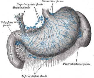
WThe gastric lymph nodes are lymph nodes which drain the stomach and consist of two sets, superior and inferior:The superior gastric lymph nodes accompany the left gastric artery and are divisible into three groups: Upper, on the stem of the artery; Lower, accompanying the descending branches of the artery along the cardiac half of the lesser curvature of the stomach, between the two layers of the lesser omentum; Paracardial outlying members of the gastric lymph nodes, disposed in a manner comparable to a chain of beads around the neck of the stomach. They receive their afferents from the stomach; their efferents pass to the celiac group of preaortic lymph nodes. The inferior gastric lymph nodes, four to seven in number, lie between the two layers of the greater omentum along the pyloric half of the greater curvature of the stomach.
WThe hepatic lymph nodes consist of the following groups:(a) hepatic, on the stem of the hepatic artery, and extending upward along the common bile duct, between the two layers of the lesser omentum, as far as the porta hepatis; the cystic gland, a member of this group, is placed near the neck of the gall-bladder; (b) subpyloric, four or five in number, in close relation to the bifurcation of the gastroduodenal artery, in the angle between the superior and descending parts of the duodenum; an outlying member of this group is sometimes found above the duodenum on the right gastric (pyloric) artery.
 W
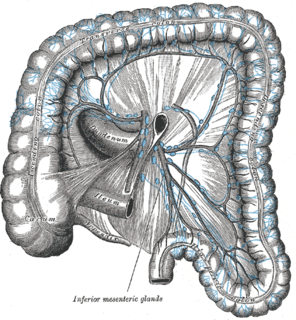
WThe inferior mesenteric lymph nodes consist of:(a) small glands on the branches of the left colic and sigmoid arteries (b) a group in the sigmoid mesocolon, around the superior hemorrhoidal artery (c) a pararectal group in contact with the muscular coat of the rectum
 W
WThe intercostal lymph nodes occupy the posterior parts of the intercostal spaces, in relation to the intercostal vessels.
 W
WThe internal iliac lymph nodes surround the internal iliac artery and its branches, and receive the lymphatics corresponding to the distribution of the branches of it, i. e., they receive lymphatics from all the pelvic viscera, from the deeper parts of the perineum, including the membranous and cavernous portions of the urethra, and from the buttock and back of the thigh. The internal iliac lymph nodes also drain the superior half of the rectum, above the pectinate line.
WThe intestinal trunk receives the lymph from the stomach and intestine, from the pancreas and spleen, and from the lower and front part of the liver, and empties lymph into the cisterna chyli, which in turn drains into the thoracic duct.
WThe lumbar trunks are formed by the union of the efferent vessels from the lateral aortic lymph nodes.
 W
WThe pararectal lymph nodes are in contact with the muscular coat of the rectum. They drain the descending iliac and sigmoid parts of the colon and the upper part of the rectum; their efferents pass to the preaortic glands.
 W
WThe parasternal lymph nodes are placed at the anterior ends of the intercostal spaces, by the side of the internal thoracic artery.
 W
WThe periaortic lymph nodes are a group of lymph nodes that lie in front of the lumbar vertebrae near the aorta. These lymph nodes receive drainage from the gastrointestinal tract and the abdominal organs.
 W
WThe preaortic lymph nodes lie in front of the aorta, and may be divided into celiac lymph nodes, superior mesenteric lymph nodes, and inferior mesenteric lymph nodes groups, arranged around the origins of the corresponding arteries.
WThe retroaortic lymph nodes are placed below the cisterna chyli, on the bodies of the third and fourth lumbar vertebrae.
 W
WThe right lymphatic duct, about 1.25 cm. in length, courses along the medial border of the anterior scalene at the root of the neck. The right lymphatic duct forms various combinations with the right subclavian vein and right internal jugular vein. A right lymphatic duct that enters directly into the junction of the internal jugular and subclavian veins is uncommon. The discovery of this structure has been credited to Niels Stensen.
 W
WThe sacral lymph nodes are placed in the concavity of the sacrum, in relation to the middle and lateral sacral arteries; they receive lymphatics from the rectum and posterior wall of the pelvis.
 W
WThe spleen is an organ found in virtually all vertebrates. Similar in structure to a large lymph node, it acts primarily as a blood filter. The word spleen comes from Ancient Greek σπλήν (splḗn).
WThe splenic lymph nodes are found at the splenic hilum and in relation to the tail of the pancreas.
 W
WThe superior mesenteric lymph nodes may be divided into three principal groups:mesenteric lymph nodes ileocolic lymph nodes mesocolic lymph nodes
WIn human anatomy, the thoracic duct is the larger of the two lymph ducts of the lymphatic system. It is also known as the left lymphatic duct, alimentary duct, chyliferous duct, and Van Hoorne's canal. The other duct is the right lymphatic duct. The thoracic duct carries chyle, a liquid containing both lymph and emulsified fats, rather than pure lymph. It also collects most of the lymph in the body other than from the right thorax, arm, head, and neck. The thoracic duct usually starts from the level of the twelfth thoracic vertebrae (T12) and extends to the root of the neck. It drains into the systemic (blood) circulation at the junction of the left subclavian and internal jugular veins, at the commencement of the brachiocephalic vein.
WThe lymph glands of the thorax may be divided into parietal and visceral — the former being situated in the thoracic wall, the latter in relation to the viscera.
 W
WThe thymus is a specialized primary lymphoid organ of the immune system. Within the thymus, Thymus cell lymphocytes or T cells mature. T cells are critical to the adaptive immune system, where the body adapts specifically to foreign invaders. The thymus is located in the upper front part of the chest, in the anterior superior mediastinum, behind the sternum, and in front of the heart. It is made up of two lobes, each consisting of a central medulla and an outer cortex, surrounded by a capsule.
 W
WThe tracheobronchial lymph nodes are lymph nodes that are located around the division of trachea and main bronchi.