 W
WIn the human brain, the anterior cingulate cortex (ACC) is the frontal part of the cingulate cortex that resembles a "collar" surrounding the frontal part of the corpus callosum. It consists of Brodmann areas 24, 32, and 33.
 W
WBrodmann area 23 (BA23) is a region in the brain that lies inside the posterior cingulate cortex. It lies between Brodmann area 30 and Brodmann area 31 and is located on the medial wall of the cingulate gyrus between the callosal sulcus and the cingulate sulcus.
 W
WBrodmann area 24 is part of the anterior cingulate in the human brain.
 W
WBrodmann area 25 (BA25) is the subgenual area, area subgenualis or subgenual cingulatea area in the cerebral cortex of the brain and delineated based on its cytoarchitectonic characteristics.
 W
WBrodmann area 26 is the name for a small part of the brain.
 W
WArea 27 of Brodmann-1909 is a cytoarchitecturally defined cortical area that is a rostral part of the parahippocampal gyrus of the guenon (Brodmann-1909). It is commonly regarded as a synonym of presubiculum (Crosby-62).
 W
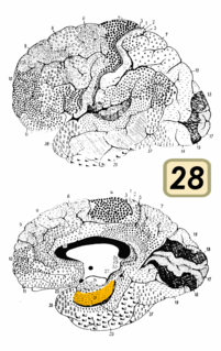
WBrodmann area 28 is a subdivision of the cerebral cortex defined on the basis of cytoarchitecture. It is located on the medial aspect of the temporal lobe and is part of the entorhinal cortex (Brodmann-1909).
 W
WBrodmann area 29, also known as granular retrolimbic area 29 or granular retrosplenial cortex, is a cytoarchitecturally defined portion of the retrosplenial region of the cerebral cortex. In the human it is a narrow band located in the isthmus of cingulate gyrus. Cytoarchitecturally it is bounded internally by the ectosplenial area 26 and externally by the agranular retrolimbic area 30 (Brodmann-1909).
 W
WBrodmann area 30, also known as agranular retrolimbic area 30, is a subdivision of the cytoarchitecturally defined retrosplenial region of the cerebral cortex. In the human it is located in the isthmus of cingulate gyrus. Cytoarchitecturally it is bounded internally by the granular retrolimbic area 29, dorsally by the ventral posterior cingulate area 23 and ventrolaterally by the ectorhinal area 36 (Brodmann-1909).
 W
WBrodmann area 31, also known as dorsal posterior cingulate area 31, is a subdivision of the cytoarchitecturally defined cingulate region of the cerebral cortex. In the human, it occupies portions of the posterior cingulate gyrus and medial aspect of the parietal lobe. Approximate boundaries are the cingulate sulcus dorsally and the parieto-occipital sulcus caudally. It partially surrounds the subparietal sulcus, the ventral continuation of the cingulate sulcus in the parietal lobe. Cytoarchitecturally it is bounded rostrally by the ventral anterior cingulate area 24, ventrally by the ventral posterior cingulate area 23, dorsally by the gigantopyramidal area 4 and preparietal area 5 and caudally by the superior parietal area 7 (H) (Brodmann-1909).
 W
WThe Brodmann area 32, also known in the human brain as the dorsal anterior cingulate area 32, refers to a subdivision of the cytoarchitecturally defined cingulate cortex. In the human it forms an outer arc around the anterior cingulate gyrus. The cingulate sulcus defines approximately its inner boundary and the superior rostral sulcus (H) its ventral boundary; rostrally it extends almost to the margin of the frontal lobe. Cytoarchitecturally it is bounded internally by the ventral anterior cingulate area 24, externally by medial margins of the agranular frontal area 6, intermediate frontal area 8, granular frontal area 9, frontopolar area 10, and prefrontal area 11-1909. (Brodmann19-09).
 W
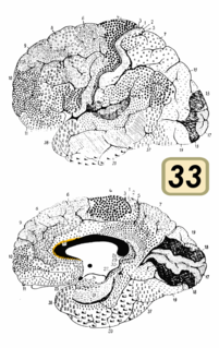
WBrodmann area 33, also known as pregenual area 33, is a subdivision of the cytoarchitecturally defined cingulate region of cerebral cortex. It is a narrow band located in the anterior cingulate gyrus adjacent to the supracallosal gyrus in the depth of the callosal sulcus, near the genu of the corpus callosum. Cytoarchitecturally it is bounded by the ventral anterior cingulate area 24 and the supracallosal gyrus (Brodmann-1909).
 W
WBrodmann area 34 is a part of the brain.
 W
WBrodmann area 35, together with Brodmann area 36, comprise the perirhinal cortex. They are cytoarchitecturally defined temporal regions of the cerebral cortex.
 W
WThe calcarine sulcus is an anatomical landmark located at the caudal end of the medial surface of the brain of humans and other primates. Its name comes from the Latin "calcar" meaning "spur". It is very deep and known as a complete sulcus.
 W
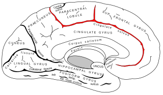
WThe cingulate sulcus is a sulcus on the cingulate cortex in the medial wall of the cerebral cortex. The frontal and parietal lobes are separated from the cingulate gyrus by the cingulate sulcus. It terminates as the marginal sulcus of the cingulate sulcus. It sends a ramus to separate the paracentral lobule from the frontal gyri, the paracentral sulcus.
 W
WThe cuneus is a smaller lobe in the occipital lobe of the brain. The cuneus is bounded anteriorly by the parieto-occipital sulcus, inferiorly by the calcarine sulcus.
 W
WThe cingulate gyrus commences below the rostrum of the corpus callosum, curves around in front of the genu, extends along the upper surface of the body, and finally turns downward behind the splenium, where it is connected by a narrow isthmus with the parahippocampal gyrus.
 W
WThe longitudinal fissure is the deep groove that separates the two cerebral hemispheres of the vertebrate brain. Lying within it is a continuation of the dura mater called the falx cerebri. The inner surfaces of the two hemispheres are convoluted by gyri and sulci just as is the outer surface of the brain.
 W
WParacentral lobule is on the medial surface of the hemisphere and is the continuation of the precentral and postcentral gyri. The paracentral lobule controls motor and sensory innervations of the contralateral lower extremity. It is also responsible for control of defecation and urination.
 W
WThe parieto-occipital sulcus is a deep fissure in the cerebral cortex that marks the boundary between the cuneus and precuneus, and also between the parietal and occipital lobes. Only a small part can be seen on the lateral surface of the hemisphere, its chief part being on the medial surface.
 W
WThe posterior cingulate cortex (PCC) is the caudal part of the cingulate cortex, located posterior to the anterior cingulate cortex. This is the upper part of the "limbic lobe". The cingulate cortex is made up of an area around the midline of the brain. Surrounding areas include the retrosplenial cortex and the precuneus.
 W
WThe precuneus is the portion of the superior parietal lobule on the medial surface of each brain hemisphere. It is located in front of the cuneus. The precuneus is bounded in front by the marginal branch of the cingulate sulcus, at the rear by the parietooccipital sulcus, and underneath by the subparietal sulcus. It is involved with episodic memory, visuospatial processing, reflections upon self, and aspects of consciousness.
 W
WThe retrosplenial cortex (RSC) is a cortical area in the brain, located posteriorly and comprising Brodmann areas 29 and 30. The region's name refers to its anatomical location immediately behind the splenium of the corpus callosum in primates, although in rodents it is located more towards the brain surface and is relatively larger. Its function is currently not well understood, but its location close to visual areas and also to the hippocampal spatial/memory system suggest it may have a role in mediating between perceptual and memory functions.
 W
WThe portion of the inferior frontal lobe immediately adjacent to the longitudinal fissure is named the straight gyrus,(or gyrus rectus) and is continuous with the superior frontal gyrus on the medial surface.
 W
WThe subparietal sulcus or suprasplenial sulcus is a sulcus, or crevice, on the medial surface of each cerebral hemisphere, above the splenium of the corpus callosum. It separates the precuneus from the posterior part of the cingulate gyrus. It is the posterior continuation of the cingulate sulcus. The cingulate sulcus actually "terminates" as the marginal sulcus of the cingulate sulcus. It extends posteriorly toward the calcarine sulcus.