 W
WAcquired brain injury (ABI) is brain damage caused by events after birth, rather than as part of a genetic or congenital disorder such as fetal alcohol syndrome, perinatal illness or perinatal hypoxia. ABI can result in cognitive, physical, emotional, or behavioural impairments that lead to permanent or temporary changes in functioning. These impairments result from either traumatic brain injury or nontraumatic injury derived from either an internal or external source. ABI does not include damage to the brain resulting from neurodegenerative disorders.
 W
WAddiction is a brain disorder characterized by compulsive engagement in rewarding stimuli despite adverse consequences. Despite the involvement of a number of psychosocial factors, a biological process—one that is induced by repeated exposure to an addictive stimulus—is the core pathology that drives the development and maintenance of an addiction, according to the "brain disease model" of addiction. However, many scholars who study addiction argue that the brain disease model is incomplete and misleading.
 W
WAnencephaly is the absence of a major portion of the brain, skull, and scalp that occurs during embryonic development. It is a cephalic disorder that results from a neural tube defect that occurs when the rostral (head) end of the neural tube fails to close, usually between the 23rd and 26th day following conception. Strictly speaking, the Greek term translates as "without a brain", but it is accepted that children born with this disorder usually only lack a telencephalon, the largest part of the brain consisting mainly of the cerebral hemispheres, including the neocortex, which is responsible for cognition. The remaining structure is usually covered only by a thin layer of membrane—skin, bone, meninges, etc. are all lacking. With very few exceptions, infants with this disorder do not survive longer than a few hours or possibly days after their birth.
 W
WAqueductal stenosis is a narrowing of the aqueduct of Sylvius which blocks the flow of cerebrospinal fluid (CSF) in the ventricular system. Blockage of the aqueduct can lead to hydrocephalus, specifically as a common cause of congenital and/or obstructive hydrocephalus.
 W
WAutosomal dominant porencephaly type I is a rare type of porencephaly that causes cysts to grow on the brain and damage to small blood vessels, which can lead to cognitive impairment, migraines, seizures, and hemiplegia or hemiparesis.
 W
WBasal ganglia disease is a group of physical problems that occur when the group of nuclei in the brain known as the basal ganglia fail to properly suppress unwanted movements or to properly prime upper motor neuron circuits to initiate motor function. Research indicates that increased output of the basal ganglia inhibits thalamocortical projection neurons. Proper activation or deactivation of these neurons is an integral component for proper movement. If something causes too much basal ganglia output, then the ventral anterior (VA) and ventral lateral (VL) thalamocortical projection neurons become too inhibited, and one cannot initiate voluntary movement. These disorders are known as hypokinetic disorders. However, a disorder leading to abnormally low output of the basal ganglia leads to reduced inhibition, and thus excitation, of the thalamocortical projection neurons which synapse onto the cortex. This situation leads to an inability to suppress unwanted movements. These disorders are known as hyperkinetic disorders.
 W
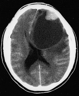
WBrain abscess is an abscess caused by inflammation and collection of infected material, coming from local or remote infectious sources, within the brain tissue. The infection may also be introduced through a skull fracture following a head trauma or surgical procedures. Brain abscess is usually associated with congenital heart disease in young children. It may occur at any age but is most frequent in the third decade of life.
 W
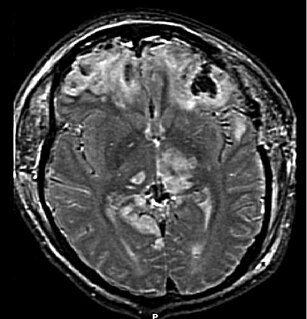
WCerebral amyloid angiopathy (CAA), is a form of angiopathy in which amyloid beta peptide deposits in the walls of small to medium blood vessels of the central nervous system and meninges. The term congophilic is sometimes used because the presence of the abnormal aggregations of amyloid can be demonstrated by microscopic examination of brain tissue after staining with Congo red. The amyloid material is only found in the brain and as such the disease is not related to other forms of amyloidosis.
 W
WCerebral folate deficiency is a condition in which concentrations of 5-methyltetrahydrofolate are low in the brain as measured in the cerebral spinal fluid despite being normal in the blood. Symptoms typically appear at about 5 to 24 months of age. Without treatment there may be poor muscle tone, trouble with coordination, trouble talking, and seizures.
 W
WCerebral hypoxia is a form of hypoxia, specifically involving the brain; when the brain is completely deprived of oxygen, it is called cerebral anoxia. There are four categories of cerebral hypoxia; they are, in order of severity: diffuse cerebral hypoxia (DCH), focal cerebral ischemia, cerebral infarction, and global cerebral ischemia. Prolonged hypoxia induces neuronal cell death via apoptosis, resulting in a hypoxic brain injury.
 W
WCerebral softening, also known as encephalomalacia, is a localized softening of the substance of the brain, due to bleeding or inflammation. Three varieties, distinguished by their color and representing different stages of the disease progress, are known respectively as red, yellow, and white softening.
 W
WChildhood acquired brain injury (ABI) is the term given to any injury to the brain that occurs during childhood but after birth and the immediate neonatal period. It excludes injuries sustained as a result of genetic or congenital disorder. It also excludes those resulting from birth traumas such as hypoxia or conditions such as foetal alcohol syndrome. It encompasses both traumatic and non-traumatic injuries.
 W
WEthylmalonic encephalopathy (EE) is a rare autosomal recessive inborn error of metabolism. Patients affected with EE are typically identified shortly after birth, with symptoms including diarrhea, petechiae and seizures. The genetic defect in EE is thought to involve an impairment in the degradation of sulfide intermediates in the body. Hydrogen sulfide then builds up to toxic levels. EE was initially described in 1994. Most cases of EE have been described in individuals of Mediterranean or Arabic origin.
WFrontotemporal dementia and parkinsonism linked to chromosome 17 (FTDP-17) is an autosomal dominant neurodegenerative tauopathy and Parkinson plus syndrome. FTDP-17 is caused by mutations in the MAPT gene located on the q arm of chromosome 17, and has three cardinal features: behavioral and personality changes, cognitive impairment, and motor symptoms. FTDP-17 was defined during the International Consensus Conference in Ann Arbor, Michigan, in 1996.
 W
WGómez–López-Hernández syndrome (GLH) or cerebellotrigeminal-dermal dysplasia is a rare neurocutaneous (Phakomatosis) disorder affecting the trigeminal nerve and causing several other neural and physical abnormalities. Gómez–López-Hernández syndrome has been diagnosed in only 34 people. Cases of Gómez–López-Hernández syndrome may be under-reported as other diseases share the characteristics of cerebellar malformation shown in Gómez–López-Hernández syndrome. Gómez–López-Hernández syndrome was first characterized in 1979.
 W
WHashimoto's encephalopathy, also known as steroid-responsive encephalopathy associated with autoimmune thyroiditis (SREAT), is a neurological condition characterized by encephalopathy, thyroid autoimmunity, and good clinical response to corticosteroids. It is associated with Hashimoto's thyroiditis, and was first described in 1966. It is sometimes referred to as a neuroendocrine disorder, although the condition's relationship to the endocrine system is widely disputed. It is recognized as a rare disease by the NIH Genetic and Rare Diseases Information Center.
 W
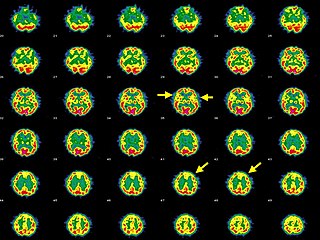
WHepatic encephalopathy (HE) is an altered level of consciousness as a result of liver failure. Its onset may be gradual or sudden. Other symptoms may include movement problems, changes in mood, or changes in personality. In the advanced stages it can result in a coma.
WLeukoencephalopathy with neuroaxonal spheroids is a special kind of leukoencephalopathy. It is a cause of severe and subacute dementia. It is inherited, following an autosomal dominant pattern.
 W
WMedial medullary syndrome, also known as inferior alternating syndrome, hypoglossal alternating hemiplegia, lower alternating hemiplegia, or Dejerine syndrome, is a type of alternating hemiplegia characterized by a set of clinical features resulting from occlusion of the anterior spinal artery. This results in the infarction of medial part of the medulla oblongata.
 W
WNeurocysticercosis is a specific form of the infectious parasitic disease cysticercosis that is caused by the infection with Taenia solium, a tapeworm found in pigs. Neurocysticercosis occurs when cysts formed by the infection take hold within the brain, causing neurologic syndromes such as epileptic seizures. It is a common cause of seizures worldwide. It has been called a "hidden epidemic" and "arguably the most common parasitic disease of the human nervous system". Common symptoms of neurocysticercosis include seizures, headaches, blindness, meningitis and dementia.
 W
WOlivopontocerebellar atrophy (OPCA) is the degeneration of neurons in specific areas of the brain – the cerebellum, pons, and inferior olivary nucleus. OPCA is present in several neurodegenerative syndromes, including inherited and non-inherited forms of ataxia and multiple system atrophy (MSA), with which it is primarily associated.
WChildhood acquired brain injury (ABI) is the term given to any injury to the brain that occurs during childhood but after birth and the immediate neonatal period. It excludes injuries sustained as a result of genetic or congenital disorder. It also excludes those resulting from birth traumas such as hypoxia or conditions such as foetal alcohol syndrome. It encompasses both traumatic and non-traumatic injuries.
 W
WPleomorphic xanthoastrocytoma (PXA) is a brain tumor that occurs most frequently in children and teenagers. At Boston Children's Hospital, the average age at diagnosis is 12 years.
 W
WProgressive multifocal leukoencephalopathy (PML) is a rare and often fatal viral disease characterized by progressive damage (-pathy) or inflammation of the white matter (leuko-) of the brain (-encephalo-) at multiple locations (multifocal). It is caused by the JC virus, which is normally present and kept under control by the immune system. The JC virus is harmless except in cases of weakened immune systems. In general, PML has a mortality rate of 30–50% in the first few months, and those who survive can be left with varying degrees of neurological disabilities.
 W
WRight hemisphere brain damage (RHD) is the result of injury to the right brain hemisphere. The right hemisphere of the brain coordinates tasks for functional communication, which include problem solving, memory, and reasoning. Deficits caused by right hemisphere brain damage vary depending on the location of the damage.
WVentriculitis is the inflammation of the ventricles in the brain. The ventricles are responsible for containing and circulating cerebrospinal fluid throughout the brain. Ventriculitis is caused by infection of the ventricles, leading to swelling and inflammation. This is especially prevalent in patients with external ventricular drains and intraventricular stents. Ventriculitis can cause a wide variety of short-term symptoms and long-term side effects ranging from headaches and dizziness to unconsciousness and death if not treated early. It is treated with some appropriate combination of antibiotics in order to rid the patient of the underlying infection. Much of the current research involving ventriculitis focuses specifically around defining the disease and what causes it. This will allow for much more advancement in the subject. There is also a lot of attention being paid to possible treatments and prevention methods to help make this disease even less prevalent and dangerous.
 W
WVisual extinction is a neurological disorder which occurs following damage to the parietal lobe of the brain. It is similar to, but distinct from, hemispatial neglect. Visual extinction has the characteristic symptom of difficulty to perceive contralesional stimuli when presented simultaneously with an ipsilesional stimulus, but the ability to correctly identify them when not presented simultaneously. Under simultaneous presentation, the contralesional stimulus is apparently ignored by the patient, or extinguished. This deficiency may lead to difficulty on behalf of the patient with processing the stimuli’s 3D position.