 W
WThe arcuate nucleus of the hypothalamus is an aggregation of neurons in the mediobasal hypothalamus, adjacent to the third ventricle and the median eminence. The arcuate nucleus includes several important and diverse populations of neurons that help mediate different neuroendocrine and physiological functions, including neuroendocrine neurons, centrally projecting neurons, and astrocytes. The populations of neurons found in the arcuate nucleus are based on the hormones they secrete or interact with and are responsible for hypothalamic function, such as regulating hormones released from the pituitary gland or secreting their own hormones. Neurons in this region are also responsible for integrating information and providing inputs to other nuclei in the hypothalamus or inputs to areas outside this region of the brain. These neurons, generated from the ventral part of the periventricular epithelium during embryonic development, locate dorsally in the hypothalamus, becoming part of the ventromedial hypothalamic region. The function of the arcuate nucleus relies on its diversity of neurons, but its central role is involved in homeostasis. The arcuate nucleus provides many physiological roles involved in feeding, metabolism, fertility, and cardiovascular regulation.
 W
WRebecca M. Calisi Rodriguez is an American neuroendocrinologist, wildlife biologist, and National Geographic Explorer. She is an Associate professor of Neurobiology, Physiology, and Behavior in the College of Biological Sciences at the University of California, Davis. Calisi leads a research team that studies how the brain controls sexual behavior, reproduction, and parental care, and how this changes under stress. As the Director for Science Communications at UC Davis, Calisi also studies science communication and is a well-known advocate for inclusivity, equity, and diversity in STEM.
 W
WThe cortisol awakening response (CAR) is an increase between 38% and 75% in cortisol levels peaking 30–45 minutes after awakening in the morning in some people. This rise is superimposed upon the late-night rise in cortisol which occurs before awakening. While its purpose is uncertain, it may be linked to the hippocampus' preparation of the hypothalamic-pituitary-adrenal axis (HPA) in order to face anticipated stress.
 W
WGigantism, also known as giantism, is a condition characterized by excessive growth and height significantly above average. In humans, this condition is caused by over-production of growth hormone in childhood, resulting in people 7 to 9 ft in height.
 W
WHashimoto's encephalopathy, also known as steroid-responsive encephalopathy associated with autoimmune thyroiditis (SREAT), is a neurological condition characterized by encephalopathy, thyroid autoimmunity, and good clinical response to corticosteroids. It is associated with Hashimoto's thyroiditis, and was first described in 1966. It is sometimes referred to as a neuroendocrine disorder, although the condition's relationship to the endocrine system is widely disputed. It is recognized as a rare disease by the NIH Genetic and Rare Diseases Information Center.
 W
WThe hypothalamic–pituitary–adrenal axis is a complex set of direct influences and feedback interactions among three components: the hypothalamus, the pituitary gland, and the adrenal glands.
 W
WThe hypothalamic–pituitary–gonadal axis refers to the hypothalamus, pituitary gland, and gonadal glands as if these individual endocrine glands were a single entity. Because these glands often act in concert, physiologists and endocrinologists find it convenient and descriptive to speak of them as a single system.
 W
WThe hypothalamic–pituitary–prolactin axis, also known as the hypothalamic–pituitary–mammary axis or hypothalamic–pituitary–breast axis, is a hypothalamic–pituitary axis which includes the secretion of prolactin from the lactotrophs of the pituitary gland into the circulation and the subsequent action of prolactin on tissues such as, particularly, the mammary glands or breasts. It is involved in lobuloalveolar maturation of the mammary glands during pregnancy and the induction and maintenance of lactation following parturition. Hormones that control the secretion of prolactin from the pituitary gland include dopamine, estradiol, progesterone, thyrotropin-releasing hormone (TRH), and vasoactive intestinal peptide (VIP).
 W
WThe hypothalamic–pituitary–somatotropic axis, or hypothalamic–pituitary–somatic axis, also known as the hypothalamic–pituitary–growth axis, is a hypothalamic–pituitary axis which includes the secretion of growth hormone from the somatotropes of the pituitary gland into the circulation and the subsequent stimulation of insulin-like growth factor 1 production by GH in tissues such as, namely, the liver. Other hypothalamic–pituitary hormones such as growth hormone-releasing hormone, growth hormone-inhibiting hormone, and ghrelin (GHS) are involved in the control of GH secretion from the pituitary gland. The HPS axis is involved in postnatal human growth. Individuals with growth hormone deficiency or Laron syndrome show symptoms like short stature, dwarfism and obesity, but are also protected from some forms of cancer. Conversely, acromegaly and gigantism are conditions of GH and IGF-1 excess usually due to a pituitary tumor, and are characterized by overgrowth and tall stature.
 W
WThe hypothalamus is a portion of the brain that contains a number of small nuclei with a variety of functions. One of the most important functions of the hypothalamus is to link the nervous system to the endocrine system via the pituitary gland. The hypothalamus is located below the thalamus and is part of the limbic system. In the terminology of neuroanatomy, it forms the ventral part of the diencephalon. All vertebrate brains contain a hypothalamus. In humans, it is the size of an almond.
WThe median eminence, part of the inferior boundary of the hypothalamus in the brain, is attached to the infundibulum. The median eminence is a small swelling on the tuber cinereum, posterior to and atop the pituitary stalk; it lies in the area roughly bounded on its posterolateral region by the cerebral peduncles, and on its anterolateral region by the optic chiasm.
 W
WNeuroendocrine tumors (NETs) are neoplasms that arise from cells of the endocrine (hormonal) and nervous systems. They most commonly occur in the intestine, where they are often called carcinoid tumors, but they are also found in the pancreas, lung and the rest of the body.
 W
WThe paraventricular nucleus is a nucleus in the hypothalamus. Anatomically, it is adjacent to the third ventricle and many of its neurons project to the posterior pituitary. These projecting neurons secrete oxytocin and a smaller amount of vasopressin, otherwise the nucleus also secretes corticotropin-releasing hormone (CRH) and thyrotropin-releasing hormone (TRH). CRH and TRH are secreted into the hypophyseal portal system and act on different targets neurons in the anterior pituitary. PVN is thought to mediate many diverse functions through these different hormones, including osmoregulation, appetite, and the response of the body to stress.
 W
WParental experience, as well as changing hormone levels during pregnancy and postpartum, cause changes in the parental brain. Displaying maternal sensitivity towards infant cues, processing those cues and being motivated to engage socially with her infant and attend to the infant's needs in any context could be described as mothering behavior and is regulated by many systems in the maternal brain. Research has shown that hormones such as oxytocin, prolactin, estradiol and progesterone are essential for the onset and the maintenance of maternal behavior in rats, and other mammals as well. Mothering behavior has also been classified within the basic drives. Less is known about the paternal brain, but changes in the father's brain occur alongside the mother once the offspring is born.
 W
WLillian Mary Pickford was a pioneering neuroendocrinologist. She was the first woman to be elected to the Pharmacological Society and the first woman appointed to a medical professorship at the University of Edinburgh.
 W
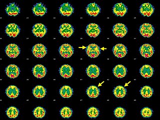
WA pineal gland cyst is a usually benign (non-malignant) cyst in the pineal gland, a small endocrine gland in the brain. Historically, these fluid-filled bodies appeared on 1-4% of magnetic resonance imaging (MRI) brain scans, but were more frequently diagnosed at death, seen in 4-11% of autopsies. A 2007 study by Pu et al. found a frequency of 23% in brain scans.
 W
WPituitary adenomas are tumors that occur in the pituitary gland. Pituitary adenomas are generally divided into three categories dependent upon their biological functioning: benign adenoma, invasive adenoma, and carcinomas. Most adenomas are benign, approximately 35% are invasive and just 0.1% to 0.2% are carcinomas. Pituitary adenomas represent from 10% to 25% of all intracranial neoplasms and the estimated prevalence rate in the general population is approximately 17%.
 W
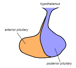
WIn vertebrate anatomy, the pituitary gland, or hypophysis, is an endocrine gland, about the size of a pea and weighing 0.5 grams (0.018 oz) in humans. It is a protrusion off the bottom of the hypothalamus at the base of the brain. The hypophysis rests upon the hypophysial fossa of the sphenoid bone in the center of the middle cranial fossa and is surrounded by a small bony cavity covered by a dural fold. The anterior pituitary is a lobe of the gland that regulates several physiological processes. The intermediate lobe synthesizes and secretes melanocyte-stimulating hormone. The posterior pituitary is a lobe of the gland that is functionally connected to the hypothalamus by the median eminence via a small tube called the pituitary stalk.
 W
WThe posterior pituitary is the posterior lobe of the pituitary gland which is part of the endocrine system. The posterior pituitary is not glandular as is the anterior pituitary. Instead, it is largely a collection of axonal projections from the hypothalamus that terminate behind the anterior pituitary, and serve as a site for the secretion of neurohypophysial hormones directly into the blood. The hypothalamic–neurohypophyseal system is composed of the hypothalamus, posterior pituitary, and these axonal projections.
 W
WPro-opiomelanocortin (POMC) is a precursor polypeptide with 241 amino acid residues. POMC is synthesized in corticotrophs of the anterior pituitary from the 285-amino-acid-long polypeptide precursor pre-pro-opiomelanocortin (pre-POMC), by the removal of a 44-amino-acid-long signal peptide sequence during translation. POMC is part of the central melanocortin system.
 W
WA Rathke's cleft cyst is a benign growth on the pituitary gland in the brain, specifically a mucin-filled cyst in the posterior portion of the anterior pituitary gland. It occurs when the Rathke's pouch does not develop properly and ranges in size from 2 to 40 mm in diameter.
 W
WSomatostatin, also known as growth hormone-inhibiting hormone (GHIH) or by several other names, is a peptide hormone that regulates the endocrine system and affects neurotransmission and cell proliferation via interaction with G protein-coupled somatostatin receptors and inhibition of the release of numerous secondary hormones. Somatostatin inhibits insulin and glucagon secretion.
 W
WThe subfornical organ (SFO) is one of the circumventricular organs of the brain. Its name comes from its location on the ventral surface of the fornix near the interventricular foramina, which interconnect the lateral ventricles and the third ventricle. Like all circumventricular organs, the subfornical organ is well-vascularized, and like all circumventricular organs except the subcommissural organ, some SFO capillaries have fenestrations, which increase capillary permeability. The SFO is considered a sensory circumventricular organ because it is responsive to a wide variety of hormones and neurotransmitters, as opposed to secretory circumventricular organs, which are specialized in the release of certain substances.
 W
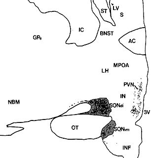
WThe supraoptic nucleus (SON) is a nucleus of magnocellular neurosecretory cells in the hypothalamus of the mammalian brain. The nucleus is situated at the base of the brain, adjacent to the optic chiasm. In humans, the SON contains about 3,000 neurons.
 W
WTestosterone is the primary sex hormone and anabolic steroid in males. In male humans, testosterone plays a key role in the development of male reproductive tissues such as testes and prostate, as well as promoting secondary sexual characteristics such as increased muscle and bone mass, and the growth of body hair. In addition, testosterone is involved in health and well-being, and the prevention of osteoporosis. Insufficient levels of testosterone in men may lead to abnormalities including frailty and bone loss.
 W
WVasopressin, also called antidiuretic hormone (ADH), arginine vasopressin (AVP) or argipressin, is a hormone synthesized as a peptide prohormone in neurons in the hypothalamus, and is converted to AVP. It then travels down the axon of that cell, which terminates in the posterior pituitary, and is released from vesicles into the circulation in response to extracellular fluid hypertonicity (hyperosmolality). AVP has two primary functions. First, it increases the amount of solute-free water reabsorbed back into the circulation from the filtrate in the kidney tubules of the nephrons. Second, AVP constricts arterioles, which increases peripheral vascular resistance and raises arterial blood pressure.
WThe ventromedial nucleus of the hypothalamus is a nucleus of the hypothalamus. "The ventromedial hypothalamus (VMH) is a distinct morphological nucleus involved in terminating hunger, fear, thermoregulation, and sexual activity." This nuclear region is involved with the recognition of the feeling of fullness.