 W
WRadiology is the medical discipline that uses medical imaging to diagnose and treat diseases within the bodies of animals and humans.
 W
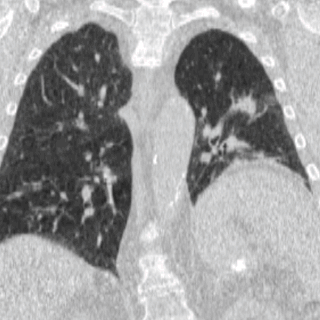
WFour-dimensional computed tomography (4DCT) is a type of CT scanning which records multiple images over time. It allows playback of the scan as a video, so that physiological processes can be observed and internal movement can be tracked. The name is derived from the addition of time to traditional 3D computed tomography. Alternatively, the phase of a particular process, such as respiration, may be considered the fourth dimension.
 W
WAcute radiation syndrome (ARS), also known as radiation sickness or radiation poisoning, is a collection of health effects that are caused by being exposed to high amounts of ionizing radiation, in a short period of time. The symptoms of ARS can start within the hour of exposure, and can last for several months. Within the first few days the symptoms are usually nausea, vomiting and a loss of appetite. In the following few hours or weeks will be a few symptoms, which later become additional symptoms, after which either recovery or death follow.
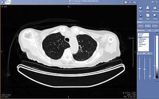
Aidoc Medical is an Israeli technology company that develops computer-aided simple triage and notification systems. Aidoc has obtained FDA and CE mark approval for its stroke, pulmonary embolism, cervical fracture, intracranial hemorrhage, intra-abdominal free gas, and incidental pulmonary embolism algorithms.
 W
WA bone scan or bone scintigraphy is a nuclear medicine imaging technique of the bone. It can help diagnose a number of bone conditions, including cancer of the bone or metastasis, location of bone inflammation and fractures, and bone infection (osteomyelitis).
 W
WCaldwell's view is a radiographic view of skull, where X-ray plate is perpendicular to the orbitomeatal line. The rays pass from behind the head and are angled at 15-20° to the radiographic plate. It is commonly used to get better view of the ethmoid and frontal sinuses. It is named after the noted American radiologist Eugene W. Caldwell, who described it in 1907.
 W
WCompanion shadow is a term used in describing radiographs that denotes the appearance of a smooth, homogenous, radiodensity with a well-defined margin that runs parallel with a bony landmark. Companion shadows represent soft tissue that overlies the respective bony landmark in profile. They are not seen in every radiograph and can be misinterpreted as pathology.
 W
WA CT scan or computed tomography scan is a medical imaging technique used in radiology to obtain detailed internal images of the body noninvasively for diagnostic purposes. The personnel that perform CT scans are called radiographers or radiology technologists.
 W
WComputer-aided detection (CADe), also called computer-aided diagnosis (CADx), are systems that assist doctors in the interpretation of medical images. Imaging techniques in X-ray, MRI, and ultrasound diagnostics yield a great deal of information that the radiologist or other medical professional has to analyze and evaluate comprehensively in a short time. CAD systems process digital images for typical appearances and to highlight conspicuous sections, such as possible diseases, in order to offer input to support a decision taken by the professional.
 W
WDual-energy X-ray absorptiometry is a means of measuring bone mineral density (BMD) using spectral imaging. Two X-ray beams, with different energy levels, are aimed at the patient's bones. When soft tissue absorption is subtracted out, the bone mineral density (BMD) can be determined from the absorption of each beam by bone. Dual-energy X-ray absorptiometry is the most widely used and most thoroughly studied bone density measurement technology.
 W
WAn echocardiography, echocardiogram, cardiac echo or simply an echo, is an ultrasound of the heart. It is a type of medical imaging of the heart, using standard ultrasound or Doppler ultrasound.
 W
WEOS imaging is a medical device company based in Paris, France that designs, develops, and markets EOSedge™ and the EOS system, innovative, orthopedic medical imaging systems, associated with several orthopedic solutions along the patient care pathway – from diagnosis to post-operative treatments. The EOS platform targets musculoskeletal disorders and orthopedic surgical care through 2D X-ray scans and 3D skeletal models from stereo-radiographic images of patients in a seated or standing position.
The European Day of Radiology (EDoR) is an annual day of action that will take place for the first time on February 10, 2011. The day is an initiative of the European Society of Radiology, an organisation that represents the interests of radiology and its practitioners throughout Europe and also hosts the European Congress of Radiology.
 W
WForensic radiology is the discipline which comprises the performance, interpretation and reportage of the radiological examinations and procedures which are needed in court procedures or law enforcement. Radiological methods are widely used in identification, age estimation and establishing cause of death. Comparison of ante mortem and post mortem radiographs is one of the means of identification. The scanning of baggage, vehicles and individuals have many applications.
 W
WThe John Thomas sign, also known as the Throckmorton sign, is a slang or joke term used in the field of radiology. It refers to the position of a penis as it relates to pathology on an X-ray of a pelvis. When the penis points towards the same side as a unilateral medical condition such as a broken bone, this is considered a "positive John Thomas sign", and if the shadow points to the other side, it is a "negative John Thomas sign."
 W
WLaboratory Unit for Computer Assisted Surgery is a system used for virtual surgical planning. Starting with 1998, LUCAS was developed at the University of Regensburg, Germany, with the support of the Carl Zeiss Company. The resulting surgical planning is then reproduced onto the patient by using a navigation system. In fact, LUCAS is integrated into the same platform together with the Surgical Segment Navigator (SSN), the Surgical Tool Navigator (STN), the Surgical Microscope Navigator (SMN) and the 6DOF Manipulator, also from the Carl Zeiss Company.
 W
WMagnetic resonance imaging (MRI) is a medical imaging technique used in radiology to form pictures of the anatomy and the physiological processes of the body. MRI scanners use strong magnetic fields, magnetic field gradients, and radio waves to generate images of the organs in the body. MRI does not involve X-rays or the use of ionizing radiation, which distinguishes it from CT and PET scans. MRI is a medical application of nuclear magnetic resonance (NMR) which can also be used for imaging in other NMR applications, such as NMR spectroscopy.
 W
WNeuroimaging or brain imaging is the use of various techniques to either directly or indirectly image the structure, function, or pharmacology of the nervous system. It is a relatively new discipline within medicine, neuroscience, and psychology. Physicians who specialize in the performance and interpretation of neuroimaging in the clinical setting are neuroradiologists. Neuroimaging falls into two broad categories:Structural imaging, which deals with the structure of the nervous system and the diagnosis of gross intracranial disease and injury. Functional imaging, which is used to diagnose metabolic diseases and lesions on a finer scale and also for neurological and cognitive psychology research and building brain–computer interfaces.
 W
WNuclear medicine is a medical specialty involving the application of radioactive substances in the diagnosis and treatment of disease. Nuclear medicine imaging, in a sense, is "radiology done inside out" or "endoradiology" because it records radiation emitting from within the body rather than radiation that is generated by external sources like X-rays. In addition, nuclear medicine scans differ from radiology, as the emphasis is not on imaging anatomy, but on the function. For such reason, it is called a physiological imaging modality. Single photon emission computed tomography (SPECT) and positron emission tomography (PET) scans are the two most common imaging modalities in nuclear medicine.
 W
WOwl's eye appearance, also known as Owl’s Eye Sign, is commonly used as a term to describe a pattern that is found in the study of histology, radiology, and pathology cases. This pattern has a resemblance of a real Owl’s Eyes Shape. Such an appearance of a pattern has been used for the analysis of symptoms on patients within the medical field.
 W
WPaleoradiology is the study of archaeological remains through the use of radiographic techniques, such as X-ray, CT and micro-CT scans. It is predominately used by archaeologists and anthropologists to examine mummified remains due to its non-invasive nature. Paleoradiologists can discover post-mortem damage to the body, or any artefacts buried with them, while still keeping the remains intact. Radiological images can also contribute evidence about the person's life, such as their age and cause of death. The first recorded use of paleoradiology was in 1896, just a year after the Rōntgen radiograph was first produced. Although this method of viewing ancient remains is advantageous due to its non-invasive manner, many radiologists lack expertise in archeology and very few radiologists can identify ancient diseases which may be present.
 W
WRadiographers, also known as radiologic technologists, diagnostic radiographers and medical radiation technologists are healthcare professionals who specialise in the imaging of human anatomy for the diagnosis and treatment of pathology. Radiographers are infrequently, and almost always erroneously, known as x-ray technicians. In countries that use the title radiologic technologist they are often informally referred to as techs in the clinical environment; this phrase has emerged in popular culture such as television programmes. The term radiographer can also refer to a therapeutic radiographer, also known as a radiation therapist.
 W
WA rectilinear scanner is an imaging device, used to capture emission from radiopharmaceuticals in nuclear medicine. The image is created by physically moving a radiation detector over the surface of a radioactive patient. It has become obsolete in medical imaging, largely replaced by the gamma camera since the late 1960s.
 W
WReid's base line is used for an unambiguous definition of the orientation of the human skull in conventional radiography, computer tomography (CT), and magnetic resonance imaging (MRI) studies. It is defined as a line drawn from the inferior margin of the orbit to the auricular point and extending backward to the center of the occipital bone. Reid's base line is used as the zero plane in computed tomography.
 W
WSchuller's view is a lateral radiographic view of skull principally used for viewing mastoid cells. The central beam of X-rays passes from one side of the head and is at angle of 25° caudad to radiographic plate. This angulation prevents overlap of images of two mastoid bones. Radiograph for each mastoid is taken separately. The Schullers view serves as an alternative view to the Law projection which uses a 15 degree angle of patient's face toward the image receptor and a 15 degree caudal angulation of the CR to achieve the same result, a lateral mastoid air cells view without overlap of the opposite side. Ear(pinna) under examination can be taped forward to avoid cartilage shadow around mastoid. Older editions of Merrill's positioning books will have detailed explanation of these and other mastoid positions. Newer texts often omit this because of the rarity of this exam in lieu of computed tomography studies.
 W
WThe surgical planning is the preoperative method of pre-visualising a surgical intervention, in order to predefine the surgical steps and furthermore the bone segment navigation in the context of computer-assisted surgery. The surgical planning is most important in neurosurgery and oral and maxillofacial surgery. The transfer of the surgical planning to the patient is generally made using a medical navigation system.
 W
WTeleradiology is the transmission of radiological patient images, such as x-rays, CTs, and MRIs, from one location to another for the purposes of sharing studies with other radiologists and physicians. Teleradiology is a growth technology given that imaging procedures are growing approximately 15% annually against an increase of only 2% in the radiologist population.
 W
WTomographic reconstruction is a type of multidimensional inverse problem where the challenge is to yield an estimate of a specific system from a finite number of projections. The mathematical basis for tomographic imaging was laid down by Johann Radon. A notable example of applications is the reconstruction of computed tomography (CT) where cross-sectional images of patients are obtained in non-invasive manner. Recent developments have seen the Radon transform and its inverse used for tasks related to realistic object insertion required for testing and evaluating computed tomography use in airport security.
 W
WWaters' view is a radiographic view, where an X-ray beam is angled at 45° to the orbitomeatal line. The rays pass from behind the head and are perpendicular to the radiographic plate. It is commonly used to get a better view of the maxillary sinuses. Another variation of the waters according to Merrill's Atlas of Radiographic Positioning and Procedures places the orbitomeatal line at a 37° angle to the image receptor. It is named after the American radiologist Charles Alexander Waters.
 W
WAn X-ray tube is a vacuum tube that converts electrical input power into X-rays. The availability of this controllable source of X-rays created the field of radiography, the imaging of partly opaque objects with penetrating radiation. In contrast to other sources of ionizing radiation, X-rays are only produced as long as the X-ray tube is energized. X-ray tubes are also used in CT scanners, airport luggage scanners, X-ray crystallography, material and structure analysis, and for industrial inspection.