 W
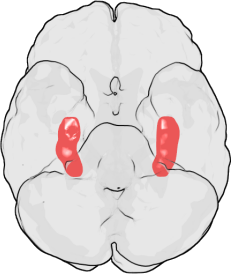
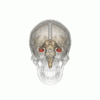
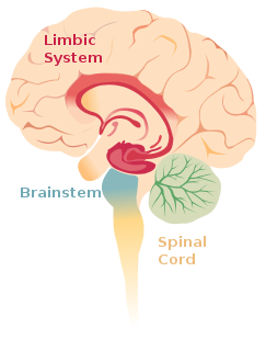
WThe hippocampus is a major component of the brain of humans and other vertebrates. Humans and other mammals have two hippocampi, one in each side of the brain. The hippocampus is part of the limbic system, and plays important roles in the consolidation of information from short-term memory to long-term memory, and in spatial memory that enables navigation. The hippocampus is located in the allocortex, with neural projections into the neocortex in humans, as well as primates. The hippocampus, as the medial pallium, is a structure found in all vertebrates. In humans, it contains two main interlocking parts: the hippocampus proper, and the dentate gyrus.
 W
WAmygdalohippocampectomy is a surgical procedure for the treatment of epilepsy. It consists of the removal of the hippocampus, which has a role in memory, spatial awareness, and navigation, and the amygdalae, which have a role in the processing and memory of emotional reactions, both structures forming part of the limbic system of the brain.
 W
WThe archicortex, or archipallium, is the phylogenetically oldest region of the brain's cerebral cortex. It is often considered contiguous with the olfactory cortex, but its extent varies among species. In older species, such as fish, the archipallium makes up most of the cerebrum. Amphibians develop an archipallium and paleopallium.
 W
WBoundary cells are neurons found in the hippocampal formation that respond to the presence of an environmental boundary at a particular distance and direction from an animal. The existence of cells with these firing characteristics were first predicted on the basis of properties of place cells. Boundary cells were subsequently discovered in several regions of the hippocampal formation: the subiculum, presubiculum and entorhinal cortex.
 W
WThe calcar avis, previously known as the hippocampus minor, is an involution of the wall of the lateral ventricle's posterior cornu produced by the calcarine fissure.
 W
WThe dentate gyrus (DG) is part of the hippocampal formation in the temporal lobe of the brain, which also includes the hippocampus and the subiculum. The dentate gyrus is part of the hippocampal trisynaptic circuit and is thought to contribute to the formation of new episodic memories, the spontaneous exploration of novel environments and other functions.
 W
WEthanol is the type of alcohol found in alcoholic beverages. It is a volatile, flammable, colorless liquid that acts as a central nervous system depressant. Ethanol can impair different types of memory.
 W
WThe fascia dentata is the earliest stage of the hippocampal circuit. Its primary input is the perforant path from the superficial layers of entorhinal cortex. Its principal neurons are tiny granule cells which give rise to unmyelinated axons called the mossy fibers which project to the hilus and CA3. The fascia dentata of the rat contains approximately 1,000,000 granule cells. It receives feedback connections from mossy cells in the hilus at distant levels in the septal and temporal directions. The fascia dentata and the hilus together make up the dentate gyrus. As with all regions of the hippocampus, the dentate gyrus also receives GABAergic and cholinergic input from the medial septum and the diagonal band of Broca.
 W
WThe fornix is a C-shaped bundle of nerve fibers in the brain that acts as the major output tract of the hippocampus. The fornix also carries some afferent fibers to the hippocampus from structures in the diencephalon and basal forebrain. The fornix is part of the limbic system. While its exact function and importance in the physiology of the brain are still not entirely clear, it has been demonstrated in humans that surgical transection – the cutting of the fornix along its body – can cause memory loss. There is some debate over what type of memory is affected by this damage, but it has been found to most closely correlate with recall memory rather than recognition memory. This means that damage to the fornix can cause difficulty in recalling long-term information such as details of past events, but it has little effect on the ability to recognize objects or familiar situations.
 W
WThe hippocampus is an area of the brain integral to learning and memory. Removal of this structure can result in the inability to form new memories as most famously demonstrated in a patient referred to as HM. The unique morphology of the hippocampus can be appreciated without the use of special stains and this distinct circuitry has helped further the understanding of neuronal signal potentiation. The following will provide an introduction to hippocampal development with particular focus on the role of glucocorticoid signaling.
 W
WHippocalcin is a protein that in humans is encoded by the HPCA gene.
WThe hippocampal formation is a compound structure in the medial temporal lobe of the brain. There is no consensus concerning which brain regions are encompassed by the term, with some authors defining it as the dentate gyrus, the hippocampus proper and the subiculum; and others including also the presubiculum, parasubiculum, and entorhinal cortex. The hippocampal formation is thought to play a role in memory, spatial navigation and control of attention. The neural layout and pathways within the hippocampal formation are very similar in all mammals.
 W
WHippocampal sclerosis (HS) or Mesial temporal sclerosis (MTS) is a neuropathological condition with severe neuronal cell loss and gliosis in the hippocampus, specifically in the CA-1 and subiculum of the hippocampus. It was first described in 1880 by Wilhelm Sommer. Hippocampal sclerosis is a frequent pathologic finding in community-based dementia. Hippocampal sclerosis can be detected with autopsy or MRI. In MRI, a decrease in signal is observed at T1 and an increase in signal at T2. Positron emission tomography is also used as an aid for diagnosis. In PET examination, glucose uptake is lower than in the normal part. The reason for this is that the sclerotic part works at a lower level than the normal part and needs less energy. Individuals with hippocampal sclerosis have similar initial symptoms and rates of dementia progression to those with Alzheimer's disease (AD) and therefore are frequently misclassified as having Alzheimer's Disease. But clinical and pathologic findings suggest that hippocampal sclerosis has characteristics of a progressive disorder although the underlying cause remains elusive. A diagnosis of hippocampal sclerosis has a significant effect on the life of patients because of the notable mortality, morbidity and social impact related to epilepsy, as well as side effects associated with antiepileptic treatments. Findings indicate that there is a strong genetic connection in the development of mesial temporal sclerosis.Mesial temporal sclerosis used to be most commonly found as a single lesion in the brains of chronic epileptics who died a natural death which was estimated to be developed as a result of continued febrile convulsions.
 W
WThe hippocampal sulcus, also known as the hippocampal fissure, is a sulcus that separates the dentate gyrus from the subiculum and the CA1 field in the hippocampus.
WHippocampus anatomy describes the physical aspects and properties of the hippocampus, a neural structure in the medial temporal lobe of the brain. It has a distinctive, curved shape that has been likened to the sea-horse monster of Greek mythology and the ram's horns of Amun in Egyptian mythology. This general layout holds across the full range of mammalian species, from hedgehog to human, although the details vary. For example, in the rat, the two hippocampi look similar to a pair of bananas, joined at the stems. In primate brains, including humans, the portion of the hippocampus near the base of the temporal lobe is much broader than the part at the top. Due to the three-dimensional curvature of this structure, two-dimensional sections such as shown are commonly seen. Neuroimaging pictures can show a number of different shapes, depending on the angle and location of the cut.
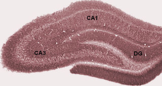
WThe hippocampus proper refers to the actual structure of the hippocampus which is made up of three regions or subfields. The subfields CA1, CA2, and CA3 use the initials of cornu Ammonis, an earlier name of the hippocampus.
 W
WIn the hippocampus, the mossy fiber pathway consists of unmyelinated axons projecting from granule cells in the dentate gyrus that terminate on modulatory hilar mossy cells and in Cornu Ammonis area 3 (CA3), a region involved in encoding short-term memory. These axons were first described as mossy fibers by Santiago Ramón y Cajal as they displayed varicosities along their lengths that gave them a mossy appearance. The axons that make up the pathway emerge from the basal portions of the granule cells and pass through the hilus of the dentate gyrus before entering the stratum lucidum of CA3. Granule cell synapses tend to be glutamatergic, though immunohistological data has indicated that some synapses contain neuropeptidergic elements including opiate peptides such as dynorphin and enkephalin. There is also evidence for co-localization of both GABAergic and glutamatergic neurotransmitters within mossy fiber terminals. GABAergic and glutamatergic co-localization in mossy fiber boutons has been observed primarily in the developing hippocampus, but in adulthood, evidence suggests that mossy fiber synapses may alternate which neurotransmitter is released through activity-dependent regulation.
 W
WNegri bodies are eosinophilic, sharply outlined, pathognomonic inclusion bodies found in the cytoplasm of certain nerve cells containing the virus of rabies, especially in pyramidal cells within Ammon's horn of the hippocampus. They are also often found in the Purkinje cells of the cerebellar cortex from postmortem brain samples of rabies victims. They consist of ribonuclear proteins produced by the virus.
 W
WIn anatomy of animals, the paleocortex, or paleopallium, is a region within the telencephalon in the vertebrate brain. This type of cortical tissue consists of three cortical laminae. In comparison, the neocortex has six layers and the archicortex has three or four layers. Because the number of laminae that compose a type of cortical tissue seems to be directly proportional to both the information-processing capabilities of that tissue and its phylogenetic age, paleocortex is thought to be an intermediate between the archicortex and the neocortex in both aspects.
 W
WThe Papez circuit, or medial limbic circuit, is a neural circuit for the control of emotional expression. In 1937, James Papez proposed that the circuit connecting the hypothalamus to the limbic lobe was the basis for emotional experiences. Paul D. MacLean reconceptualized Papez's proposal and coined the term limbic system. MacLean redefined the circuit as the "visceral brain" which consisted of the limbic lobe and its major connections in the forebrain – hypothalamus, amygdala, and septum. Over time, the concept of a forebrain circuit for the control of emotional expression has been modified to include the prefrontal cortex.
 W
WThe parahippocampal gyrus is a grey matter cortical region of the brain that surrounds the hippocampus and is part of the limbic system. The region plays an important role in memory encoding and retrieval. It has been involved in some cases of hippocampal sclerosis. Asymmetry has been observed in schizophrenia.
WThe paralimbic cortex is an area of three-layered cortex that includes the following regions: the piriform cortex, entorhinal cortex, the parahippocampal cortex on the medial surface of the temporal lobe, and the cingulate cortex just above the corpus callosum.
WIn the brain, the perforant path or perforant pathway provides a connectional route from the entorhinal cortex to all fields of the hippocampal formation, including the dentate gyrus, all CA fields, and the subiculum.
 W
WThe lower end of the hippocampus is enlarged, and presents two or three rounded elevations or digitations which give it a paw-like appearance, and hence it is named the pes hippocampi or pes hippocampi major
 W
WPhase precession is a neurophysiological process in which the time of firing of action potentials by individual neurons occurs progressively earlier in relation to the phase of the local field potential oscillation with each successive cycle. In place cells, a type of neuron found in the hippocampal region of the brain, phase precession is believed to play a major role in the neural coding of information. John O'Keefe, who later shared the 2014 Nobel Prize in Physiology or Medicine for his discovery that place cells help form a "map" of the body's position in space, co-discovered phase precession with Michael Recce in 1993.
 W
WPhase resetting in neurons is a behavior observed in different biological oscillators and plays a role in creating neural synchronization as well as different processes within the body. Phase resetting in neurons is when the dynamical behavior of an oscillation is shifted. This occurs when a stimulus perturbs the phase within an oscillatory cycle and a change in period occurs. The periods of these oscillations can vary depending on the biological system, with examples such as: (1) neural responses can change within a millisecond to quickly relay information; (2) In cardiac and respiratory changes that occur throughout the day, could be within seconds; (3) circadian rhythms may vary throughout a series of days; (4) rhythms such as hibernation may have periods that are measured in years. This activity pattern of neurons is a phenomenon seen in various neural circuits throughout the body and is seen in single neuron models and within clusters of neurons. Many of these models utilize phase response (resetting) curves where the oscillation of a neuron is perturbed and the effect the perturbation has on the phase cycle of a neuron is measured.
WA place cell is a kind of pyramidal neuron in the hippocampus that becomes active when an animal enters a particular place in its environment, which is known as the place field. Place cells are thought to act collectively as a cognitive representation of a specific location in space, known as a cognitive map. Place cells work with other types of neurons in the hippocampus and surrounding regions to perform this kind of spatial processing. They have been found in a variety of animals, including rodents, bats, monkeys and humans.
 W
WPyramidal cells, or pyramidal neurons, are a type of multipolar neuron found in areas of the brain including the cerebral cortex, the hippocampus, and the amygdala. Pyramidal neurons are the primary excitation units of the mammalian prefrontal cortex and the corticospinal tract. Pyramidal neurons are also one of two cell types where the characteristic sign, Negri bodies, are found in post-mortem rabies infection. Pyramidal neurons were first discovered and studied by Santiago Ramón y Cajal. Since then, studies on pyramidal neurons have focused on topics ranging from neuroplasticity to cognition.
 W
WThe subgranular zone (SGZ) is a brain region in the hippocampus where adult neurogenesis occurs. The other major site of adult neurogenesis is the subventricular zone (SVZ) in the brain.
WThe subiculum is the most inferior component of the hippocampal formation. It lies between the entorhinal cortex and the CA1 subfield of the hippocampus proper.
 W
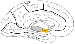
WThe uncus is an anterior extremity of the parahippocampal gyrus. It is separated from the apex of the temporal lobe by a slight fissure called the incisura temporalis.