 W
WAmetropic amblyopia, is a medical condition in which the retina cannot focus on the image of a distant object, a condition often described as reduced visual acuity. This is due to large uncorrected refractive errors in the patient's optic system of the eyes. Astigmatism is one of the most frequent causes of ametropic amblyopia.
 W
WBietti's crystalline dystrophy (BCD), is a rare autosomal recessive eye disease named after Dr. G. B. Bietti.
 W
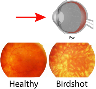
WBirdshot chorioretinopathy now commonly named "birdshot uveitis" or "HLA-A29 uveitis" is a rare form of bilateral posterior uveitis affecting both eyes. It causes severe, progressive inflammation of both the choroid and retina.
 W
WBranch retinal artery occlusion (BRAO) is a rare retinal vascular disorder in which one of the branches of the central retinal artery is obstructed.
 W
WBranch retinal vein occlusion is a common retinal vascular disease of the elderly. It is caused by the occlusion of one of the branches of central retinal vein.
 W
WCentral retinal vein occlusion, also CRVO, is when the central retinal vein becomes occluded, usually through thrombosis. The central retinal vein is the venous equivalent of the central retinal artery and both may become occluded. Since the central retinal artery and vein are the sole source of blood supply and drainage for the retina, such occlusion can lead to severe damage to the retina and blindness, due to ischemia and edema (swelling).
 W
WCentral serous retinopathy (CSR), also known as central serous chorioretinopathy, is an eye disease that causes visual impairment, often temporary, usually in one eye. When the disorder is active it is characterized by leakage of fluid under the retina that has a propensity to accumulate under the central macula. This results in blurred or distorted vision (metamorphopsia). A blurred or gray spot in the central visual field is common when the retina is detached. Reduced visual acuity may persist after the fluid has disappeared.
 W
WChloroquine retinopathy, is a form of toxic retinopathy caused by the drugs chloroquine or hydroxychloroquine, which are sometimes used in the treatment of autoimmune disorders such as rheumatoid arthritis and systemic lupus erythematosus. This eye toxicity limits long-term use of the drugs.
 W
WChorioretinitis is an inflammation of the choroid and retina of the eye. It is a form of posterior uveitis. If only the choroid is inflamed, not the retina, the condition is termed choroiditis. The ophthalmologist's goal in treating these potentially blinding conditions is to eliminate the inflammation and minimize the potential risk of therapy to the patient.
 W
WChoroideremia is a rare, X-linked recessive form of hereditary retinal degeneration that affects roughly 1 in 50,000 males. The disease causes a gradual loss of vision, starting with childhood night blindness, followed by peripheral vision loss and progressing to loss of central vision later in life. Progression continues throughout the individual's life, but both the rate of change and the degree of visual loss are variable among those affected, even within the same family.
 W
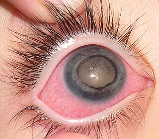
WCoats' disease, is a rare congenital, nonhereditary eye disorder, causing full or partial blindness, characterized by abnormal development of blood vessels behind the retina. Coats' disease can also fall under glaucoma.
 W
WA cone dystrophy is an inherited ocular disorder characterized by the loss of cone cells, the photoreceptors responsible for both central and color vision.
 W
WCytomegalovirus retinitis, also known as CMV retinitis, is an inflammation of the retina of the eye that can lead to blindness. Caused by human cytomegalovirus, it occurs predominantly in people whose immune system has been compromised, 15-40% of those with AIDS. There are different types of retinitis, such as retinitis pigmentosa.
 W
WDiabetic retinopathy, also known as diabetic eye disease, is a medical condition in which damage occurs to the retina due to diabetes mellitus. It is a leading cause of blindness in developed countries.
 W
WDiffuse unilateral subacute neuroretinitis (DUSN) is a rare condition that occurs in otherwise healthy, often young patients and is due to the presence of a subretinal nematode.
WEpiretinal membrane is a disease of the eye in response to changes in the vitreous humor or more rarely, diabetes. Sometimes, as a result of immune system response to protect the retina, cells converge in the macular area as the vitreous ages and pulls away in posterior vitreous detachment (PVD). PVD can create minor damage to the retina, stimulating exudate, inflammation, and leucocyte response. These cells can form a transparent layer gradually and, like all scar tissue, tighten to create tension on the retina which may bulge and pucker, or even cause swelling or macular edema. Often this results in distortions of vision that are clearly visible as bowing and blurring when looking at lines on chart paper within the macular area, or central 1.0 degree of visual arc. Usually it occurs in one eye first, and may cause binocular diplopia or double vision if the image from one eye is too different from the image of the other eye. The distortions can make objects look different in size, especially in the central portion of the visual field, creating a localized or field dependent aniseikonia that cannot be fully corrected optically with glasses. Partial correction often improves the binocular vision considerably though. In the young, these cells occasionally pull free and disintegrate on their own; but in the majority of sufferers the condition is permanent. The underlying photoreceptor cells, rod cells and cone cells, are usually not damaged unless the membrane becomes quite thick and hard; so usually there is no macular degeneration.
 W
WFamilial exudative vitreoretinopathy is a genetic disorder affecting the growth and development of blood vessels in the retina of the eye. This disease can lead to visual impairment and sometimes complete blindness in one or both eyes. FEVR is characterized by exudative leakage and hemorrhage of the blood vessels in the retina, along with incomplete vascularization of the peripheral retina. The disease process can lead to retinal folds, tears, and detachments.
 W
WHypertensive retinopathy is damage to the retina and retinal circulation due to high blood pressure.
 W
WMacular degeneration, also known as age-related macular degeneration, is a medical condition which may result in blurred or no vision in the center of the visual field. Early on there are often no symptoms. Over time, however, some people experience a gradual worsening of vision that may affect one or both eyes. While it does not result in complete blindness, loss of central vision can make it hard to recognize faces, drive, read, or perform other activities of daily life. Visual hallucinations may also occur but these do not represent a mental illness.
 W
WMacular edema occurs when fluid and protein deposits collect on or under the macula of the eye and causes it to thicken and swell (edema). The swelling may distort a person's central vision, because the macula holds tightly packed cones that provide sharp, clear, central vision to enable a person to see detail, form, and color that is directly in the centre of the field of view.
 W
WA macular hole is a small break in the macula, located in the center of the eye's light-sensitive tissue called the retina.
 W
WMacular telangiectasia a type of eye disorder of the macula, the area near the center of the retina, causing a gradual deterioration of central vision, and interfering with tasks such as reading and driving.
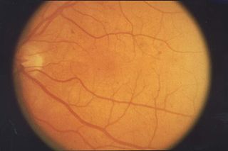
WPurtscher's retinopathy is a disease where part of the eye (retina) is damaged. Usually associated with severe head injuries, it may also occur with other types of trauma, such as long bone fractures, or with several non-traumatic systemic diseases. However, the exact cause of the disease is not well understood. There are no treatments specific for Purtscher's retinopathy, and the prognosis varies. The disease can threaten vision, sometimes causing temporary or permanent blindness.
 W
WRadiation retinopathy is damage to retina due to exposure to ionizing radiation. Radiation retinopathy has a delayed onset, typically after months or years of radiation, and is slowly progressive. In general, radiation retinopathy is seen around 18 months after treatment with external-beam radiation and with brachytherapy. The time of onset of radiation retinopathy is between 6 months to 3 years.
 W
WRetinal detachment is a disorder of the eye in which the retina separates from the layer underneath. Symptoms include an increase in the number of floaters, flashes of light, and worsening of the outer part of the visual field. This may be described as a curtain over part of the field of vision. In about 7% of cases both eyes are affected. Without treatment permanent loss of vision may occur.
WRetinitis is inflammation of the retina in the eye, which can permanently damage the retina and lead to blindness. The retina is the part of your eye that is also known as the "sensing tissue." Retinitis may be caused by a number of different infectious agents. Retinitis, also called Retinitis pigmentosa, has a prevalence of one in every 2,500–7,000 people. This condition is one of the leading causes that leads to blindness in patients in the age range of 20–60 years old.
 W
WRetinitis pigmentosa (RP) is a genetic disorder of the eyes that causes loss of vision. Symptoms include trouble seeing at night and decreased peripheral vision. As peripheral vision worsens, people may experience "tunnel vision". Complete blindness is uncommon. Onset of symptoms is generally gradual and often in childhood.
 W
WRetinopathy is any damage to the retina of the eyes, which may cause vision impairment. Retinopathy often refers to retinal vascular disease, or damage to the retina caused by abnormal blood flow. Age-related macular degeneration is technically included under the umbrella term retinopathy but is often discussed as a separate entity. Retinopathy, or retinal vascular disease, can be broadly categorized into proliferative and non-proliferative types. Frequently, retinopathy is an ocular manifestation of systemic disease as seen in diabetes or hypertension. Diabetes is the most common cause of retinopathy in the U.S. as of 2008. Diabetic retinopathy is the leading cause of blindness in working-aged people. It accounts for about 5% of blindness worldwide and is designated a priority eye disease by the World Health Organization.
 W
WRetinoschisis is an eye disease characterized by the abnormal splitting of the retina's neurosensory layers, usually in the outer plexiform layer. Most common forms are asymptomatic, some rarer forms result in a loss of vision in the corresponding visual field.
WA cone dystrophy is an inherited ocular disorder characterized by the loss of cone cells, the photoreceptors responsible for both central and color vision.
 W
WScleral reinforcement is a surgical procedure used to reduce or stop further macular damage caused by high myopia, which can be degenerative.
 W
WVitelliform macular dystrophy, is an irregular autosomal dominant eye disorder which can cause progressive vision loss. This disorder affects the retina, specifically cells in a small area near the center of the retina called the macula. The macula is responsible for sharp central vision, which is needed for detailed tasks such as reading, driving, and recognizing faces. The condition is characterized by yellow, slightly elevated, round structures similar to the yolk of an egg.