 W
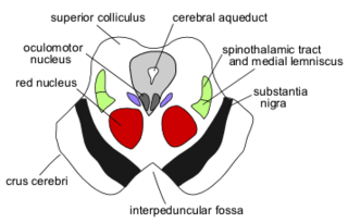
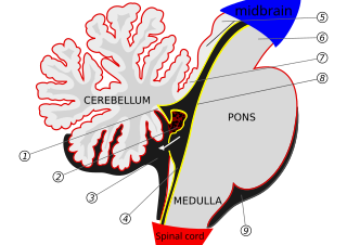
WThe brainstem is the posterior part of the brain, continuous with the spinal cord. In the human brain the brainstem is composed of the midbrain, the pons, and the medulla oblongata. The midbrain is continuous with the thalamus of the diencephalon through the tentorial notch, and sometimes the diencephalon is included in the brainstem.
 W
WThe central tegmental tract is a structure in the midbrain and pons.The central tegmental tract includes ascending axonal fibers that arise from the rostral nucleus solitarius and terminate in the ventral posteromedial nucleus (VPM) of thalamus. Information from the thalamus will go to cortical taste area, namely the insula and frontal operculum. It also contains descending axonal fibers from the parvocellular red nucleus. The descending axons will project to the inferior olivary nucleus. This latter pathway will be used to connect the contralateral cerebellum.
 W
WThe cerebral crus is the anterior portion of the cerebral peduncle which contains the motor tracts, travelling from the cerebral cortex to the pons and spine. The plural of which is cerebral crura. It forms the majority of the basis pedunculi in the midbrain.
 W
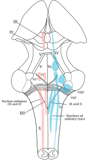
WA cranial nerve nucleus is a collection of neurons in the brain stem that is associated with one or more cranial nerves. Axons carrying information to and from the cranial nerves form a synapse first at these nuclei. Lesions occurring at these nuclei can lead to effects resembling those seen by the severing of nerve(s) they are associated with. All the nuclei except that of the trochlear nerve supply nerves of the same side of the body.
 W
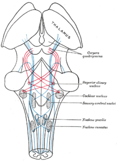
WIn neuroanatomy, the dorsal column nuclei are a pair of nuclei in the dorsal columns in the brainstem. The name refers collectively to the cuneate nucleus and gracile nucleus, which are present at the junction between the spinal cord and the medulla oblongata. Both nuclei contain second-order neurons of the dorsal column-medial lemniscus pathway, which carries fine touch and proprioceptive information from the body to the brain. Each nucleus has an associated nerve tract, the gracile fasciculus and the cuneate fasciculus.
 W
WThe fourth ventricle is one of the four connected fluid-filled cavities within the human brain. These cavities, known collectively as the ventricular system, consist of the left and right lateral ventricles, the third ventricle, and the fourth ventricle. The fourth ventricle extends from the cerebral aqueduct to the obex, and is filled with cerebrospinal fluid (CSF).
 W
WThe hindbrain or rhombencephalon is a developmental categorization of portions of the central nervous system in vertebrates. It includes the medulla, pons, and cerebellum. Together they support vital bodily processes.
 W
WHSD2 neurons are a small group of neurons in the brainstem which are uniquely sensitive to the mineralocorticosteroid hormone aldosterone, through expression of HSD11B2. They are located within the caudal medulla oblongata, in the nucleus of the solitary tract (NTS). HSD2 neurons are activated during a prolonged deficit in body sodium or fluid volume, as occurs after dietary sodium deprivation or during frank hypovolemia. They are also activated by supraphysiologic stimulation of the mineralocorticoid receptor. They are inactivated when salt is ingested. To date, HSD2 neurons have been identified and studied only in rats and mice.
 W
WThe upper part of the posterior district of the medulla oblongata is occupied by the inferior cerebellar peduncle, a thick rope-like strand situated between the lower part of the fourth ventricle and the roots of the glossopharyngeal and vagus nerves.
 W
WThe internal arcuate fibers or internal arcuate tract are the axons of second-order sensory neurons that compose the gracile and cuneate nuclei of the medulla oblongata. These second-order neurons begin in the gracile and cuneate nuclei in the medulla. They receive input from first-order sensory neurons, which provide sensation to many areas of the body and have cell bodies in the dorsal root ganglia of the dorsal root of the spinal nerves. Upon decussation from one side of the medulla to the other, also known as the sensory decussation, they are then called the medial lemniscus.
 W
WThe lateral lemniscus is a tract of axons in the brainstem that carries information about sound from the cochlear nucleus to various brainstem nuclei and ultimately the contralateral inferior colliculus of the midbrain. Three distinct, primarily inhibitory, cellular groups are located interspersed within these fibers, and are thus named the nuclei of the lateral lemniscus.
 W
WThe lenticular fasciculus is a tract connecting the globus pallidus (internus) to the thalamus and is a part of the thalamic fasciculus. It is synonymous with field H2 of Forel. The thalamic fasciculus (composed of both the lenticular fasciculus and ansa lenticularis) runs to the thalamus. Basically, it is part of a pathway that connects the globus pallidus and the thalamus.
 W
WThe medial lemniscus, also known as Reil's band or Reil's ribbon, is a large ascending bundle of heavily myelinated axons that decussate in the brainstem, specifically in the medulla oblongata. The medial lemniscus is formed by the crossings of the internal arcuate fibers. The internal arcuate fibers are composed of axons of nucleus gracilis and nucleus cuneatus. The axons of the nucleus gracilis and nucleus cuneatus in the medial lemniscus have cell bodies that lie contralaterally.
 W
WThe medial longitudinal fasciculus (MLF) is one of a pair of crossed over tracts, on each side of the brainstem. These bundles of axons are situated near the midline of the brainstem and are made up of both ascending and descending fibers that arise from a number of sources and terminate in different areas. The MLF is the main central connection for the oculomotor nerve, trochlear nerve, and abducens nerve. The vertical gaze center is at the rostral interstitial nucleus (riMLF).
 W
WThe medullary pyramids are paired white matter structures of the brainstem's medulla oblongata that contain motor fibers of the corticospinal and corticobulbar tracts – known together as the pyramidal tracts. The lower limit of the pyramids is marked when the fibers cross (decussate).
 W
WThe mesencephalic locomotor region (MLR) is a functionally defined area of the midbrain that is associated with the initiation and control of locomotor movements in vertebrate species.
WThe metencephalon is the embryonic part of the hindbrain that differentiates into the pons and the cerebellum. It contains a portion of the fourth ventricle and the trigeminal nerve, abducens nerve, facial nerve, and a portion of the vestibulocochlear nerve.
 W
WThe myoclonic triangle is an important feedback circuit of the brainstem and deep cerebellar nuclei which is responsible for modulating spinal cord motor activity.
 W
WLocated in the caudal pons and upper medulla oblongata, the nucleus prepositus is part of the horizontal gaze holding system. It functions as a neural integrator.
 W
WThe olivospinal fasciculus (Helweg) was thought to arise in the vicinity of the inferior olivary nucleus in the medulla oblongata, and was thought to be seen only in the cervical region of the medulla spinalis, where it forms a small triangular area at the periphery, close to the most lateral of the anterior nerve roots. Its existence is now strongly doubted.
WThe pallidothalamic tracts are a part of the basal ganglia. They provide connectivity between the internal globus pallidus (GPi) and the thalamus, primarily the ventral anterior nucleus and the ventral lateral nucleus.
 W
WThe spinocerebellar tract is a nerve tract originating in the spinal cord and terminating in the same side (ipsilateral) of the cerebellum.
 W
WThe pre-Bötzinger complex (preBötC) is a cluster of interneurons in the ventral respiratory group of the medulla of the brainstem. This complex has been proven to be essential for the generation of the respiratory rhythm in mammals. The exact mechanism of the rhythm generation and transmission to motor nuclei remains controversial and the topic of much research.
 W
WThe respiratory center is located in the medulla oblongata and pons, in the brainstem. The respiratory center is made up of three major respiratory groups of neurons, two in the medulla and one in the pons. In the medulla they are the dorsal respiratory group, and the ventral respiratory group. In the pons, the pontine respiratory group includes two areas known as the pneumotaxic centre and the apneustic centre.
 W
WThe reticular formation is a set of interconnected nuclei that are located throughout the brainstem. It is not anatomically well defined, because it includes neurons located in different parts of the brain. The neurons of the reticular formation make up a complex set of networks in the core of the brainstem that extend from the upper part of the midbrain to the lower part of the medulla oblongata. The reticular formation includes ascending pathways to the cortex in the ascending reticular activating system (ARAS) and descending pathways to the spinal cord via the reticulospinal tracts.
 W
WThe roof of fourth ventricle is the dorsal surface of the fourth ventricle.
 W
WThe rostral interstitial nucleus of medial longitudinal fasciculus (riMLF) is a portion of the medial longitudinal fasciculus which controls vertical gaze.
WThe sensory decussation or decussation of the lemnisci is a decussation or crossover of axons from the gracile nucleus and cuneate nucleus, which are responsible for fine touch, vibration, proprioception and two-point discrimination of the body. The fibres of this decussation are called the internal arcuate fibres and are found at the superior aspect of the closed medulla superior to the motor decussation. It is part of the second neuron in the posterior column–medial lemniscus pathway.
 W
WThe sulcus limitans is found in the fourth ventricle of the brain. It separates the cranial nerve motor nuclei (medial) from the sensory nuclei (lateral). It can also be located by searching laterally from the medial eminence. It is parallel to the median sulcus.
 W
WThe superior olivary complex (SOC) or superior olive is a collection of brainstem nuclei that functions in multiple aspects of hearing and is an important component of the ascending and descending auditory pathways of the auditory system. The SOC is intimately related to the trapezoid body: most of the cell groups of the SOC are dorsal to this axon bundle while a number of cell groups are embedded in the trapezoid body. Overall, the SOC displays a significant interspecies variation, being largest in bats and rodents and smaller in primates.
 W
WThe tegmentum is a general area within the brainstem. The tegmentum is the ventral part of the midbrain and the tectum is the dorsal part of the midbrain. It is located between the ventricular system and distinctive basal or ventral structures at each level. It forms the floor of the midbrain (mesencephalon) whereas the tectum forms the ceiling.It is a multisynaptic network of neurons that is involved in many subconscious homeostatic and reflexive pathways. It is a motor center that relays inhibitory signals to the thalamus and basal nuclei preventing unwanted body movement.
 W
WUnipolar brush cells (UBCs) are a class of excitatory glutamatergic interneuron found in the granular layer of the cerebellar cortex and also in the granule cell domain of the cochlear nucleus.
 W
WThe cells of the dorsal nucleus of vagus nerve are spindle-shaped, like those of the posterior column of the spinal cord, and the nucleus is usually considered as representing the base of the posterior column. It measures about 2 cm. in length, and in the lower, closed part of the medulla oblongata is situated behind the dorsal motor nucleus of the vagus; whereas in the upper, open part it lies lateral to that nucleus, and corresponds to an eminence, named the vagal trigone, in the rhomboid fossa.