 W
WThe alveolar ridge is one of the two jaw ridges, extensions of the mandible or maxilla, either on the roof of the mouth between the upper teeth and the hard palate or on the bottom of the mouth behind the lower teeth. Most of the roof of one's mouth is the hard palate and the soft palate. The alveolar ridges contain the sockets of the teeth. They can be felt with the tongue in the area right above the top teeth or below the bottom teeth. Its surface is covered with little ridges.The [upper] alveolar ridge is a small protuberance just behind the upper front teeth that can easily be felt with the tongue.
 W
WThe cementoenamel junction, frequently abbreviated as the CEJ, is a slightly visible anatomical border identified on a tooth. It is the location where the enamel, which covers the anatomical crown of a tooth, and the cementum, which covers the anatomical root of a tooth, meet. Informally it is known as the neck of the tooth. The border created by these two dental tissues has much significance as it is usually the location where the gingiva attaches to a healthy tooth by fibers called the gingival fibers.
 W
WCementum is a specialized calcified substance covering the root of a tooth. The cementum is the part of the periodontium that attaches the teeth to the alveolar bone by anchoring the periodontal ligament.
 W
WIn dentistry, crown refers to the anatomical area of teeth, usually covered by enamel. The crown is usually visible in the mouth after developing below the gingiva and then erupting into place. If part of the tooth gets chipped or broken, a dentist can apply an artificial crown. Crowns are used most commonly to entirely cover a damaged tooth or cover an implant. Bridges are also used to cover a space if one or more teeth is missing. They are cemented to natural teeth or implants surrounding the space where the tooth once stood. There are various materials that can be used including a type of cement or stainless steel. The cement crowns look like regular teeth while the stainless steel crowns are silver or gold.
 W
WThe facial nerve is the seventh cranial nerve, or simply CN VII. It emerges from the pons of the brainstem, controls the muscles of facial expression, and functions in the conveyance of taste sensations from the anterior two-thirds of the tongue. The nerves typically travels from the pons through the facial canal in the temporal bone and exits the skull at the stylomastoid foramen. It arises from the brainstem from an area posterior to the cranial nerve VI and anterior to cranial nerve VIII.
WThe gums or gingiva, consist of the mucosal tissue that lies over the mandible and maxilla inside the mouth. Gum health and disease can have an effect on general health.
 W
WIn human anatomy, the mouth is the first portion of the alimentary canal that receives food and produces saliva. The oral mucosa is the mucous membrane epithelium lining the inside of the mouth.
 W
WThe jaw is any opposable articulated structure at the entrance of the mouth, typically used for grasping and manipulating food. The term jaws is also broadly applied to the whole of the structures constituting the vault of the mouth and serving to open and close it and is part of the body plan of humans and most animals.
 W
WLips are a visible body part at the mouth of many animals, including humans.
 W
WThe mandibular canine is the tooth located distally from both mandibular lateral incisors of the mouth but mesially from both mandibular first premolars. Both the maxillary and mandibular canines are called the "cornerstone" of the mouth because they are all located three teeth away from the midline, and separate the premolars from the incisors. The location of the canines reflect their dual function as they complement both the premolars and incisors during mastication, commonly known as chewing. Nonetheless, the most common action of the canines is tearing of food. The canine teeth are able to withstand the tremendous lateral pressures from chewing. There is a single cusp on canines, and they resemble the prehensile teeth found in carnivorous animals. Though relatively the same, there are some minor differences between the deciduous (baby) mandibular canine and that of the permanent mandibular canine.
 W
WThe mandibular central incisor is the tooth located on the jaw, adjacent to the midline of the face. It is mesial from both mandibular lateral incisors. As with all incisors, its function includes shearing or cutting food during mastication, commonly known as chewing. There are no cusps on the tooth. Instead, the surface area of the tooth used in eating is called an incisal ridge or incisal edge. Though the two are similar, there are some minor differences between the deciduous (baby) mandibular central incisor and that of the permanent mandibular central incisor. The mandibular central incisors are usually the first teeth to appear in the mouth, typically around the age of 6-8 months.
 W
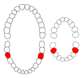
WThe mandibular first molar or six-year molar is the tooth located distally from both the mandibular second premolars of the mouth but mesial from both mandibular second molars. It is located on the mandibular (lower) arch of the mouth, and generally opposes the maxillary (upper) first molars and the maxillary 2nd premolar in normal class I occlusion. The function of this molar is similar to that of all molars in regard to grinding being the principal action during mastication, commonly known as chewing. There are usually five well-developed cusps on mandibular first molars: two on the buccal, two lingual, and one distal. The shape of the developmental and supplementary grooves, on the occlusal surface, are describes as being 'M' shaped. There are great differences between the deciduous (baby) mandibular molars and those of the permanent mandibular molars, even though their function are similar. The permanent mandibular molars are not considered to have any teeth that precede it. Despite being named molars, the deciduous molars are followed by permanent premolars.
 W
WThe mandibular first premolar is the tooth located laterally from both the mandibular canines of the mouth but mesial from both mandibular second premolars. The function of this premolar is similar to that of canines in regard to tearing being the principal action during mastication, commonly known as chewing. Mandibular first premolars have two cusps. The one large and sharp is located on the buccal side of the tooth. Since the lingual cusp is small and nonfunctional, the mandibular first premolar resembles a small canine. There are no deciduous (baby) mandibular premolars. Instead, the teeth that precede the permanent mandibular premolars are the deciduous mandibular molars.
 W
WThe mandibular lateral incisor is the tooth located distally from both mandibular central incisors of the mouth and mesially from both mandibular canines. As with all incisors, their function is for shearing or cutting food during mastication, commonly known as chewing. There are no cusps on the teeth. Instead, the surface area of the tooth used in eating is called an incisal ridge or incisal edge. Though relatively the same, there are some minor differences between the deciduous (baby) mandibular lateral incisor and that of the permanent mandibular lateral incisor.
 W
WThe mandibular second molar is the tooth located distally from both the mandibular first molars of the mouth but mesial from both mandibular third molars. This is true only in permanent teeth. The function of this molar is similar to that of all molars in regard to grinding being the principal action during mastication, commonly known as chewing. Though there is more variation between individuals to that of the first mandibular molar, there are usually four cusps on mandibular second molars: two on the buccal and two palatal. There are great differences between the deciduous (baby) mandibular molars and those of the permanent mandibular molars, even though their function are similar. The permanent mandibular molars are not considered to have any teeth that precede it. Despite being named molars, the deciduous molars are followed by permanent premolars.
 W
WThe mandibular second premolar is the tooth located distally from both the mandibular first premolars of the mouth but mesial from both mandibular first molars. The function of this premolar is assist the mandibular first molar during mastication, commonly known as chewing. Mandibular second premolars have three cusps. There is one large cusp on the buccal side of the tooth. The lingual cusps are well developed and functional. Therefore, whereas the mandibular first premolar resembles a small canine, the mandibular second premolar is more alike to the first molar. There are no deciduous (baby) mandibular premolars. Instead, the teeth that precede the permanent mandibular premolars are the deciduous mandibular molars.
 W
WThe maxilla in vertebrates is the upper fixed bone of the jaw formed from the fusion of two maxillary bones. In humans, the upper jaw includes the hard palate in the front of the mouth. The two maxillary bones are fused at the intermaxillary suture, forming the anterior nasal spine. This is similar to the mandible, which is also a fusion of two mandibular bones at the mandibular symphysis. The mandible is the movable part of the jaw.
 W
WIn human dentistry, the maxillary canine is the tooth located laterally from both maxillary lateral incisors of the mouth but mesial from both maxillary first premolars. Both the maxillary and mandibular canines are called the "cornerstone" of the mouth because they are all located three teeth away from the midline, and separate the premolars from the incisors. The location of the canines reflect their dual function as they complement both the premolars and incisors during mastication, commonly known as chewing. Nonetheless, the most common action of the canines is tearing of food. The canines often erupt in the upper gums several millimeters above the gum line. The canine teeth are able to withstand the tremendous lateral pressure caused by chewing. There is a single cusp on canines, and they resemble the prehensile teeth found in carnivorous animals such as the extinct Saber-toothed cat. Though relatively the same, there are some minor differences between the deciduous (baby) maxillary canine and that of the permanent maxillary canine.
 W
WThe maxillary central incisor is a human tooth in the front upper jaw, or maxilla, and is usually the most visible of all teeth in the mouth. It is located mesial to the maxillary lateral incisor. As with all incisors, their function is for shearing or cutting food during mastication (chewing). There is typically a single cusp on each tooth, called an incisal ridge or incisal edge. Formation of these teeth begins at 14 weeks in utero for the deciduous (baby) set and 3–4 months of age for the permanent set.
 W
W W
WThe maxillary first premolar is one of two teeth located in the upper jaw, laterally from both the maxillary canines of the mouth but mesial from both maxillary second premolars. The function of this premolar is similar to that of canines in regard to tearing being the principal action during mastication, commonly known as chewing. There are two cusps on maxillary first premolars, and the buccal cusp is sharp enough to resemble the prehensile teeth found in carnivorous animals. There are no deciduous maxillary premolars. Around 10-11 years of age, the primary molars are shed and the permanent premolars erupt in their place. It takes about 3 years for the adult premolar and its root to fully calcify.
 W
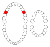
WThe maxillary lateral incisors are a pair of upper (maxillary) teeth that are located laterally from both maxillary central incisors of the mouth and medially from both maxillary canines. As with all incisors, their function is for shearing or cutting food during mastication, commonly known as chewing. There are generally no cusps on the teeth, but the rare condition known as talon cusps are most prevalent on the maxillary lateral incisors. The surface area of the tooth used in eating is called an incisal ridge or incisal edge. Though relatively the same, there are some minor differences between the deciduous (baby) maxillary lateral incisor and that of the permanent maxillary lateral incisor. The maxillary lateral incisors occlude in opposition to the mandibular lateral incisors.
 W
WThe maxillary second molar is the tooth located distally from both the maxillary first molars of the mouth but mesial from both maxillary third molars. This is true only in permanent teeth. In deciduous (baby) teeth, the maxillary second molar is the last tooth in the mouth and does not have a third molar behind it. The function of this molar is similar to that of all molars in regard to grinding being the principal action during mastication, commonly known as chewing. There are usually four cusps on maxillary molars, two on the buccal and two palatal.
 W
WThe maxillary second premolar is one of two teeth located in the upper jaw, laterally from both the maxillary first premolars of the mouth but mesial from both maxillary first molars. The function of this premolar is similar to that of first molars in regard to grinding being the principal action during mastication, commonly known as chewing. There are two cusps on maxillary second premolars, but both of them are less sharp than those of the maxillary first premolars. There are no deciduous (baby) maxillary premolars. Instead, the teeth that precede the permanent maxillary premolars are the deciduous maxillary molars.
 W
WThe hard palate is a thin horizontal bony plate made up of two bones of the facial skeleton, located in the roof of the mouth. The bones are the palatine process of the maxilla and the horizontal plate of palatine bone. The hard palate spans the alveolar arch formed by the alveolar process that holds the upper teeth.
 W
WThe soft palate is, in mammals, the soft tissue constituting the back of the roof of the mouth. The soft palate is part of the palate of the mouth; the other part is the hard palate. The soft palate is distinguished from the hard palate at the front of the mouth in that it does not contain bone.
WThe periodontium is the specialized tissues that both surround and support the teeth, maintaining them in the maxillary and mandibular bones. The word comes from the Greek terms περί peri-, meaning "around" and -odont, meaning "tooth". Literally taken, it means that which is "around the tooth". Periodontics is the dental specialty that relates specifically to the care and maintenance of these tissues. It provides the support necessary to maintain teeth in function. It consists of four principal components, namely:Gingiva Periodontal ligament (PDL) Cementum Alveolar bone proper
 W
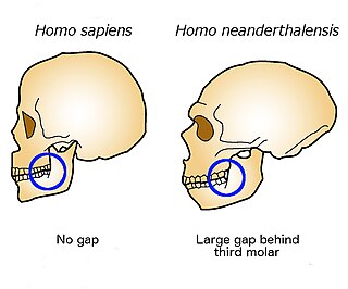
WThe retromolar space or retromolar gap is a space at the rear of the mandible, between the back of the last molar and the anterior edge of the ascending ramus where it crosses the alveolar margin.
 W
WA root canal is the naturally occurring anatomic space within the root of a tooth. It consists of the pulp chamber, the main canal(s), and more intricate anatomical branches that may connect the root canals to each other or to the surface of the root.
 W
WThe tongue is a muscular organ in the mouth of most vertebrates that manipulates food for mastication and is used in the act of swallowing. It has importance in the digestive system and is the primary organ of taste in the gustatory system. The tongue's upper surface (dorsum) is covered by taste buds housed in numerous lingual papillae. It is sensitive and kept moist by saliva and is richly supplied with nerves and blood vessels. The tongue also serves as a natural means of cleaning the teeth. A major function of the tongue is the enabling of speech in humans and vocalization in other animals.
 W
WThe Universal Numbering System also called the "American System", is a dental notation system for associating information to a specific tooth, commonly used in the United States.
 W
WWhite roll is the white line that borders the top of the upper lip. It's an adnexal mass of specialized glands and fat. White roll occurs naturally for nearly everyone, although it can be not white and less visible for dark skinned individuals. Well defined white roll indicates youthfulness and is considered aesthetically pleasing.
 W
WA wisdom tooth or third molar is one of the three molars per quadrant of the human dentition. It is the most posterior of the three. The age at which wisdom teeth come through (erupt) is variable, but generally occurs between late teens and early twenties. Most adults have four wisdom teeth, one in each of the four quadrants, but it is possible to have none, fewer, or more, in which case the extras are called supernumerary teeth. Wisdom teeth may get stuck (impacted) against other teeth if there is not enough space for them to come through normally. While the impaction does not cause movement of other teeth, it can cause tooth decay if oral hygiene becomes more difficult. Wisdom teeth which are partially erupted through the gum may also cause inflammation and infection in the surrounding gum tissues, termed pericoronitis. Wisdom teeth are often extracted when or even before these problems occur. However, some, including the National Institute for Health and Care Excellence in the UK, recommend against the prophylactic extraction of disease-free impacted wisdom teeth.