 W
WMedical ultrasound includes diagnostic techniques using ultrasound, as well as therapeutic applications of ultrasound. In diagnosis, it is used to create an image of internal body structures such as tendons, muscles, joints, blood vessels, and internal organs, to measure some characteristics or to generate an informative audible sound. Its aim is usually to find a source of disease or to exclude pathology. The usage of ultrasound to produce visual images for medicine is called medical ultrasonography or simply sonography. The practice of examining pregnant women using ultrasound is called obstetric ultrasonography, and was an early development of clinical ultrasonography.
 W
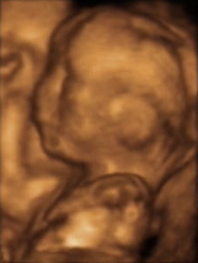
W3D ultrasound is a medical ultrasound technique, often used in fetal, cardiac, trans-rectal and intra-vascular applications. 3D ultrasound refers specifically to the volume rendering of ultrasound data. When involving a series of 3D volumes collected over time, it can also be referred to as 4D ultrasound or real-time 3D ultrasound.
 W
WAbdominal ultrasonography is a form of medical ultrasonography to visualise abdominal anatomical structures. It uses transmission and reflection of ultrasound waves to visualise internal organs through the abdominal wall. For this reason, the procedure is also called a transabdominal ultrasound, in contrast to endoscopic ultrasound, the latter combining ultrasound with endoscopy through visualize internal structures from within hollow organs.
 W
WThe ankle-brachial pressure index (ABPI) or ankle-brachial index (ABI) is the ratio of the blood pressure at the ankle to the blood pressure in the upper arm (brachium). Compared to the arm, lower blood pressure in the leg suggests blocked arteries due to peripheral artery disease (PAD). The ABPI is calculated by dividing the systolic blood pressure at the ankle by the systolic blood pressure in the arm.
 W
WThe arterial resistivity index, developed by Leandre Pourcelot, is a measure of pulsatile blood flow that reflects the resistance to blood flow caused by microvascular bed distal to the site of measurement.
 W
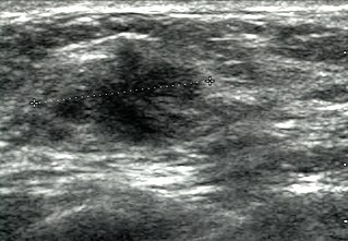
WBreast ultrasound is the use of medical ultrasonography to perform imaging of the breast.
 W
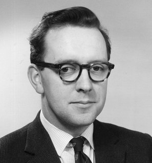
WThomas Graham Brown was a Scottish engineer who was most notable for collaborating in the design of the first medical ultrasound machine along with the obstetrician and designer Ian Donald, who would later be the Regius Professor of Obstetrics and Gynaecology at the University of Glasgow and industrial designer and obstetrician John MacVicar.
 W
WCarotid ultrasonography is an ultrasound-based diagnostic imaging technique to evaluate structural details of the carotid arteries. Carotid ultrasound is used to diagnose carotid artery stenosis (CAS) and can assess atherosclerotic plaque morphology and characteristics. Carotid duplex and contrast-enhanced ultrasound are two of the most common imaging techniques used to evaluate carotid artery disease.
 W
WContrast-enhanced ultrasound (CEUS) is the application of ultrasound contrast medium to traditional medical sonography. Ultrasound contrast agents rely on the different ways in which sound waves are reflected from interfaces between substances. This may be the surface of a small air bubble or a more complex structure. Commercially available contrast media are gas-filled microbubbles that are administered intravenously to the systemic circulation. Microbubbles have a high degree of echogenicity. There is a great difference in echogenicity between the gas in the microbubbles and the soft tissue surroundings of the body. Thus, ultrasonic imaging using microbubble contrast agents enhances the ultrasound backscatter, (reflection) of the ultrasound waves, to produce a sonogram with increased contrast due to the high echogenicity difference. Contrast-enhanced ultrasound can be used to image blood perfusion in organs, measure blood flow rate in the heart and other organs, and for other applications.
 W
WDiagnostic medical sonography (DMS), a branch of diagnostic medical imaging, is the use of imaging by medical ultrasound for medical diagnosis. DMS uses non-ionizing ultrasound to produce 2D and 3D images of the body. In Canada, the credentialing for diagnostic medical sonography is the Canadian Association of Registered Ultrasound Professionals. In the United States, the credentialing body is the American Registry for Diagnostic Medical Sonography.
 W
WIan Donald was an English physician who was most notable for pioneering the diagnostic use of ultrasound in obstetrics, enabling the visual discovery of abnormalities in pregnancy. Donald was Regius Professor of Obstetrics and Gynaecology at the University of Glasgow. Donald's work was characterised by a series of collaborations between clinicians and engineers that led to the designing and building of a series of instruments that enabled the examination of the unborn and that eventually enabled him to build the world's first obstetric ultrasound machine, the Diasonograph in 1963. His other great achievement was to secure the construction of the Queen Mother's Maternity Hospital that was built next to the Royal Hospital for Children in Glasgow.
 W
WDoppler echocardiography is a procedure that uses Doppler ultrasonography to examine the heart. An echocardiogram uses high frequency sound waves to create an image of the heart while the use of Doppler technology allows determination of the speed and direction of blood flow by utilizing the Doppler effect.
 W
WDoppler ultrasonography is medical ultrasonography that employs the Doppler effect to generate imaging of the movement of tissues and body fluids, and their relative velocity to the probe. By calculating the frequency shift of a particular sample volume, for example, flow in an artery or a jet of blood flow over a heart valve, its speed and direction can be determined and visualized. Color Doppler or color flow Doppler is the presentation of the velocity by color scale. Color Doppler images are generally combined with grayscale (B-mode) images to display duplex ultrasonography images, allowing for simultaneous visualization of the anatomy of the area.
 W
WAn echocardiography, echocardiogram, cardiac echo or simply an echo, is an ultrasound of the heart. It is a type of medical imaging of the heart, using standard ultrasound or Doppler ultrasound.
 W
WEchogenicity or echogeneity is the ability to bounce an echo, e.g. return the signal in ultrasound examinations. In other words, echogenicity is higher when the surface bouncing the sound echo reflects increased sound waves. Tissues that have higher echogenicity are called "hyperechogenic" and are usually represented with lighter colors on images in medical ultrasonography. In contrast, tissues with lower echogenicity are called "hypoechogenic" and are usually represented with darker colors. Areas that lack echogenicity are called "anechogenic" and are usually displayed as completely dark.
 W
WEndoscopic ultrasound (EUS) or echo-endoscopy is a medical procedure in which endoscopy is combined with ultrasound to obtain images of the internal organs in the chest, abdomen and colon. It can be used to visualize the walls of these organs, or to look at adjacent structures. Combined with Doppler imaging, nearby blood vessels can also be evaluated.
 W
WGynecologic ultrasonography or gynecologic sonography refers to the application of medical ultrasonography to the female pelvic organs as well as the bladder, the adnexa, and the recto-uterine pouch. The procedure may lead to other medically relevant findings in the pelvis.
 W
WHigh-intensity focused ultrasound (HIFU) is a non-invasive therapeutic technique that uses non-ionizing ultrasonic waves to heat or ablate tissue. HIFU can be used to increase the flow of blood or lymph, or to destroy tissue, such as tumors, via thermal and mechanical mechanisms. Given the prevalence and relatively low cost of ultrasound, HIFU has been subject to much research and development. The premise of HIFU is that it is a non-invasive low cost therapy that can at minimum outperform the current standard of care.
 W
WLiposuction, or simply lipo, is a type of fat-removal procedure used in plastic surgery. Evidence does not support an effect on weight beyond a couple of months and does not appear to affect obesity-related problems. In the United States, liposuction is the most common cosmetic surgery.
 W
WJohn MacVicar was a British physician who was most notable for pioneering the diagnostic use of ultrasound in obstetrics as well as later, being a clinical educator. MacVicar was part of a team along with physician Ian Donald and engineer Tom Brown, who developed the worlds first obstetric ultrasound machine in 1963. Using the new technique of ultrasound, MacVicar's research transformed the treatment of gynaecological conditions in pregnant women, through the use of clinical trials.
 W
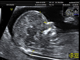
WA nuchal scan or nuchal translucency (NT) scan/procedure is a sonographic prenatal screening scan (ultrasound) to detect chromosomal abnormalities in a fetus, though altered extracellular matrix composition and limited lymphatic drainage can also be detected.
 W
WObstetric ultrasonography, or prenatal ultrasound, is the use of medical ultrasonography in pregnancy, in which sound waves are used to create real-time visual images of the developing embryo or fetus in the uterus (womb). The procedure is a standard part of prenatal care in many countries, as it can provide a variety of information about the health of the mother, the timing and progress of the pregnancy, and the health and development of the embryo or fetus.
 W
WOCT Biomicroscopy is the use of optical coherence tomography (OCT) in place of slit lamp biomicroscopy to examine the transparent axial tissues of the eye. Traditionally, ophthalmic biomicroscopy has been completed with a slit lamp biomicroscope that uses slit beam illumination and an optical microscope to enable stereoscopic, magnified, cross-sectional views of transparent tissues in the eye, with or without the aid of an additional lens. Like slit lamp biomicroscopy, OCT does not penetrate opaque tissues well but enables detailed, cross-sectional views of transparent tissues, often with greater detail than is possible with a slit lamp. Ultrasound biomicroscopy (UBM) is much better at imaging through opaque tissues since it uses high energy sound waves. Because of its limited depth of penetration, UBM's main use within ophthalmology has been to visualize anterior structures such as the angle and ciliary body. Both ultrasound and OCT biomicroscopy produce an objective image of ocular tissues from which measurements can be made. Unlike UBM, OCT biomicroscopy can image tissues with high axial resolution as far posteriorly as the choroid.
 W
WPhased array ultrasonics (PA) is an advanced method of ultrasonic testing that has applications in medical imaging and industrial nondestructive testing. Common applications are to noninvasively examine the heart or to find flaws in manufactured materials such as welds. Single-element probes, known technically as monolithic probes, emit a beam in a fixed direction. To test or interrogate a large volume of material, a conventional probe must be physically scanned to sweep the beam through the area of interest. In contrast, the beam from a phased array probe can be focused and swept electronically without moving the probe. The beam is controllable because a phased array probe is made up of multiple small elements, each of which can be pulsed individually at a computer-calculated timing. The term phased refers to the timing, and the term array refers to the multiple elements. Phased array ultrasonic testing is based on principles of wave physics, which also have applications in fields such as optics and electromagnetic antennae.
 W
WRenal ultrasonography is the examination of one or both kidneys using medical ultrasound.
 W
WScrotal ultrasound is a medical ultrasound examination of the scrotum. It is used in the evaluation of testicular pain, and can help identify solid masses.
 W
WSonication is the act of applying sound energy to agitate particles in a sample, for various purposes such as the extraction of multiple compounds from plants, microalgae and seaweeds. Ultrasonic frequencies (>20 kHz) are usually used, leading to the process also being known as ultrasonication or ultra-sonication.
WA sonographer is a healthcare professional who specialises in the use of ultrasonic imaging devices to produce diagnostic images, scans, videos or three-dimensional volumes of anatomy and diagnostic data. The requirements for clinical practice vary greatly by country. Sonography requires specialised education and skills to acquire, analyze and optimize information in the image. Due to the high levels of decisional latitude and diagnostic input, sonographers have a high degree of responsibility in the diagnostic process. Many countries require medical sonographers to have professional certification. Sonographers have core knowledge in ultrasound physics, cross-sectional anatomy, physiology, and pathology.
 W
WTranscranial Doppler (TCD) and transcranial color Doppler (TCCD) are types of Doppler ultrasonography that measure the velocity of blood flow through the brain's blood vessels by measuring the echoes of ultrasound waves moving transcranially. These modes of medical imaging conduct a spectral analysis of the acoustic signals they receive and can therefore be classified as methods of active acoustocerebrography. They are used as tests to help diagnose emboli, stenosis, vasospasm from a subarachnoid hemorrhage, and other problems. These relatively quick and inexpensive tests are growing in popularity. The tests are effective for detecting sickle cell disease, ischemic cerebrovascular disease, subarachnoid hemorrhage, arteriovenous malformations, and cerebral circulatory arrest. The tests are possibly useful for perioperative monitoring and meningeal infection. The equipment used for these tests is becoming increasingly portable, making it possible for a clinician to travel to a hospital, to a doctor's office, or to a nursing home for both inpatient and outpatient studies. The tests are often used in conjunction with other tests such as MRI, MRA, carotid duplex ultrasound and CT scans. The tests are also used for research in cognitive neuroscience.
 W
WA transesophageal echocardiogram, or TEE, is an alternative way to perform an echocardiogram. A specialized probe containing an ultrasound transducer at its tip is passed into the patient's esophagus. This allows image and Doppler evaluation which can be recorded. It is commonly used during cardiac surgery and is an excellent modality for assessing the aorta, although there are some limitations.
 W
WTransrectal ultrasonography, or TRUS in short, is a method of creating an image of organs in the pelvis, most commonly used to perform an ultrasound-guided needle biopsy evaluation of the prostate gland in men with elevated prostate-specific antigen or prostatic nodules on digital rectal exam. TRUS--guided biopsy may reveal prostate cancer, benign prostatic hypertrophy, or prostatitis. TRUS may also detect other diseases of the lower rectum and can be used to stage primary rectal cancer.
 W
WUltrasonography of suspected or previously confirmed chronic venous insufficiency of leg veins is a risk-free, non-invasive procedure. It gives information about the anatomy, physiology and pathology of mainly superficial veins. As with heart ultrasound (echocardiography) studies, venous ultrasonography requires an understanding of hemodynamics in order to give useful examination reports. In chronic venous insufficiency, sonographic examination is of most benefit; in confirming varicose disease, making an assessment of the hemodynamics, and charting the progression of the disease and its response to treatment. It has become the reference standard for examining the condition and hemodynamics of the lower limb veins. Particular veins of the deep venous system (DVS), and the superficial venous system (SVS) are looked at. The great saphenous vein (GSV), and the small saphenous vein (SSV) are superficial veins which drain into respectively, the common femoral vein and the popliteal vein. These veins are deep veins. Perforator veins drain superficial veins into the deep veins. Three anatomic compartments are described, (N1) containing the deep veins, (N2) containing the perforator veins, and (N3) containing the superficial veins, known as the saphenous compartment. This compartmentalisation makes it easier for the examiner to systematize and map. The GSV can be located in the saphenous compartment where together with the Giacomini vein and the accessory saphenous vein (ASV) an image resembling an eye, known as the 'eye sign' can be seen. The ASV which is often responsible for varicose veins, can be located at the 'alignment sign', where it is seen to align with the femoral vessels.
 W
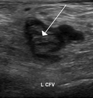
WUltrasonography in suspected deep vein thrombosis focuses primarily on the femoral vein and the popliteal vein, because thrombi in these veins are associated with the greatest risk of harmful pulmonary embolism.
 W
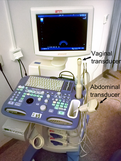
WVaginal ultrasonography is a medical ultrasonography that applies an ultrasound transducer in the vagina to visualize organs within the pelvic cavity. It is also called transvaginal ultrasonography because the ultrasound waves go across the vaginal wall to study tissues beyond it.