 W
WThe spinal cord is a long, thin, tubular structure made up of nervous tissue, which extends from the medulla oblongata in the brainstem to the lumbar region of the vertebral column. It encloses the central canal of the spinal cord, which contains cerebrospinal fluid. The brain and spinal cord together make up the central nervous system (CNS). In humans, the spinal cord begins at the occipital bone, passing through the foramen magnum and entering the spinal canal at the beginning of the cervical vertebrae. The spinal cord extends down to between the first and second lumbar vertebrae, where it ends. The enclosing bony vertebral column protects the relatively shorter spinal cord. It is around 45 cm (18 in) in men and around 43 cm (17 in) long in women. The diameter of the spinal cord ranges from 13 mm in the cervical and lumbar regions to 6.4 mm in the thoracic area.
 W
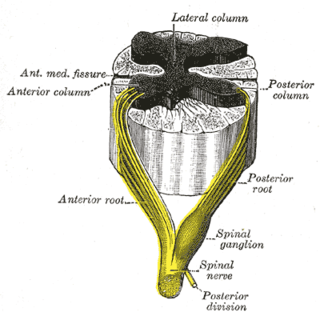
WThe anterior grey column is the front column of grey matter in the spinal cord. It is one of the three grey columns. The anterior grey column contains motor neurons that affect the skeletal muscles while the posterior grey column receives information regarding touch and sensation. The anterior grey column is the column where the cell bodies of alpha motor neurons are located.
 W
WThe anterior median fissure of the spinal cord has an average depth of about 3 mm, but this is increased in the lower part of the spinal cord.
 W
WThe anterior white commissure is a bundle of nerve fibers which cross the midline of the spinal cord just anterior to the gray commissure. A delta fibers and C fibers carrying pain sensation in the spinothalamic tract contribute to this commissure, as do fibers of the anterior corticospinal tract, which carry motor signals from the primary motor cortex.
 W
WThe Anterolateral sulcus of spinal cord is a landmark on the anterior side of the spinal cord. It denotes the location at which the ventral fibers leave the spinal cord.
WThe central canal, also known as ependymal canal, is the cerebrospinal fluid-filled space that runs through the spinal cord. The central canal below at the ventricular system of the brain, from which it receives cerebrospinal fluid, and shares the same ependymal lining. The central canal helps to transport nutrients to the spinal cord as well as protect it by cushioning the impact of a force when the spine is affected.
 W
WThe ciliospinal center is a structure which receives input from the pretectum, and has output to the superior cervical ganglion.
 W
WThe conus medullaris or conus terminalis is the tapered, lower end of the spinal cord. It occurs near lumbar vertebral levels 1 (L1) and 2 (L2), occasionally lower. The upper end of the conus medullaris is usually not well defined, however, its corresponding spinal cord segments are usually S1-S5.
 W
WDural ectasia is widening or ballooning of the dural sac surrounding the spinal cord. This usually occurs in the lumbosacral region, as this is where the cerebrospinal fluid pressure is greatest, but the spinal canal can be affected in any plane.
 W
WIn anatomy, the epidural space is the potential space between the two layers of the dura mater.
 W
WThe general somatic afferent fibers afferent fibers arise from neurons in sensory ganglia and are found in all the spinal nerves, except occasionally the first cervical, and conduct impulses of pain, touch and temperature from the surface of the body through the dorsal roots to the spinal cord and impulses of muscle sense, tendon sense and joint sense from the deeper structures.
WThe grey column refers to a somewhat ridge-shaped mass of grey matter in the spinal cord. This presents as three columns: the anterior grey column, the posterior grey column, and the lateral grey column, all of which are visible in cross-section of the spinal cord.
 W
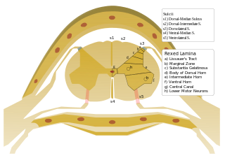
WThe grey commissure is a thin strip of grey matter that surrounds the central canal of the spinal cord and, along with the anterior white commissure, connects the two halves of the cord. It comprises lamina X in the Rexed classification.
WThe lateral grey column is one of the three grey columns of the spinal cord ; the others being the anterior and posterior grey columns. The lateral grey column is primarily involved with activity in the sympathetic division of the autonomic motor system. It projects to the side as a triangular field in the thoracic and upper lumbar regions of the postero-lateral part of the anterior grey column.
WThe intermediolateral nucleus (IML) is a region of grey matter found in one of the three grey columns of the spinal cord, the lateral grey column. This is Rexed lamina VII.
WThe lateral grey column is one of the three grey columns of the spinal cord ; the others being the anterior and posterior grey columns. The lateral grey column is primarily involved with activity in the sympathetic division of the autonomic motor system. It projects to the side as a triangular field in the thoracic and upper lumbar regions of the postero-lateral part of the anterior grey column.
WThe marginal nucleus of spinal cord, or posteromarginal nucleus, Rexed lamina I, is located at the most dorsal aspect of the dorsal horn of the spinal cord. The neurons located here receive input primarily from Lissauer's tract and relay information related to pain and temperature sensation. Pain sensation relayed here cannot be modulated, e.g. pain from burning the skin The axons of neurons contribute to the lateral spinothalamic tract.
 W
WGood posture refers to the "three natural curves [that] are present in a healthy spine.".It is also called neutral spine. Looking directly at the front or back of the body, the 33 vertebrae in the spinal column should appear completely vertical. From a side view, the cervical (neck) region of the spine (C1–C7) is bent inward, the thoracic region (T1–T12) bends outward, and the lumbar region (L1–L5) bends inward. The sacrum and coccyx rest between the pelvic bones. A neutral pelvis indicates the anterior superior iliac spines and pubic symphysis fall in the same vertical line.
WThe nucleus proprius is a layer of the spinal cord adjacent to the substantia gelatinosa. The nucleus proprius can be found in the gray matter in all levels of the spinal cord. It constitutes the first synapse of the spinothalamic tract carrying pain and temperature sensations from peripheral nerves. Cells in this nucleus project to deeper laminae of the spinal cord, to the posterior column nuclei, and to other supraspinal relay centers including the midbrain, thalamus, and hypothalamus. Rexed laminae III and IV make up the nucleus proprius.
WOnuf's nucleus is a distinct group of neurons located in the ventral part of the anterior horn of the sacral region of the human spinal cord involved in the maintenance of micturition and defecatory continence, as well as muscular contraction during orgasm. It contains motor neurons, and is the origin of the pudendal nerve. The sacral region of the spinal cord is the fourth segment of vertebrae in the spinal cord which consists of the vertebrae 26-30. While working in New York City in 1899, Bronislaw Onuf-Onufrowicz discovered this group of unique cells and originally identified it as “Group X.” “Group X” was considered distinct by Onufrowicz because the cells were different in size from the surrounding neurons in the anterolateral group, suggesting that they were independent.
WThe posterior grey column of the spinal cord is one of the three grey columns of the spinal cord. It receives several types of sensory information from the body, including fine touch, proprioception, and vibration. This information is sent from receptors of the skin, bones, and joints through sensory neurons whose cell bodies lie in the dorsal root ganglion.
WThe posterior median sulcus is the posterior end of the posterior median septum of neuroglia of the spinal cord. The septum varies in depth from 4 to 6 mm, but diminishes considerably in the lower part of the spinal cord.
 W
WThe posterior thoracic nucleus, is a group of interneurons found in the medial part of lamina VII, also known as the intermediate zone, of the spinal cord. It is mainly located from the cervical vertebra C7 to lumbar L3-L4 levels and is an important structure for proprioception of the lower limb.
WOn either side of the posterior median sulcus of the spinal cord, and at a short distance from it, the posterior nerve roots are attached along a vertical furrow named the posterolateral sulcus. The portion of the medulla spinalis which lies between this and the posterior median sulcus is named the posterior funiculus.
 W
WThe Prince Henry Hospital site, formerly known as the Prince Henry Hospital, Sydney, is a heritage-listed former teaching hospital and infectious diseases hospital and now UNSW teaching hospital and spinal rehabilitation unit located at 1430 Anzac Parade, Little Bay, City of Randwick, New South Wales, Australia. It was designed by NSW NSW Colonial Architect and NSW Government Architect and built from 1881 by NSW Public Works Department. It is also known as Prince Henry Hospital and The Coast Hospital. The property is owned by Landcom, an agency of the Government of New South Wales. It was added to the New South Wales State Heritage Register on 2 May 2003.
 W
WThe proper fasciculi, or spinospinal fasciculi, or propriospinal tracts, are groups of short fibres, ascending and descending, and crossed and uncrossed, within the spinal cord. These fibres are grouped into anterior, posterior, and lateral regions and make up a spinal pathway. Descending dorsal root collaterals are often included in the pathway.
WThe Rexed laminae comprise a system of ten layers of grey matter (I–X), identified in the early 1950s by Bror Rexed to label portions of the grey columns of the spinal cord.
 W
WSpinal locomotion results from intricate dynamic interactions between a central program in lower thoracolumbar spine and proprioceptive feedback from body in the absence of central control by brain as in complete spinal cord injury (SCI). Following SCI, the spinal circuitry below the lesion site does not become silent rather it continues to maintain active and functional neuronal properties although in a modified manner.
 W
WA spinal neuron is a neuron in the spinal cord.
 W
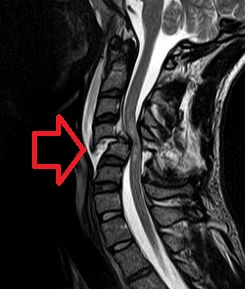
WA spinal cord injury (SCI) is damage to the spinal cord that causes temporary or permanent changes in its function. Symptoms may include loss of muscle function, sensation, or autonomic function in the parts of the body served by the spinal cord below the level of the injury. Injury can occur at any level of the spinal cord and can be complete, with a total loss of sensation and muscle function at lower sacral segments, or incomplete, meaning some nervous signals are able to travel past the injured area of the cord up to the Sacral S4-5 spinal cord segments. Depending on the location and severity of damage, the symptoms vary, from numbness to paralysis, including bowel or bladder incontinence. Long term outcomes also range widely, from full recovery to permanent tetraplegia or paraplegia. Complications can include muscle atrophy, loss of voluntary motor control, spasticity, pressure sores, infections, and breathing problems.
 W
WA spontaneous cerebrospinal fluid leak is a cerebrospinal fluid leak – a leak of cerebrospinal fluid that surrounds the brain and spinal cord from the protective dural sac for no apparent reason. The dura mater is the tough, outermost of layer of the meninges, the membranes surrounding the brain and spinal cord.
WThe apex of the posterior grey column, one of the three grey columns of the spinal cord, is capped by a V-shaped or crescentic mass of translucent, gelatinous neuroglia, termed the substantia gelatinosa of Rolando, which contains both neuroglia cells, and small nerve cells. The gelatinous appearance is due to a very low concentration of myelinated fibers. It extends the entire length of the spinal cord and into the medulla oblongata where it becomes the spinal nucleus of the trigeminal nerve.
 W
WThe thecal sac or dural sac is the membranous sheath (theca) or tube of dura mater that surrounds the spinal cord and the cauda equina. The thecal sac contains the cerebrospinal fluid which provides nutrients and buoyancy to the spinal cord. From the skull the tube adheres to bone at the foramen magnum and extends down to the second sacral vertebra where it tapers to cover over the filum terminale. Along most of the spinal canal it is separated from the inner surface by the epidural space. The sac has projections that follow the spinal nerves along their paths out of the vertebral canal which become the dural root sheaths.
 W
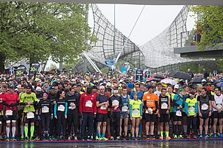
WThe Wings for Life World Run is a running competition held on the first weekend of May since 2014 to collect funds for the not-for-profit foundation Wings for Life. The entry fee goes completely to the foundation.