 W
WAcute disseminated encephalomyelitis (ADEM), or acute demyelinating encephalomyelitis, is a rare autoimmune disease marked by a sudden, widespread attack of inflammation in the brain and spinal cord. As well as causing the brain and spinal cord to become inflamed, ADEM also attacks the nerves of the central nervous system and damages their myelin insulation, which, as a result, destroys the white matter. It is often triggered by a viral infection or specific non-routine vaccinations.
 W
WAcute flaccid myelitis (AFM) is a serious condition of the spinal cord. Symptoms include rapid onset of arm or leg weakness and decreased reflexes. Difficulty moving the eyes, speaking, or swallowing may also occur. Occasionally, numbness or pain may be present. Complications can include trouble breathing.
 W
WAn altered level of consciousness is any measure of arousal other than normal. Level of consciousness (LOC) is a measurement of a person's arousability and responsiveness to stimuli from the environment. A mildly depressed level of consciousness or alertness may be classed as lethargy; someone in this state can be aroused with little difficulty. People who are obtunded have a more depressed level of consciousness and cannot be fully aroused. Those who are not able to be aroused from a sleep-like state are said to be stuporous. Coma is the inability to make any purposeful response. Scales such as the Glasgow coma scale have been designed to measure the level of consciousness.
 W
WAnterior horn disease is one of a number of medical disorders affecting the anterior horn of the spinal cord. Anterior horn diseases include spinal muscular atrophy, poliomyelitis and amyotrophic lateral sclerosis.
 W
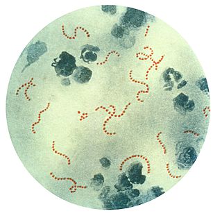
WCavernous sinus thrombosis (CST) is the formation of a blood clot within the cavernous sinus, a cavity at the base of the brain which drains deoxygenated blood from the brain back to the heart. This is a rare disorder and can be of two types–septic cavernous thrombosis and aseptic cavernous thrombosis. Most commonly the form is of septic cavernous sinus thrombosis. The cause is usually from a spreading infection in the nose, sinuses, ears, or teeth. Staphylococcus aureus and Streptococcus are often the associated bacteria.
 W
WCentral nervous system diseases, also known as central nervous system disorders, are a group of neurological disorders that affect the structure or function of the brain or spinal cord, which collectively form the central nervous system (CNS).
 W
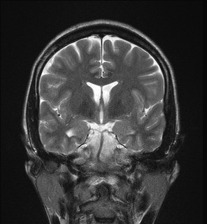
WChoroid plexus cysts (CPCs) are cysts that occur within choroid plexus of the brain. They are the most common type of intraventricular cyst. The brain contains pockets or spaces called ventricles with a spongy layer of cells and blood vessels called the choroid plexus. This is in the middle of the fetal brain. The choroid plexus has the important function of producing cerebrospinal fluid. The fluid produced by the cells of the choroid plexus fills the ventricles and then flows around the brain and the spinal cord to provide a cushion of fluid around these structures.
 W
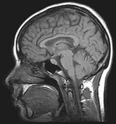
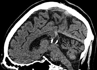
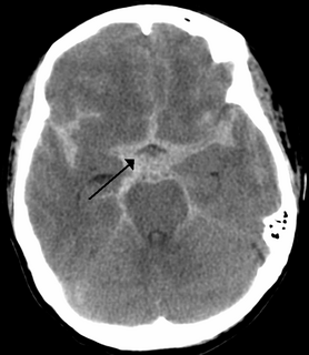
WA colloid cyst is a non-cancerous tumor in the brain. It consists of a gelatinous material contained within a membrane of epithelial tissue. It is almost always found just posterior to the foramen of Monro in the anterior aspect of the third ventricle, originating from the roof of the ventricle. Because of its location, it can cause obstructive hydrocephalus and increased intracranial pressure. Colloid cysts represent 0.5–1.0% of intracranial tumors.
 W
WDentatorubral–pallidoluysian atrophy (DRPLA) is an autosomal dominant spinocerebellar degeneration caused by an expansion of a CAG repeat encoding a polyglutamine tract in the atrophin-1 protein. It is also known as Haw River Syndrome and Naito–Oyanagi disease. Although this condition was perhaps first described by Smith et al. in 1958, and several sporadic cases have been reported from Western countries, this disorder seems to be very rare except in Japan.
 W
WFoix-Chavany-Marie Syndrome (FCMS), also known as Bilateral Opercular Syndrome, is a neuropathological disorder characterized by paralysis of the facial, tongue, pharynx, and masticatory muscles of the mouth that aid in chewing. The disorder is primarily caused by thrombotic and embolic strokes, which cause a deficiency of oxygen in the brain. As a result, bilateral lesions may form in the junctions between the frontal lobe and temporal lobe, the parietal lobe and cortical lobe, or the subcortical region of the brain. FCMS may also arise from defects existing at birth that may be inherited or nonhereditary. Symptoms of FCMS can be present in a person of any age and it is diagnosed using automatic-voluntary dissociation assessment, psycholinguistic testing, neuropsychological testing, and brain scanning. Treatment for FCMS depends on the onset, as well as on the severity of symptoms, and it involves a multidisciplinary approach.
 W
WGray matter heterotopias are neurological disorders caused by clumps of gray matter located in the wrong part of the brain. A grey matter heterotopia is characterized as a type of focal cortical dysplasia. The neurons in heterotopia appear to be normal, except for their mislocation; nuclear studies have shown glucose metabolism equal to that of normally positioned gray matter. The condition causes a variety of symptoms, but usually includes some degree of epilepsy or recurring seizures, and often affects the brain's ability to function on higher levels. Symptoms range from nonexistent to profound; the condition is occasionally discovered as an incidentaloma when brain imaging performed for an unrelated problem and has no apparent ill effect on the patient. At the other extreme, heterotopia can result in severe seizure disorder, loss of motor skills, and mental retardation. Fatalities are practically unknown, other than the death of unborn male fetuses with a specific genetic defect.
WHereditary diffuse leukoencephalopathy with spheroids (HDLS) is a rare adult onset autosomal dominant disorder characterized by cerebral white matter degeneration with demyelination and axonal spheroids leading to progressive cognitive and motor dysfunction. Spheroids are axonal swellings with discontinuous or absence of myelin sheaths. It is believed that the disease arises from primary microglial dysfunction that leads to secondary disruption of axonal integrity, neuroaxonal damage, and focal axonal spheroids leading to demyelination. Spheroids in HDLS resemble to some extent those produced by shear stress in a closed head injury with damage to axons, causing them to swell due to blockage of axoplasmic transport. In addition to trauma, axonal spheroids can be found in aged brain, stroke, and in other degenerative diseases. In HDLS, it is uncertain whether demyelination occurs prior to the axonal spheroids or what triggers neurodegeneration after apparently normal brain and white matter development, although genetic deficits suggest that demyelination and axonal pathology may be secondary to microglial dysfunction. The clinical syndrome in patients with HDLS is not specific and it can be mistaken for Alzheimer's disease, frontotemporal dementia, atypical Parkinsonism, multiple sclerosis, or corticobasal degeneration.
 W
WHippocampal sclerosis (HS) is a neuropathological condition with severe neuronal cell loss and gliosis in the hippocampus, specifically in the CA-1 and subiculum of the hippocampus. It was first described in 1880 by Wilhelm Sommer. Hippocampal sclerosis is a frequent pathologic finding in community-based dementia. Hippocampal sclerosis can be detected with autopsy or MRI. Individuals with hippocampal sclerosis have similar initial symptoms and rates of dementia progression to those with Alzheimer's disease (AD) and therefore are frequently misclassified as having Alzheimer's Disease. But clinical and pathologic findings suggest that hippocampal sclerosis has characteristics of a progressive disorder although the underlying cause remains elusive. A diagnosis of hippocampal sclerosis has a significant effect on the life of patients because of the notable mortality, morbidity and social impact related to epilepsy, as well as side effects associated with antiepileptic treatments.
 W
WIdiopathic intracranial hypertension (IIH), previously known as pseudotumor cerebri and benign intracranial hypertension, is a condition characterized by increased intracranial pressure without a detectable cause. The main symptoms are headache, vision problems, ringing in the ears with the heartbeat, and shoulder pain. Complications may include vision loss.
 W
WThe term dolichoectasia means elongation and distension. It is used to characterize arteries throughout the human body which have shown significant deterioration of their tunica intima, weakening the vessel walls and causing the artery to elongate and distend.
 W
WLeukodystrophies are a group of usually inherited disorders characterized by degeneration of the white matter in the brain. The word leukodystrophy comes from the Greek roots leuko, "white", dys, "abnormal" and troph, "growth". The leukodystrophies are caused by imperfect growth or development of the myelin sheath, the fatty insulating covering around nerve fibers. Leukodystrophies may be classified as hypomyelinating or demyelinating diseases, depending on whether the damage is present before birth or occurs after. Other demyelinating diseases are usually not congenital and have a toxic or autoimmune cause.
 W
WLhermitte–Duclos disease (LDD), also called dysplastic gangliocytoma of the cerebellum, is a rare, slowly growing tumor of the cerebellum, a gangliocytoma sometimes considered to be a hamartoma, characterized by diffuse hypertrophy of the granular layer of the cerebellum. It is often associated with Cowden syndrome. It was described by Jacques Jean Lhermitte and P. Duclos in 1920.
 W
WLow-pressure hydrocephalus (LPH) is a condition whereby ventricles are enlarged and the individual experiences severe dementia, inability to walk, and incontinence – despite very low intracranial pressure (ICP). Low pressure hydrocephalus appears to be a more acute form of normal pressure hydrocephalus. If not diagnosed in a timely fashion, the individual runs the risk of remaining in the low pressure hydrocephalic state or LPHS. Shunt revisions, even when they are set to drain at a low ICP, are not always effective. The pressure in the brain does not get high enough to allow the cerebrospinal fluid to drain in a shunt system, therefore the shunt is open, but malfunctioning in LPH. In cases of LPH, chronic infarcts can also develop along the corona radiata in response to the tension in the brain as the ventricles increase in size. Certain causes of LPH include trauma, tumor, bleeding, inflammation of the lining of the brain, whole brain radiation, as well as other brain pathology that affects the compliance of the brain parenchyma. One treatment for the LPHS is an external ventricular drain (EVD) set at negative pressures. According to Pang & Altschuler et al., a controlled, steady, negative pressure siphoning with EVD, carefully monitored with partial computer tomography scans is a safe and effective way of treating LPH. In their experience, this approach helps restore the brain mantle. They caution against dropping or raising the pressure of the EVD too quickly as it increases risk and also destabilizes the ventricles. Getting the ventricles smaller, is the initial step, stabilising them is the second step before placing a shunt – which is the final step in therapy. Any variation from this formula can lead to an ineffective, yet patent shunt system, despite a low-pressure setting. Care should be taken with EVD therapy, as mismanagement of the EVD can lead to long-term permanent complications and brain injury.
 W
WMeningoencephalitis, also known as herpes meningoencephalitis, is a medical condition that simultaneously resembles both meningitis, which is an infection or inflammation of the meninges, and encephalitis, which is an infection or inflammation of the brain.
 W
WA minimally conscious state (MCS) is a disorder of consciousness distinct from persistent vegetative state and locked-in syndrome. Unlike persistent vegetative state, patients with MCS have partial preservation of conscious awareness. MCS is a relatively new category of disorders of consciousness. The natural history and longer term outcome of MCS have not yet been thoroughly studied. The prevalence of MCS was estimated to be 112,000 to 280,000 adult and pediatric cases.
 W
WMyelomalacia is a pathological term referring to the softening of the spinal cord. Possible causes of myelomalacia include cervical myelopathy, hemorrhagic infarction, or acute injury, such as that caused by intervertebral disc extrusion.
 W
WPediatric autoimmune neuropsychiatric disorders associated with streptococcal infections (PANDAS) is a hypothesis that there exists a subset of children with rapid onset of obsessive-compulsive disorder (OCD) or tic disorders and these symptoms are caused by group A beta-hemolytic streptococcal (GABHS) infections. The proposed link between infection and these disorders is that an initial autoimmune reaction to a GABHS infection produces antibodies that interfere with basal ganglia function, causing symptom exacerbations. It has been proposed that this autoimmune response can result in a broad range of neuropsychiatric symptoms. The PANDAS hypothesis was based on observations in clinical case studies at the US National Institutes of Health and in subsequent clinical trials where children appeared to have dramatic and sudden OCD exacerbations and tic disorders following infections.
 W
WPeriventricular leukomalacia (PVL) is a form of white-matter brain injury, characterized by the necrosis of white matter near the lateral ventricles. It can affect newborns and fetuses; premature infants are at the greatest risk of neonatal encephalopathy which may lead to this condition. Affected individuals generally exhibit motor control problems or other developmental delays, and they often develop cerebral palsy or epilepsy later in life.
 W
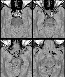
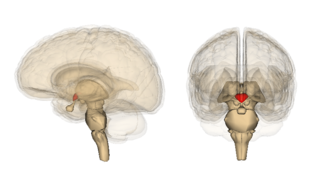
WA pineal gland cyst is a usually benign (non-malignant) cyst in the pineal gland, a small endocrine gland in the brain. Historically, these fluid-filled bodies appeared on 1-4% of magnetic resonance imaging (MRI) brain scans, but were more frequently diagnosed at death, seen in 4-11% of autopsies. A 2007 study by Pu et al. found a frequency of 23% in brain scans.
 W
WPoliomyelitis, commonly shortened to polio, is an infectious disease caused by the poliovirus. In about 0.5 percent of cases, it moves from the gut to affect the central nervous system and there is muscle weakness resulting in a flaccid paralysis. This can occur over a few hours to a few days. The weakness most often involves the legs, but may less commonly involve the muscles of the head, neck and diaphragm. Many people fully recover. In those with muscle weakness, about 2 to 5 percent of children and 15 to 30 percent of adults die. For all those infected, in up to 70 percent of infections there are no symptoms. Another 25 percent of people have minor symptoms such as fever and a sore throat, and up to 5 percent have headache, neck stiffness and pains in the arms and legs. These people are usually back to normal within one or two weeks. Years after recovery, post-polio syndrome may occur, with a slow development of muscle weakness similar to that which the person had during the initial infection.
 W
WPosterior cortical atrophy (PCA), also called Benson's syndrome, is a form of dementia which is usually considered an atypical variant of Alzheimer's disease (AD). The disease causes atrophy of the posterior part of the cerebral cortex, resulting in the progressive disruption of complex visual processing. PCA was first described by D. Frank Benson in 1988.
 W
WPosterior reversible encephalopathy syndrome (PRES), also known as reversible posterior leukoencephalopathy syndrome (RPLS), is a rare condition in which parts of the brain are affected by swelling, usually as a result of an underlying cause. Someone with PRES may experience headache, changes in vision, and seizures, with some developing other neurological symptoms such as confusion or weakness of one or more limbs. The name of the condition includes the word "posterior" because it predominantly though not exclusively affects the back of the brain. Common underlying causes are severely elevated blood pressure, kidney failure, severe infections, certain medications, some autoimmune diseases, and pre-eclampsia. The diagnosis is usually made by brain scan (MRI) on which areas of swelling can be identified.
 W
WRaymond-Céstan syndrome is caused by blockage of the long circumferential branches of the basilar artery. It was described by Fulgence Raymond and Étienne Jacques Marie Raymond Céstan. Along with other related syndromes such as Millard-Gubler syndrome, Foville's syndrome, and Weber's syndrome, the description was instrumental in establishing important principles in brain-stem localization.
WSpinocerebellar ataxia type 13 (SCA13) is a rare autosomal dominant disorder, which, like other types of SCA, is characterized by dysarthria, nystagmus, and ataxia of gait, stance and the limbs due to cerebellar dysfunction. Patients with SCA13 also tend to present with epilepsy, an inability to run, and increased reflexes. This cerebellar dysfunction is permanent and progressive. SCA13 is caused by mutations in KCNC3, a gene encoding a voltage-gated potassium channel KV3.3. There are two known mutations in this gene causative for SCA13. Unlike many other types of SCA, these are not polyglutamine expansions but, rather, point mutations resulting in channels with no current or altered kinetics.
WA spontaneous cerebrospinal fluid leak is a cerebrospinal fluid leak – a leak of cerebrospinal fluid that surrounds the brain and spinal cord from the protective dural sac for no apparent reason. The dura mater is the tough, outermost of layer of the meninges, the membranes surrounding the brain and spinal cord.
 W
WSubarachnoid hemorrhage (SAH) is bleeding into the subarachnoid space—the area between the arachnoid membrane and the pia mater surrounding the brain. Symptoms may include a severe headache of rapid onset, vomiting, decreased level of consciousness, fever, and sometimes seizures. Neck stiffness or neck pain are also relatively common. In about a quarter of people a small bleed with resolving symptoms occurs within a month of a larger bleed.
 W
WSuperficial hemosiderosis of the central nervous system is a disease of the brain resulting from chronic iron deposition in neuronal tissues associated with cerebrospinal fluid. This occurs via the deposition of hemosiderin in neuronal tissue, and is associated with neuronal loss, gliosis, and demyelination of neuronal cells. This disease was first discovered in 1908 by R.C. Hamill after performing an autopsy. Detection of this disease was largely post-mortem until the advent of MRI technology, which made diagnosis far easier. Superficial siderosis is largely considered a rare disease, with less than 270 total reported cases in scientific literature as of 2006, and affects people of a wide range of ages with men being approximately three times more frequently affected than women. The number of reported cases of superficial siderosis has increased with advances in MRI technology, but it remains a rare disease.
 W
WTransverse myelitis (TM) is a rare neurological condition in which the spinal cord is inflamed. Transverse implies that the inflammation extends horizontally across the spinal cord. Partial transverse myelitis and partial myelitis are terms sometimes used to specify inflammation that only affects part of the width of the spinal cord. TM is characterized by weakness and numbness of the limbs, deficits in sensation and motor skills, dysfunctional urethral and anal sphincter activities, and dysfunction of the autonomic nervous system that can lead to episodes of high blood pressure. Signs and symptoms vary according to the affected level of the spinal cord. The underlying cause of TM is unknown. The spinal cord inflammation seen in TM has been associated with various infections, immune system disorders, or damage to nerve fibers, by loss of myelin. As opposed to leukomyelitis which affects only the white matter, it affects the entire cross-section of the spinal cord. Decreased electrical conductivity in the nervous system can result.
 W
WWernicke encephalopathy (WE), also Wernicke's encephalopathy is the presence of neurological symptoms caused by biochemical lesions of the central nervous system after exhaustion of B-vitamin reserves, in particular thiamine. The condition is part of a larger group of thiamine deficiency disorders, that includes beriberi in all its forms, and alcoholic Korsakoff syndrome. When it occurs simultaneously with alcoholic Korsakoff syndrome it is known as Wernicke–Korsakoff syndrome.
 W
WWernicke–Korsakoff syndrome (WKS) is the combined presence of Wernicke encephalopathy (WE) and alcoholic Korsakoff syndrome. Due to the close relationship between these two disorders, people with either are usually diagnosed with WKS as a single syndrome. It mainly causes vision changes, ataxia and impaired memory.