 W
WThe cerebral cortex, also known as the cerebral mantle, is the outer layer of neural tissue of the cerebrum of the brain in humans and other mammals. The cerebral cortex mostly consists of the six-layered neocortex, with just ten per cent consisting of allocortex. It is separated into two cortices, by the longitudinal fissure that divides the cerebrum into the left and right cerebral hemispheres. The two hemispheres are joined beneath the cortex by the corpus callosum. The cerebral cortex is the largest site of neural integration in the central nervous system. It plays a key role in attention, perception, awareness, thought, memory, language, and consciousness.
 W
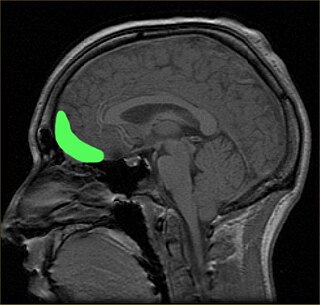
WIn the human brain, the anterior cingulate cortex (ACC) is the frontal part of the cingulate cortex that resembles a "collar" surrounding the frontal part of the corpus callosum. It consists of Brodmann areas 24, 32, and 33.
 W
WThe archicortex, or archipallium, is the phylogenetically oldest region of the brain's cerebral cortex. It is often considered contiguous with the olfactory cortex, but its extent varies among species. In older species, such as fish, the archipallium makes up most of the cerebrum. Amphibians develop an archipallium and paleopallium.
 W
WThe auditory cortex is the part of the temporal lobe that processes auditory information in humans and many other vertebrates. It is a part of the auditory system, performing basic and higher functions in hearing, such as possible relations to language switching. It is located bilaterally, roughly at the upper sides of the temporal lobes – in humans, curving down and onto the medial surface, on the superior temporal plane, within the lateral sulcus and comprising parts of the transverse temporal gyri, and the superior temporal gyrus, including the planum polare and planum temporale.
 W
WBetz cells are giant pyramidal cells (neurons) located within the fifth layer of the grey matter in the primary motor cortex. These neurons are the largest in the central nervous system, sometimes reaching 100 μm in diameter.
 W
WThe cingulate cortex is a part of the brain situated in the medial aspect of the cerebral cortex. The cingulate cortex includes the entire cingulate gyrus, which lies immediately above the corpus callosum, and the continuation of this in the cingulate sulcus. The cingulate cortex is usually considered part of the limbic lobe.
 W
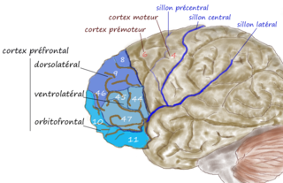
WThe dorsolateral prefrontal cortex is an area in the prefrontal cortex of the brain of humans and other primates. It is one of the most recently derived parts of the human brain. It undergoes a prolonged period of maturation which lasts until adulthood. The DLPFC is not an anatomical structure, but rather a functional one. It lies in the middle frontal gyrus of humans. In macaque monkeys, it is around the principal sulcus. Other sources consider that DLPFC is attributed anatomically to BA 9 and 46 and BA 8, 9 and 10.
 W
WThe dorsomedial prefrontal cortex is a section of the prefrontal cortex in some species' brain anatomy. It includes portions of Brodmann areas BA8, BA9, BA10, BA24 and BA32, although some authors identify it specifically with BA8 and BA9 Some notable sub-components include the dorsal anterior cingulate cortex, the prelimbic cortex, and the infralimbic cortex.
 W
WThe entorhinal cortex (EC) is an area of the brain located in the medial temporal lobe and functions as a hub in a widespread network for memory, navigation and the perception of time. The EC is the main interface between the hippocampus and neocortex. The EC-hippocampus system plays an important role in declarative (autobiographical/episodic/semantic) memories and in particular spatial memories including memory formation, memory consolidation, and memory optimization in sleep. The EC is also responsible for the pre-processing (familiarity) of the input signals in the reflex nictitating membrane response of classical trace conditioning; the association of impulses from the eye and the ear occurs in the entorhinal cortex.
 W
WThe frontal eye fields (FEF) are a region located in the frontal cortex, more specifically in Brodmann area 8 or BA8, of the primate brain. In humans, it can be more accurately said to lie in a region around the intersection of the middle frontal gyrus with the precentral gyrus, consisting of a frontal and parietal portion. The FEF is responsible for saccadic eye movements for the purpose of visual field perception and awareness, as well as for voluntary eye movement. The FEF communicates with extraocular muscles indirectly via the paramedian pontine reticular formation. Destruction of the FEF causes deviation of the eyes to the ipsilateral side.
 W
WThe insular cortex is a portion of the cerebral cortex folded deep within the lateral sulcus within each hemisphere of the mammalian brain.
 W
WThe line of Gennari is a band of myelinated axons that run parallel to the surface of the cerebral cortex on the banks of the calcarine fissure in the occipital lobe. This formation is visible to the naked eye as a white strip running through the cortical grey matter, and is the reason the V1 in primates is also referred to as "striate cortex." The line of Gennari is due to dense axonal input from the thalamus to layer IV of visual cortex. The structure is named for its discoverer, Francesco Gennari, who first observed it in 1776 as a medical student at the University of Parma. He described it in a book which he published six years later. Although non-primate species have areas that are designated primary visual cortex, some lack a stria of Gennari.
 W
WThe motor cortex is the region of the cerebral cortex involved in the planning, control, and execution of voluntary movements. Classically, the motor cortex is an area of the frontal lobe located in the posterior precentral gyrus immediately anterior to the central sulcus.
WThe neocortex, also called the neopallium, isocortex, or the six-layered cortex, is a set of layers of the mammalian cerebral cortex involved in higher-order brain functions such as sensory perception, cognition, generation of motor commands, spatial reasoning and language. The neocortex is further subdivided into the true isocortex and the proisocortex.
 W
WThe olfactory tubercle (OT), also known as the tuberculum olfactorium, is a multi-sensory processing center that is contained within the olfactory cortex and ventral striatum and plays a role in reward cognition. The OT has also been shown to play a role in locomotor and attentional behaviors, particularly in relation to social and sensory responsiveness, and it may be necessary for behavioral flexibility. The OT is interconnected with numerous brain regions, especially the sensory, arousal, and reward centers, thus making it a potentially critical interface between processing of sensory information and the subsequent behavioral responses.
 W
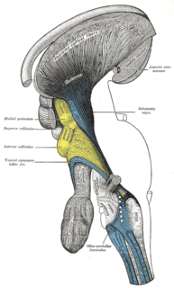
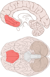
WThe orbitofrontal cortex (OFC) is a prefrontal cortex region in the frontal lobes of the brain which is involved in the cognitive process of decision-making. In non-human primates it consists of the association cortex areas Brodmann area 11, 12 and 13; in humans it consists of Brodmann area 10, 11 and 47.
 W
WOrientation columns are organized regions of neurons that are excited by visual line stimuli of varying angles. These columns are located in the primary visual cortex (V1) and span multiple cortical layers. The geometry of the orientation columns are arranged in slabs that are perpendicular to the surface of the primary visual cortex.
 W
WIn anatomy of animals, the paleocortex, or paleopallium, is a region within the telencephalon in the vertebrate brain. This type of cortical tissue consists of three cortical laminae. In comparison, the neocortex has six layers and the archicortex has three or four layers. Because the number of laminae that compose a type of cortical tissue seems to be directly proportional to both the information-processing capabilities of that tissue and its phylogenetic age, paleocortex is thought to be an intermediate between the archicortex and the neocortex in both aspects.
WThe paralimbic cortex is an area of three-layered cortex that includes the following regions: the piriform cortex, entorhinal cortex, the parahippocampal cortex on the medial surface of the temporal lobe, and the cingulate cortex just above the corpus callosum.
WThe piriform cortex, or pyriform cortex, is a region in the brain, part of the rhinencephalon situated in the cerebrum. The function of the piriform cortex relates to the sense of smell.
 W
WThe planum temporale is the cortical area just posterior to the auditory cortex within the Sylvian fissure. It is a triangular region which forms the heart of Wernicke's area, one of the most important functional areas for language. Original studies on this area found that the planum temporale was one of the most asymmetric regions in the brain, with this area being up to ten times larger in the left cerebral hemisphere than the right.
 W
WThe posterior parietal cortex plays an important role in planned movements, spatial reasoning, and attention.
 W
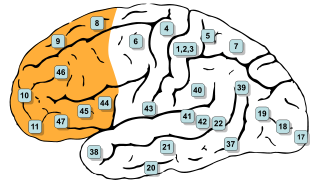
WIn mammalian brain anatomy, the prefrontal cortex (PFC) is the cerebral cortex which covers the front part of the frontal lobe. The PFC contains the Brodmann areas BA8, BA9, BA10, BA11, BA12, BA13, BA14, BA24, BA25, BA32, BA44, BA45, BA46, and BA47.
WThe premotor cortex is an area of motor cortex lying within the frontal lobe of the brain just anterior to the primary motor cortex. It occupies part of Brodmann's area 6. It has been studied mainly in primates, including monkeys and humans. The functions of the premotor cortex are diverse and not fully understood. It projects directly to the spinal cord and therefore may play a role in the direct control of behavior, with a relative emphasis on the trunk muscles of the body. It may also play a role in planning movement, in the spatial guidance of movement, in the sensory guidance of movement, in understanding the actions of others, and in using abstract rules to perform specific tasks. Different subregions of the premotor cortex have different properties and presumably emphasize different functions. Nerve signals generated in the premotor cortex cause much more complex patterns of movement than the discrete patterns generated in the primary motor cortex.
 W
WThe primary motor cortex is a brain region that in humans is located in the dorsal portion of the frontal lobe. It is the primary region of the motor system and works in association with other motor areas including premotor cortex, the supplementary motor area, posterior parietal cortex, and several subcortical brain regions, to plan and execute movements. Primary motor cortex is defined anatomically as the region of cortex that contains large neurons known as Betz cells. Betz cells, along with other cortical neurons, send long axons down the spinal cord to synapse onto the interneuron circuitry of the spinal cord and also directly onto the alpha motor neurons in the spinal cord which connect to the muscles.
 W
WThe primary somatosensory cortex is located in the postcentral gyrus, and is part of the somatosensory system. It was initially defined from surface stimulation studies of Wilder Penfield, and parallel surface potential studies of Bard, Woolsey, and Marshall. Although initially defined to be roughly the same as Brodmann areas 3, 1 and 2, more recent work by Kaas has suggested that for homogeny with other sensory fields only area 3 should be referred to as "primary somatosensory cortex", as it receives the bulk of the thalamocortical projections from the sensory input fields.
WPyramidal cells, or pyramidal neurons, are a type of multipolar neuron found in areas of the brain including the cerebral cortex, the hippocampus, and the amygdala. Pyramidal neurons are the primary excitation units of the mammalian prefrontal cortex and the corticospinal tract. Pyramidal neurons are also one of two cell types where the characteristic sign, Negri bodies, are found in post-mortem rabies infection. Pyramidal neurons were first discovered and studied by Santiago Ramón y Cajal. Since then, studies on pyramidal neurons have focused on topics ranging from neuroplasticity to cognition.
 W
WThe Radial Unit Hypothesis (RUH) is a conceptual theory of cerebral cortex development, first described by Pasko Rakic. The RUH states that the cerebral cortex develops during embryogenesis as an array of interacting cortical columns, or 'radial units', each of which originates from a transient stem cell layer called the ventricular zone, which contains neural stem cells known as radial glial cells.
 W
WThe retrosplenial cortex (RSC) is a cortical area in the brain, located posteriorly and comprising Brodmann areas 29 and 30. The region's name refers to its anatomical location immediately behind the splenium of the corpus callosum in primates, although in rodents it is located more towards the brain surface and is relatively larger. Its function is currently not well understood, but its location close to visual areas and also to the hippocampal spatial/memory system suggest it may have a role in mediating between perceptual and memory functions.
 W
WThe human secondary somatosensory cortex is a region of cortex in the parietal operculum on the ceiling of the lateral sulcus.
 W
WThe subplate, also called the subplate zone, together with the marginal zone and the cortical plate, in the fetus represents the developmental anlage of the mammalian cerebral cortex. It was first described, as a separate transient fetal zone by Ivica Kostović and Mark E. Molliver in 1974.
 W
WThe supplementary motor area (SMA) is a part of the primate cerebral cortex that contributes to the control of movement. It is located on the midline surface of the hemisphere just in front of the primary motor cortex leg representation. In monkeys the SMA contains a rough map of the body. In humans the body map is not apparent. Neurons in the SMA project directly to the spinal cord and may play a role in the direct control of movement. Possible functions attributed to the SMA include the postural stabilization of the body, the coordination of both sides of the body such as during bimanual action, the control of movements that are internally generated rather than triggered by sensory events, and the control of sequences of movements. All of these proposed functions remain hypotheses. The precise role or roles of the SMA is not yet known.
 W
WThalamocortical radiations are the fibers between the thalamus and the cerebral cortex.
 W
WThe ventrolateral prefrontal cortex (VLPFC) is a part of the prefrontal cortex located on the inferior frontal gyrus, bounded superiorly by the inferior frontal sulcus and inferiorly by the lateral sulcus. It is attributed to the anatomical structures of Brodmann's area (BA) 47, 45 and 44.
 W
WThe ventromedial prefrontal cortex (vmPFC) is a part of the prefrontal cortex in the mammalian brain. The ventral medial prefrontal is located in the frontal lobe at the bottom of the cerebral hemispheres and is implicated in the processing of risk and fear, as it is critical in the regulation of amygdala activity in humans. It also plays a role in the inhibition of emotional responses, and in the process of decision making and self control. It is also involved in the cognitive evaluation of morality.
 W
WVestibular cortex is the portion of the cerebrum which responds to input from the vestibular system.
WWernicke's area, also called Wernicke's speech area, is one of the two parts of the cerebral cortex that are linked to speech, the other being Broca's area. It is involved in the comprehension of written and spoken language, in contrast to Broca's area, which is involved in the production of language. It is traditionally thought to reside in Brodmann area 22, which is located in the superior temporal gyrus in the dominant cerebral hemisphere, which is the left hemisphere in about 95% of right-handed individuals and 60% of left-handed individuals.