 W
WThe abdominal external oblique muscle is the largest and outermost of the three flat abdominal muscles of the lateral anterior abdomen.
 W
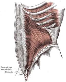
WThe abdominal internal oblique muscle, also internal oblique muscle or interior oblique, is an abdominal muscle in the abdominal wall that lies below the external oblique muscle and just above the transverse abdominal muscle.
 W
WThe aponeurosis of the abdominal external oblique muscle is a thin but strong membranous structure, the fibers of which are directed downward and medially.
 W
WThe bulbospongiosus muscle is one of the superficial muscles of the perineum. It has a slightly different origin, insertion and function in males and females. In males, it covers the bulb of the penis. In females, it covers the vestibular bulb.
 W
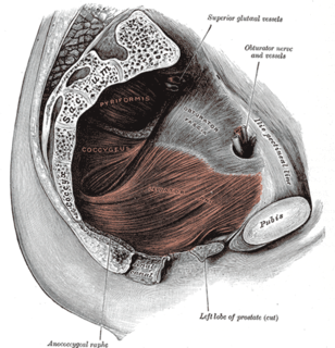
WThe Coccygeus or ischiococcygeus is a muscle of the pelvic floor, located posterior to levator ani and anterior to the sacrospinous ligament.
 W
WThe cremaster muscle is a muscle that covers the testis and the spermatic cord.
 W
WThe detrusor muscle, also detrusor urinae muscle, muscularis propria of the urinary bladder and muscularis propria, is smooth muscle found in the wall of the bladder. The detrusor muscle remains relaxed to allow the bladder to store urine, and contracts during urination to release urine. Related are the urethral sphincter muscles which envelop the urethra to control the flow of urine when they contract.
 W
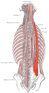
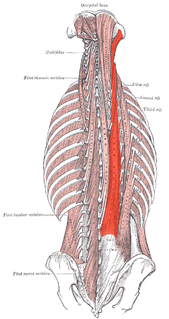
WThe erector spinae or spinal erectors is a set of muscles that straighten and rotate the back.
 W
WThe external intercostal muscles, or external intercostals are eleven in number on both sides.
 W
WThe iliocostalis is the muscle immediately lateral to the longissimus that is the nearest to the furrow that separates the epaxial muscles from the hypaxial. It lies very deep to the fleshy portion of the serratus posterior muscle.
 W
WThe innermost intercostal muscle is a layer of intercostal muscles deep to the plane that contains the intercostal nerves and intercostal vessels and the internal intercostal muscles.
 W
WIntercostal muscles are several groups of muscles that run between the ribs, and help form and move the chest wall. The intercostal muscles are mainly involved in the mechanical aspect of breathing. These muscles help expand and shrink the size of the chest cavity to facilitate breathing.
 W
WThe internal intercostal muscles are a group of skeletal muscles located between the ribs. They are eleven in number on either side. They commence anteriorly at the sternum, in the intercostal spaces between the cartilages of the true ribs, and at the anterior extremities of the cartilages of the false ribs, and extend backward as far as the angles of the ribs, hence they are continued to the vertebral column by thin aponeuroses, the posterior intercostal membranes.
 W
WThe intertransversarii are small muscles placed between the transverse processes of the vertebrae.
 W
WThe ischiocavernosus muscle is a muscle just below the surface of the perineum, present in both men and women.
WThe levator ani is a broad, thin muscle group, situated on either side of the pelvis. It is formed from three muscle components: the pubococcygeus, the iliococcygeus, and the puborectalis.
 W
WThe Levatores costarum, twelve in number on either side, are small tendinous and fleshy bundles, which arise from the ends of the transverse processes of the seventh cervical and upper eleven thoracic vertebrae
 W
WThe longissimus is the muscle lateral to the semispinalis muscles. It is the longest subdivision of the erector spinae muscles that extends forward into the transverse processes of the posterior cervical vertebrae.
 W
WThe multifidus muscle consists of a number of fleshy and tendinous fasciculi, which fill up the groove on either side of the spinous processes of the vertebrae, from the sacrum to the axis. While very thin, the Multifidus muscle plays an important role in stabilizing the joints within the spine. The multifidus is one of the transversospinales.
 W
WPectoral muscles are the muscles that connect the front of the human chest with the bones of the upper arm and shoulder.
 W
WThe pelvic floor or pelvic diaphragm is composed of muscle fibers of the levator ani, the coccygeus muscle, and associated connective tissue which span the area underneath the pelvis. The pelvic diaphragm is a muscular partition formed by the levatores ani and coccygei, with which may be included the parietal pelvic fascia on their upper and lower aspects. The pelvic floor separates the pelvic cavity above from the perineal region below. Because, to accommodate the birth canal, a female's pelvic cavity is larger than a male's, the pelvic floor tends to be considered a part of female anatomy, but males have an equivalent pelvic floor.
 W
WThe pyramidalis muscle is a small triangular muscle, anterior to the rectus abdominis muscle, and contained in the rectus sheath.
 W
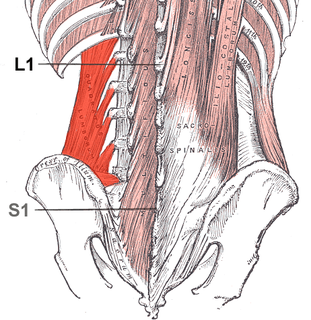
WThe quadratus lumborum muscle, informally called the QL, is a paired muscle of the left and right posterior abdominal wall. It is the deepest abdominal muscle, and commonly referred to as a back muscle. Each is irregular and quadrilateral in shape.
 W
WThe rectococcygeal muscles are two bands of smooth muscle tissue arising from the 2nd and 3rd coccygeal vertebrae, and passing downward and forward to blend with the rectal longitudinal smooth muscle fibers on the posterior wall of the anal canal.
 W
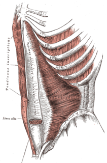
WThe rectus abdominis muscle, also known as the "abdominal muscle", is a paired muscle running vertically on each side of the anterior wall of the human abdomen, as well as that of some other mammals. There are two parallel muscles, separated by a midline band of connective tissue called the linea alba. It extends from the pubic symphysis, pubic crest and pubic tubercle inferiorly, to the xiphoid process and costal cartilages of ribs V to VII superiorly. The proximal attachments are the pubic crest and the pubic symphysis. It attaches distally at the costal cartilages of ribs 5-7 and the xiphoid process of the sternum.
WThe rectus sheath, also called the rectus fascia, is formed by the aponeuroses of the transverse abdominal and the internal and external oblique muscles. It contains the rectus abdominis and pyramidalis muscles.
 W
WThe rhomboid muscles, often simply called the rhomboids, are rhombus-shaped muscles associated with the scapula. There are two rhomboid muscles on each side of the upper back:Rhomboid major muscle Rhomboid minor muscle
 W
WThe rotatores muscles lie beneath the multifidus and are present in all spinal regions but are most prominent in the thoracic region; they are eleven in number on either side.
 W
WThe semispinalis muscles are a group of three muscles belonging to the transversospinales. These are the semispinalis capitis, the semispinalis cervicis and the semispinalis thoracis.
 W
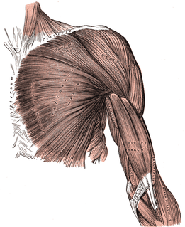
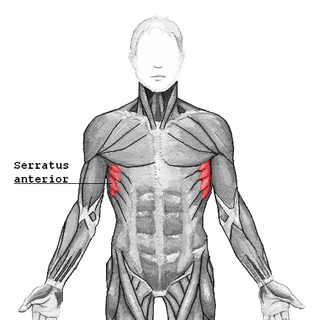
WThe serratus anterior is a muscle that originates on the surface of the 1st to 8th ribs at the side of the chest and inserts along the entire anterior length of the medial border of the scapula. The serratus anterior acts to pull the scapula forward around the thorax. The muscle is named from Latin: serrare = to saw, referring to the shape, anterior = on the front side of the body.
 W
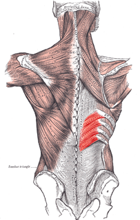
WThe Serratus posterior inferior muscle is a muscle of the human body.
 W
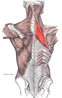
WThe serratus posterior superior is a thin, quadrilateral muscle, situated at the upper and back part of the thorax, deep to the rhomboid muscles.
 W
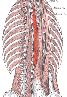
WThe spinalis is a portion of the erector spinae, a bundle of muscles and tendons, located nearest to the spine. It is divided into three parts: Spinalis dorsi, spinalis cervicis, and spinalis capitis.
 W
WThe splenius capitis is a broad, straplike muscle in the back of the neck. It pulls on the base of the skull from the vertebrae in the neck and upper thorax. It is involved in movements such as shaking the head.
 W
WThe splenius cervicis is a muscle in the back of the neck. It arises by a narrow tendinous band from the spinous processes of the third to the sixth thoracic vertebrae; it is inserted, by tendinous fasciculi, into the posterior tubercles of the transverse processes of the upper two or three cervical vertebrae.
 W
WThe splenius muscles are:Splenius capitis muscle Splenius cervicis muscle
 W
WThe thoracic diaphragm, or simply the diaphragm, is a sheet of internal skeletal muscle in humans and other mammals that extends across the bottom of the thoracic cavity. The diaphragm separates the thoracic cavity, containing the heart and lungs, from the abdominal cavity and performs an important function in respiration: as the diaphragm contracts, the volume of the thoracic cavity increases, creating a negative pressure there, which draws air into the lungs.
WThe transverse abdominal muscle (TVA), also known as the transverse abdominis, transversalis muscle and transversus abdominis muscle, is a muscle layer of the anterior and lateral abdominal wall which is deep to the internal oblique muscle. It is thought by most fitness instructors to be a significant component of the core.
 W
WThe transversospinales are a group of muscles of the human back. Their combined action is rotation and extension of the vertebral column. These muscles are small and have a poor mechanical advantage for contributing to motion. They include:the three semispinalis muscles, spanning 4-6 vertebral segments semispinalis thoracis semispinalis cervicis semispinalis capitis multifidus, spanning 2-4 vertebral segments rotatores, spanning 1-2 vertebral segments rotatores cervicis rotatores thoracis rotatores lumborum
 W
WThe transversus thoracis muscle lies internal to the thoracic cage, anteriorly. It is a thin plane of muscular and tendinous fibers, situated upon the inner surface of the front wall of the chest. It is in the same layer as the subcostal muscles and the innermost intercostal muscles.