 W
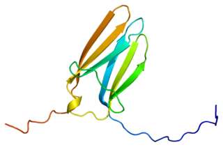
W14-3-3 proteins are a family of conserved regulatory molecules that are expressed in all eukaryotic cells. 14-3-3 proteins have the ability to bind a multitude of functionally diverse signaling proteins, including kinases, phosphatases, and transmembrane receptors. More than 200 signaling proteins have been reported as 14-3-3 ligands.
 W
WApoptotic protease activating factor 1, also known as APAF1, is a human homolog of C. elegans CED-4 gene.
 W
WApoptosis is a form of programmed cell death that occurs in multicellular organisms. Biochemical events lead to characteristic cell changes (morphology) and death. These changes include blebbing, cell shrinkage, nuclear fragmentation, chromatin condensation, chromosomal DNA fragmentation, and global mRNA decay. The average adult human loses between 50 and 70 billion cells each day due to apoptosis. For an average human child between the ages of 8 and 14, approximately 20–30 billion cells die per day.
 W
WApoptosis inducing factor is involved in initiating a caspase-independent pathway of apoptosis by causing DNA fragmentation and chromatin condensation. Apoptosis inducing factor is a flavoprotein. It also acts as an NADH oxidase. Another AIF function is to regulate the permeability of the mitochondrial membrane upon apoptosis. Normally it is found behind the outer membrane of the mitochondrion and is therefore secluded from the nucleus. However, when the mitochondrion is damaged, it moves to the cytosol and to the nucleus. Inactivation of AIF leads to resistance of embryonic stem cells to death following the withdrawal of growth factors indicating that it is involved in apoptosis.
 W
WThe apoptosome is a large quaternary protein structure formed in the process of apoptosis. Its formation is triggered by the release of cytochrome c from the mitochondria in response to an internal (intrinsic) or external (extrinsic) cell death stimulus. Stimuli can vary from DNA damage and viral infection to developmental cues such as those leading to the degradation of a tadpole's tail.
 W
WApoptosis signal-regulating kinase 1 (ASK1) also known as mitogen-activated protein kinase 5 (MAP3K5) is a member of MAP kinase family and as such a part of mitogen-activated protein kinase pathway. It activates c-Jun N-terminal kinase (JNK) and p38 mitogen-activated protein kinases in a Raf-independent fashion in response to an array of stresses such as oxidative stress, endoplasmic reticulum stress and calcium influx. ASK1 has been found to be involved in cancer, diabetes, rheumatoid arthritis, cardiovascular and neurodegenerative diseases.
 W
WAutophagy is the natural, regulated mechanism of the cell that removes unnecessary or dysfunctional components. It allows the orderly degradation and recycling of cellular components. Although initially characterised as a primordial degradation pathway induced to protect against starvation, it has become increasingly clear that autophagy also plays a major role in the homeostasis of non-starved cells. Defects in autophagy have been linked to various human diseases, including neurodegeneration and cancer, and interest in modulating autophagy as a potential treatment for these diseases has grown rapidly.
 W
WBcl-2, encoded in humans by the BCL2 gene, is the founding member of the Bcl-2 family of regulator proteins that regulate cell death (apoptosis), by either inhibiting (anti-apoptotic) or inducing (pro-apoptotic) apoptosis. It was the first apoptosis regulator identified in any organism.
 W
WBcl-2 homologous antagonist/killer is a protein that in humans is encoded by the BAK1 gene on chromosome 6. The protein encoded by this gene belongs to the BCL2 protein family. BCL2 family members form oligomers or heterodimers and act as anti- or pro-apoptotic regulators that are involved in a wide variety of cellular activities. This protein localizes to mitochondria, and functions to induce apoptosis. It interacts with and accelerates the opening of the mitochondrial voltage-dependent anion channel, which leads to a loss in membrane potential and the release of cytochrome c. This protein also interacts with the tumor suppressor P53 after exposure to cell stress.
 W
WThe BCL2 associated agonist of cell death (BAD) protein is a pro-apoptotic member of the Bcl-2 gene family which is involved in initiating apoptosis. BAD is a member of the BH3-only family, a subfamily of the Bcl-2 family. It does not contain a C-terminal transmembrane domain for outer mitochondrial membrane and nuclear envelope targeting, unlike most other members of the Bcl-2 family. After activation, it is able to form a heterodimer with anti-apoptotic proteins and prevent them from stopping apoptosis.
 W
WApoptosis regulator BAX, also known as bcl-2-like protein 4, is a protein that in humans is encoded by the BAX gene. BAX is a member of the Bcl-2 gene family. BCL2 family members form hetero- or homodimers and act as anti- or pro-apoptotic regulators that are involved in a wide variety of cellular activities. This protein forms a heterodimer with BCL2, and functions as an apoptotic activator. This protein is reported to interact with, and increase the opening of, the mitochondrial voltage-dependent anion channel (VDAC), which leads to the loss in membrane potential and the release of cytochrome c. The expression of this gene is regulated by the tumor suppressor P53 and has been shown to be involved in P53-mediated apoptosis.
 W
WThe BH3 interacting-domain death agonist, or BID, gene is a pro-apoptotic member of the Bcl-2 protein family. Bcl-2 family members share one or more of the four characteristic domains of homology entitled the Bcl-2 homology (BH) domains, and can form hetero- or homodimers. Bcl-2 proteins act as anti- or pro-apoptotic regulators that are involved in a wide variety of cellular activities.
 W
WBcl10-interacting CARD protein, also known as BinCARD, is a protein that in humans is encoded by the C9orf89 gene on chromosome 9. BinCARD is a member of the death-domain superfamily and contains a caspase recruitment domain (CARD). This protein regulates apoptosis and the immune response by inhibiting Bcl10, thus implicating it in diseases stemming from Bcl10 dysfunction.
 W
WIn cell biology, a bleb is a bulge of the plasma membrane of a cell, human bioparticulate or abscess with an internal environment synonymous to that of a simple cell, characterized by a spherical, bulky morphology. It is characterized by the decoupling of the cytoskeleton from the plasma membrane, degrading the internal structure of the cell, allowing the flexibility required for the cell to separate into individual bulges or pockets of the intercellular matrix. Most commonly, blebs are seen in apoptosis but are also seen in other non-apoptotic functions. Blebbing, or zeiosis, is the formation of blebs.
 W
WCaspase recruitment domains, or caspase activation and recruitment domains (CARDs), are interaction motifs found in a wide array of proteins, typically those involved in processes relating to inflammation and apoptosis. These domains mediate the formation of larger protein complexes via direct interactions between individual CARDs. CARD domains are found on a strikingly wide range of proteins, including helicases, kinases, mitochondrial proteins, caspases, and other cytoplasmic factors.
 W
WCaspases are a family of protease enzymes playing essential roles in programmed cell death. They are named caspases due to their specific cysteine protease activity – a cysteine in its active site nucleophilically attacks and cleaves a target protein only after an aspartic acid residue. As of 2009, there are 12 confirmed caspases in humans and 10 in mice, carrying out a variety of cellular functions.
 W
WThe cytochrome complex, or cyt c, is a small hemeprotein found loosely associated with the inner membrane of the mitochondrion. It belongs to the cytochrome c family of proteins and plays a major role in cell apoptosis. Cytochrome c is highly water-soluble, unlike other cytochromes, and is an essential component of the electron transport chain, where it carries one electron. It is capable of undergoing oxidation and reduction as its iron atom converts between the ferrous and ferric forms, but does not bind oxygen. It transfers electrons between Complexes III and IV. In humans, cytochrome c is encoded by the CYCS gene.
 W
WThe death-inducing signaling complex or DISC is a multi-protein complex formed by members of the "death receptor" family of apoptosis-inducing cellular receptors. A typical example is FasR, which forms the DISC upon trimerization as a result of its ligand (FasL) binding. The DISC is composed of the death receptor, FADD, and caspase 8. It transduces a downstream signal cascade resulting in apoptosis.
 W
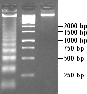
WDNA laddering is a feature that can be observed when DNA fragments, resulting from apoptotic DNA fragmentation, are visualized after separation by gel electrophoresis. It was first described in 1980 by Andrew Wyllie at the University of Edinburgh Medical School. DNA fragments can also be detected in cells that underwent necrosis, but when these DNA fragments after separation are subjected to gel electrophoresis no clear "ladder" pattern is apparent.
 W
WFas-associated protein with death domain (FADD), also called MORT1, is encoded by the FADD gene on the 11q13.3 region of chromosome 11 in humans.
 W
WFas ligand is a type-II transmembrane protein that belongs to the tumor necrosis factor (TNF) family. Its binding with its receptor induces apoptosis. Fas ligand/receptor interactions play an important role in the regulation of the immune system and the progression of cancer.
 W
WThe Fas receptor, also known as Fas, FasR, apoptosis antigen 1, cluster of differentiation 95 (CD95) or tumor necrosis factor receptor superfamily member 6 (TNFRSF6), is a protein that in humans is encoded by the FAS gene. Fas was first identified using a monoclonal antibody generated by immunizing mice with the FS-7 cell line. Thus, the name Fas is derived from FS-7-associated surface antigen.
 W
WIKK-β also known as inhibitor of nuclear factor kappa-B kinase subunit beta is a protein that in humans is encoded by the IKBKB gene.
 W
WKaryolysis, and λύσις lysis from λύειν lyein, "to separate") is the complete dissolution of the chromatin of a dying cell due to the enzymatic degradation by endonucleases. The whole cell will eventually stain uniformly with eosin after karyolysis. It is usually associated with karyorrhexis and occurs mainly as a result of necrosis, while in apoptosis after karyorrhexis the nucleus usually dissolves into apoptotic bodies.
WKaryorrhexis is the destructive fragmentation of the nucleus of a dying cell whereby its chromatin is distributed irregularly throughout the cytoplasm. It is usually preceded by pyknosis and can occur as a result of either programmed cell death (apoptosis), cellular senescence, or necrosis.
 W
WThe mitochondrial apoptosis-induced channel, is an early marker of the onset of apoptosis. This ion channel is formed on the outer mitochondrial membrane in response to certain apoptotic stimuli. MAC activity is detected by patch clamping mitochondria from apoptotic cells at the time of cytochrome c release.
 W
WNeurotrophins are a family of proteins that induce the survival, development, and function of neurons.
 W
WNF-κB is a protein complex that controls transcription of DNA, cytokine production and cell survival. NF-κB is found in almost all animal cell types and is involved in cellular responses to stimuli such as stress, cytokines, free radicals, heavy metals, ultraviolet irradiation, oxidized LDL, and bacterial or viral antigens. NF-κB plays a key role in regulating the immune response to infection. Incorrect regulation of NF-κB has been linked to cancer, inflammatory and autoimmune diseases, septic shock, viral infection, and improper immune development. NF-κB has also been implicated in processes of synaptic plasticity and memory.
 W
WTumor protein P53, also known as p53, cellular tumor antigen p53, the Guardian of the Genome, phosphoprotein p53, tumor suppressor p53, antigen NY-CO-13, or transformation-related protein 53 (TRP53), is any isoform of a protein encoded by homologous genes in various organisms, such as TP53 (humans) and Trp53 (mice). This homolog is crucial in multicellular vertebrates, where it prevents cancer formation, and thus functions as a tumor suppressor. As such, p53 has been described as "the guardian of the genome" because of its role in conserving stability by preventing genome mutation. Hence TP53 is classified as a tumor suppressor gene.
 W
WThe p53 upregulated modulator of apoptosis (PUMA) also known as Bcl-2-binding component 3 (BBC3), is a pro-apoptotic protein, member of the Bcl-2 protein family. In humans, the Bcl-2-binding component 3 protein is encoded by the BBC3 gene.The expression of PUMA is regulated by the tumor suppressor p53. PUMA is involved in p53-dependent and -independent apoptosis induced by a variety of signals, and is regulated by transcription factors, not by post-translational modifications. After activation, PUMA interacts with antiapoptotic Bcl-2 family members, thus freeing Bax and/or Bak which are then able to signal apoptosis to the mitochondria. Following mitochondrial dysfunction, the caspase cascade is activated ultimately leading to cell death.
 W
WThis article refers to the anti-tumor molecule, and not the a2iib3 integrin activation specific antibody of the same name
 W
WParaptosis is a type of programmed cell death, morphologically distinct from apoptosis and necrosis. The defining features of paraptosis are cytoplasmic vacuolation, independent of caspase activation and inhibition, and lack of apoptotic morphology. Paraptosis lacks several of the hallmark characteristics of apoptosis, such as membrane blebbing, chromatin condensation, and nuclear fragmentation. Like apoptosis and other types of programmed cell death, the cell is involved in causing its own death, and gene expression is required. This is in contrast to necrosis, which is non-programmed cell death that results from injury to the cell.
 W
WThe pathophysiology of Parkinson's disease is death of dopaminergic neurons as a result of changes in biological activity in the brain with respect to Parkinson's disease (PD). There are several proposed mechanisms for neuronal death in PD; however, not all of them are well understood. Five proposed major mechanisms for neuronal death in Parkinson's Disease include protein aggregation in Lewy bodies, disruption of autophagy, changes in cell metabolism or mitochondrial function, neuroinflammation, and blood-brain barrier (BBB) breakdown resulting in vascular leakiness.
 W
WPerforin-1 is a protein that in humans is encoded by the PRF1 gene and the Prf1 gene in mice.
 W
WPhorbol-12-myristate-13-acetate-induced protein 1 is a protein that in humans is encoded by the PMAIP1 gene, and is also known as Noxa.
 W
WPoly (ADP-ribose) polymerase (PARP) is a family of proteins involved in a number of cellular processes such as DNA repair, genomic stability, and programmed cell death.
WPyknosis, or karyopyknosis, is the irreversible condensation of chromatin in the nucleus of a cell undergoing necrosis or apoptosis. It is followed by karyorrhexis, or fragmentation of the nucleus. Pyknosis is also observed in the maturation of erythrocytes and the neutrophil. The maturing metarubricyte will condense its nucleus before expelling it to become a reticulocyte. The maturing neutrophil will condense its nucleus into several connected lobes that stay in the cell until the end of its cell life.
 W
WTropomyosin receptor kinase C (TrkC), also known as NT-3 growth factor receptor, neurotrophic tyrosine kinase receptor type 3, or TrkC tyrosine kinase is a protein that in humans is encoded by the NTRK3 gene.
 W
WTerminal deoxynucleotidyl transferase dUTP nick end labeling (TUNEL) is a method for detecting DNA fragmentation by labeling the 3′- hydroxyl termini in the double-strand DNA breaks generated during apoptosis.
 W
WX-linked inhibitor of apoptosis protein (XIAP), also known as inhibitor of apoptosis protein 3 (IAP3) and baculoviral IAP repeat-containing protein 4 (BIRC4), is a protein that stops apoptotic cell death. In humans, this protein (XIAP) is produced by a gene named XIAP gene located on the X chromosome.