 W
WThe adductor tubercle is a tubercle on the Lower extremity of the femur.
 W
WIn humans and many other primates, the calcaneus or heel bone is a bone of the tarsus of the foot which constitutes the heel. In some other animals, it is the point of the hock.
 W
WIn the human body, the cuboid bone is one of the seven tarsal bones of the foot.
 W
WThe femoral head is the highest part of the thigh bone (femur). It is supported by the femoral neck.
WThe femoral neck is a flattened pyramidal process of bone, connecting the femoral head with the femoral shaft, and forming with the latter a wide angle opening medialward.
 W
WThe femur, or thigh bone, is the proximal bone of the hindlimb in tetrapod vertebrates. The head of the femur articulates with the acetabulum in the pelvic bone forming the hip joint, while the distal part of the femur articulates with the tibia and kneecap, forming the knee joint. By most measures the two femurs are the strongest bones of the body, and in humans, the longest.
 W
WThe fibula or calf bone is a leg bone on the lateral side of the tibia, to which it is connected above and below. It is the smaller of the two bones and, in proportion to its length, the slenderest of all the long bones. Its upper extremity is small, placed toward the back of the head of the tibia, below the knee joint and excluded from the formation of this joint. Its lower extremity inclines a little forward, so as to be on a plane anterior to that of the upper end; it projects below the tibia and forms the lateral part of the ankle joint.
 W
WThe fibular notch of the tibia is an indentation at the inferior portion of the tibia where it articulates with the fibula to form the inferior tibiofibular articulation.
 W
WThe fifth metatarsal bone is a long bone in the foot, and is palpable along the distal outer edges of the feet. It is the second smallest of the five metatarsal bones. The fifth metatarsal is analogous to the fifth metacarpal bone in the hand
 W
WThe first metatarsal bone is the bone in the foot just behind the big toe. The first metatarsal bone is the shortest of the metatarsal bones and by far the thickest and strongest of them.
 W
WThe fourth metatarsal bone is a long bone in the foot. It is smaller in size than the third metatarsal bone and is the third longest of the five metatarsal bones. The fourth metatarsal is analogous to the fourth metacarpal bone in the hand
WThe greater trochanter of the femur is a large, irregular, quadrilateral eminence and a part of the skeletal system.
 W
WIn vertebrate anatomy, hip refers to either an anatomical region or a joint.
 W
WThe hip bone is a large irregular bone, constricted in the center and expanded above and below. In some vertebrates it is composed of three parts: the ilium, ischium, and the pubis.
 W
WThe intercondylar area is the separation between the medial and lateral condyle on the upper extremity of the tibia. The anterior and posterior cruciate ligaments and the menisci attach to the intercondylar area.
 W
WThe intercondylar fossa of femur is a deep notch between the rear surfaces of the medial and lateral epicondyle of the femur, two protrusions on the distal end of the femur that joins the knee. On the front of the femur, the condyles are but much less prominent and are separated from one another by a smooth shallow articular depression called the patellar surface because it articulates with the posterior surface of the patella (kneecap).
WThe intertrochanteric crest is a bony ridge located on the posterior side of the head of the femur, stretching obliquely downward and medially from the summit of the greater trochanter to the lesser trochanter.
 W
WThe intertrochanteric line is a line located on the anterior side of the proximal end of the femur.
 W
WThe lateral condyle is one of the two projections on the lower extremity of the femur. The other one is the medial condyle. The lateral condyle is the more prominent and is broader both in its front-to-back and transverse diameters.
WThe lateral condyle is the lateral portion of the upper extremity of tibia.
WThe lateral epicondyle of the femur, smaller and less prominent than the medial epicondyle, gives attachment to the fibular collateral ligament of the knee-joint. Directly below it is a small depression from which a smooth well-marked groove curves obliquely upward and backward to the posterior extremity of the condyle.
 W
WA leg bone is a bone found in the leg. These can include any the following:Femur – the bone in the thigh Patella – the knee cap Tibia – the shin bone, the larger of the two leg bones located below the knee cap Fibula – the smaller of the two leg bones located below the knee cap Tarsals Metatarsels Phalenges
 W
WThe lesser trochanter of the femur is a conical eminence, which varies in size in different species.
WThe lower extremity of femur is the lower end of the femur in human and other animals, closer to the knee. It is larger than the upper extremity of femur, is somewhat cuboid in form, but its transverse diameter is greater than its antero-posterior; it consists of two oblong eminences known as the lateral condyle and medial condyle.
 W
WA malleolus is the bony prominence on each side of the human ankle.
WThe medial condyle is one of the two projections on the lower extremity of femur, the other being the lateral condyle.
WThe medial condyle is the medial portion of the upper extremity of tibia.
 W
WThe medial epicondyle of the femur is an epicondyle, a bony protrusion, located on the medial side of the femur at its distal end.
 W
WThe metatarsal bones, or metatarsus are a group of five long bones in the foot, located between the tarsal bones of the hind- and mid-foot and the phalanges of the toes. Lacking individual names, the metatarsal bones are numbered from the medial side : the first, second, third, fourth, and fifth metatarsal. The metatarsals are analogous to the metacarpal bones of the hand. The lengths of the metatarsal bones in humans are, in descending order: second, third, fourth, fifth and first.
 W
WThe patella, also known as the kneecap, is a flat, circular-triangular bone which articulates with the femur and covers and protects the anterior articular surface of the knee joint. The patella is found in many tetrapods, such as mice, cats and birds, but not in whales, or most reptiles.
 W
WThe quadrate tubercle is a small tubercle found upon the upper part of the femur, that serves as a point of insertion of the quadratus femoris along with the intertrochanteric crest and the linea quadrata.
 W
WThe second metatarsal bone is a long bone in the foot. It is the longest of the metatarsal bones, being prolonged backward and held firmly into the recess formed by the three cuneiform bones. The second metatarsal forms joints with the second proximal phalanx through the metatarsophalangeal joint, the cuneiform bones, third metatarsal and occasionally the first metatarsal bone.
 W
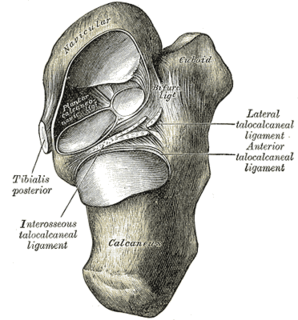
WThe talocalcaneonavicular joint is a ball and socket joint: the rounded head of the talus being received into the concavity formed by the posterior surface of the navicular, the anterior articular surface of the calcaneus, and the upper surface of the plantar calcaneonavicular ligament.
WThe talus, talus bone, astragalus, or ankle bone is one of the group of foot bones known as the tarsus. The tarsus forms the lower part of the ankle joint. It transmits the entire weight of the body from the lower legs to the foot.
 W
WThe tarsometatarsus is a bone that is only found in the lower leg of birds and some non-avian dinosaurs. It is formed from the fusion of several bones found in other types of animals, and homologous to the mammalian tarsus and metatarsal bones (foot). Despite this, the tarsometatarsus of birds is often referred to as just the tarsus or metatarsus.
 W
WThe third metatarsal bone is a long bone in the foot. It is the second longest metatarsal. The longest being the second metatarsal. The third metatarsal is analogous to the third metacarpal bone in the hand
 W
WIn human anatomy, the third trochanter is a bony projection occasionally present on the proximal femur near the superior border of the gluteal tuberosity. When present, it is oblong, rounded, or conical in shape and sometimes continuous with the gluteal ridge. It generally occurs bilaterally without significant side to side dimorphism. A structure of minor importance in humans, the incidence of the third trochanter varies from 17–72% between ethnic groups and it is frequently reported as more common in females than in males. Structures analogous to the third trochanter are present in other mammals, including some primates. It is called the third trochanter in reference to the greater and lesser trochanters that are always present on the femur.
 W
WThe tibia, also known as the shinbone or shankbone, is the larger, stronger, and anterior (frontal) of the two bones in the leg below the knee in vertebrates, and it connects the knee with the ankle bones. The tibia is found on the medial side of the leg next to the fibula and closer to the median plane or centre-line. The tibia is connected to the fibula by the interosseous membrane of the leg, forming a type of fibrous joint called a syndesmosis with very little movement. The tibia is named for the flute tibia. It is the second largest bone in the human body next to the femur. The leg bones are the strongest long bones as they support the rest of the body.
WIn mammals including humans, the medial surface of the greater trochanter has at its base a deep depression bounded posteriorly by the intertrochanteric crest, called the trochanteric fossa. This fossa is the point of insertion of four muscles. Moving from the inferior-most to the superior-most, they are: the tendon of the obturator externus muscle, the obturator internus, the superior gemellus and inferior gemellus. The width and depth of the trochanteric fossa varies taxonomically.
 W
WThe tuberosity of the tibia or tibial tuberosity or tibial tubercle is an elevation on the proximal, anterior aspect of the tibia, just below where the anterior surfaces of the lateral and medial tibial condyles end.
 W
WThe upper extremity, proximal extremity or superior epiphysis of the femur is the part of the femur closest to the pelvic bone and the trunk. It contains the following structures:Femoral head including the fovea Femur neck Greater trochanter Lesser trochanter Intertrochanteric line Intertrochanteric crest Trochanteric fossa Linea quadrata Quadrate tubercle