 W
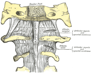
WIn anatomy, the alar ligaments are ligaments which connect the dens to tubercles on the medial side of the occipital condyle.
 W
WThe anterior atlantoaxial ligament is a strong membrane, fixed, above, to the lower border of the anterior arch of the atlas; below, to the front of the body of the axis.
 W
WThe anterior longitudinal ligament is a ligament that runs down the anterior surface of the spine. It traverses all of the vertebral bodies and intervertebral discs on their ventral side.
 W
WThe anterior sacrococcygeal ligament or ventral sacrococcygeal ligament consists of a few irregular fibers, which descend from the anterior surface of the sacrum to the front of the coccyx, blending with the periosteum.
WThe ligament of apex dentis is a ligament that spans between the second cervical vertebra in the neck and the skull. It lies as a fibrous cord in the triangular interval between the alar ligaments, which extends from the tip of the odontoid process on the axis to the anterior margin of the foramen magnum, being intimately blended with the deep portion of the anterior atlantooccipital membrane and superior crus of the transverse ligament of the atlas.
 W
WThe atlantoaxial joint is a joint in the upper part of the neck between the first and second cervical vertebrae; the atlas and axis. It is a pivot joint.
 W
WIn anatomy, the atlas (C1) is the most superior (first) cervical vertebra of the spine and is located in the neck. It is named for Atlas of Greek mythology because, just as Atlas supported the globe, it supports the entire head.
 W
WBack pain, also known as backache, is pain felt in the back. The back is divided into neck pain (cervical), middle back pain (thoracic), lower back pain (lumbar) or coccydynia based on the segment affected. The lumbar area is the most common area affected. Episodes of back pain may be acute, sub-acute, or chronic depending on the duration. The pain may be characterized as a dull ache, shooting or piercing pain, or a burning sensation. Discomfort can radiate into the arms and hands as well as the legs or feet, and may include numbness, or weakness in the legs and arms.
 W
WThe cauda equina is a bundle of spinal nerves and spinal nerve rootlets, consisting of the second through fifth lumbar nerve pairs, the first through fifth sacral nerve pairs, and the coccygeal nerve, all of which arise from the lumbar enlargement and the conus medullaris of the spinal cord. The cauda equina occupies the lumbar cistern, a subarachnoid space inferior to the conus medullaris. The nerves that compose the cauda equina innervate the pelvic organs and lower limbs to include motor innervation of the hips, knees, ankles, feet, internal anal sphincter and external anal sphincter. In addition, the cauda equina extends to sensory innervation of the perineum and, partially, parasympathetic innervation of the bladder.
 W
WIn tetrapods, cervical vertebrae are the vertebrae of the neck, immediately below the skull. Truncal vertebrae lie caudal of cervical vertebrae. In sauropsid species, the cervical vertebrae bear cervical ribs. In lizards and saurischian dinosaurs, the cervical ribs are large; in birds, they are small and completely fused to the vertebrae. The vertebral transverse processes of mammals are homologous to the cervical ribs of other amniotes. Most mammals have seven cervical vertebrae, with the only three known exceptions being the manatee with six, the two-toed sloth with five or six, and the three-toed sloth with nine.
 W
WThe coccyx, commonly referred to as the tailbone, is the final segment of the vertebral column in all apes, and analogous structures in certain other mammals such as horses. In tailless primates since Nacholapithecus, the coccyx is the remnant of a vestigial tail. In animals with bony tails, it is known as tailhead or dock, in bird anatomy as tailfan. It comprises three to five separate or fused coccygeal vertebrae below the sacrum, attached to the sacrum by a fibrocartilaginous joint, the sacrococcygeal symphysis, which permits limited movement between the sacrum and the coccyx.
WThe cruciform ligament of atlas is a cruciate ligament in the neck forming part of the atlanto-axial joint. The ligament is named as such because it is in the shape of a cross.
 W
WDegenerative disc disease (DDD) is a medical condition in which there are anatomic changes and a loss of function of varying degrees of one or more intervertebral discs of the spine of sufficient magnitude as to cause symptoms. The root cause is thought to be loss of soluble proteins within the fluid contained in the disc with resultant reduction of the oncotic pressure, which in turn causes loss of fluid volume. Normal downward forces cause the affected disc to lose height, and the distance between vertebrae is reduced. The anulus fibrosus, the rigid outer shell of a disc, also weakens. This loss of height causes laxity of the longitudinal ligaments, which may allow anterior, posterior, or lateral shifting of the vertebral bodies, causing facet joint malalignment and arthritis; scoliosis; cervical hyperlordosis; thoracic hyperkyphosis; lumbar hyperlordosis; narrowing of the space available for the spinal tract within the vertebra ; or narrowing of the space through which a spinal nerve exits with resultant inflammation and impingement of a spinal nerve, causing a radiculopathy.
 W
WA disc protrusion is a disease condition that can occur in some vertebrates, including humans, in which the outermost layers of the anulus fibrosus of the intervertebral discs of the spine are intact but bulge when one or more of the discs are under pressure.
 W
WEpidural steroid injection (ESI) is a technique in which corticosteroids and a local anesthetic are injected into the epidural space around the spinal cord in an effort to improve spinal stenosis, spinal disc herniation, or both. It is of benefit with a rare rate of major side effects.
 W
WThe facet joints, are a set of synovial, plane joints between the articular processes of two adjacent vertebrae. There are two facet joints in each spinal motion segment and each facet joint is innervated by the recurrent meningeal nerves.
WThe interspinous ligaments are thin and membranous ligaments, that connect adjoining spinous processes of the vertebra in the spine. They extend from the root to the apex of each spinous process. They meet the ligamenta flava in front and blend with the supraspinous ligament behind.
 W
WAn intervertebral disc lies between adjacent vertebrae in the vertebral column. Each disc forms a fibrocartilaginous joint, to allow slight movement of the vertebrae, to act as a ligament to hold the vertebrae together, and to function as a shock absorber for the spine.
 W
WThe intervertebral foramen, is a foramen between two spinal vertebrae. Cervical, thoracic, and lumbar vertebrae all have intervertebral foramina.
 W
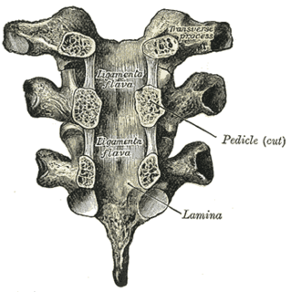
WThe ligamenta flava are a series of ligaments that connect the ventral parts of the laminae of adjacent vertebrae. Each ligamentum flavum connects two adjacent vertebrae, beginning with the junction of the axis and third cervical vertebra, continuing down to the junction of the fifth lumbar vertebra and the sacrum. They are best seen from the interior of the vertebral canal; when looked at from the outer surface they appear short, being overlapped by the lamina of the vertebral arch.
 W
WLumbar provocative discography is an invasive diagnostic procedure for evaluation for intervertebral disc pathology. It is usually reserved for persons with persistent, severe low back pain (LBP) who have abnormal spaces between vertebrae on magnetic resonance imaging (MRI), where other diagnostic tests have failed to reveal clear confirmation of a suspected disc as the source of pain, and surgical intervention is being considered.
 W
WLumbar spinal stenosis (LSS) is a medical condition in which the spinal canal narrows and compresses the nerves and blood vessels at the level of the lumbar vertebrae. Spinal stenosis may also affect the cervical or thoracic region, in which case it is known as cervical spinal stenosis or thoracic spinal stenosis. Lumbar spinal stenosis can cause pain in the low back or buttocks, abnormal sensations, and the absence of sensation (numbness) in the legs, thighs, feet, or buttocks, or loss of bladder and bowel control.
 W
WThe lumbar vertebrae are, in human anatomy, the five vertebrae between the rib cage and the pelvis. They are the largest segments of the vertebral column and are characterized by the absence of the foramen transversarium within the transverse process and by the absence of facets on the sides of the body. They are designated L1 to L5, starting at the top. The lumbar vertebrae help support the weight of the body, and permit movement.
 W
WThe nuchal ligament is a ligament at the back of the neck that is continuous with the supraspinous ligament.
WThe posterior atlantoaxial ligament is a broad, thin membrane attached, above, to the lower border of the posterior arch of the atlas; below, to the upper edges of the laminæ of the axis.
 W
WThe posterior longitudinal ligament is situated within the vertebral canal, and extends along the posterior surfaces of the bodies of the vertebrae, from the body of the axis, where it is continuous with the tectorial membrane of atlanto-axial joint, to the sacrum. The ligament is thicker in the thoracic than in the cervical and lumbar regions. In the thoracic and lumbar regions, it presents a series of dentations with intervening concave margins.
 W
WThe posterior sacrococcygeal ligament or dorsal sacrococcygeal ligament is a ligament which stretches from the sacrum to the coccyx and thus dorsally across the sacrococcygeal symphysis shared by these two bones.
WThe sacrococcygeal symphysis is an amphiarthrodial joint, formed between the oval surface at the apex of the sacrum, and the base of the coccyx.
 W
WThe sacrum, in human anatomy, is a large, triangular bone at the base of the spine that forms by the fusing of sacral vertebrae S1–S5 between 18 and 30 years of age.
 W
WScheuermann's disease is a self-limiting skeletal disorder of childhood. Scheuermann's disease describes a condition where the vertebrae grow unevenly with respect to the sagittal plane; that is, the posterior angle is often greater than the anterior. This uneven growth results in the signature "wedging" shape of the vertebrae, causing kyphosis. It is named after Danish surgeon Holger Scheuermann.
 W
WSchmorl's nodes are protrusions of the nucleus pulposus of the intervertebral disc through the vertebral body endplate and into the adjacent vertebra.
 W
WScoliosis is a medical condition in which a person's spine has a sideways curve. The curve is usually "S"- or "C"-shaped over three dimensions. In some, the degree of curve is stable, while in others, it increases over time. Mild scoliosis does not typically cause problems, but more severe cases can affect breathing and movement. Pain is usually present in adults, and can worsen with age.
 W
WThe spinal canal is the canal that contains the spinal cord within the vertebral column. The spinal canal is formed by the vertebrae through which the spinal cord passes. It is a process of the dorsal body cavity. This canal is enclosed within the foramen of the vertebrae. In the intervertebral spaces, the canal is protected by the ligamentum flavum posteriorly and the posterior longitudinal ligament anteriorly.
 W
WThe spinal cord is a long, thin, tubular structure made up of nervous tissue, which extends from the medulla oblongata in the brainstem to the lumbar region of the vertebral column. It encloses the central canal of the spinal cord, which contains cerebrospinal fluid. The brain and spinal cord together make up the central nervous system (CNS). In humans, the spinal cord begins at the occipital bone, passing through the foramen magnum and entering the spinal canal at the beginning of the cervical vertebrae. The spinal cord extends down to between the first and second lumbar vertebrae, where it ends. The enclosing bony vertebral column protects the relatively shorter spinal cord. It is around 45 cm (18 in) in men and around 43 cm (17 in) long in women. The diameter of the spinal cord ranges from 13 mm in the cervical and lumbar regions to 6.4 mm in the thoracic area.
 W
WSpinal disc herniation is an injury to the cushioning and connective tissue between vertebrae, usually caused by excessive strain or trauma to the spine. It may result in back pain, pain or sensation in different parts of the body, and physical disability. The most conclusive diagnostic tool for disc herniation is MRI, and treatment may range from painkillers to surgery. Protection from disc herniation is best provided by core strength and an awareness of body mechanics including posture.
WThe supraspinous ligament, also known as the supraspinal ligament, is a ligament found along the vertebral column.
WThe tectorial membrane of atlanto-axial joint is situated within the vertebral canal.
 W
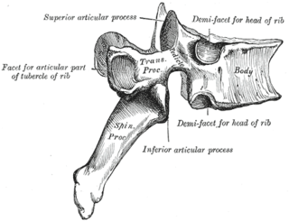
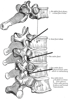
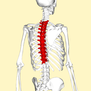
WIn vertebrates, thoracic vertebrae compose the middle segment of the vertebral column, between the cervical vertebrae and the lumbar vertebrae. In humans, there are twelve thoracic vertebrae and they are intermediate in size between the cervical and lumbar vertebrae; they increase in size going towards the lumbar vertebrae, with the lower ones being a lot larger than the upper. They are distinguished by the presence of facets on the sides of the bodies for articulation with the heads of the ribs, as well as facets on the transverse processes of all, except the eleventh and twelfth, for articulation with the tubercles of the ribs. By convention, the human thoracic vertebrae are numbered T1–T12, with the first one (T1) located closest to the skull and the others going down the spine toward the lumbar region.
 W
WIn anatomy, the transverse ligament of the atlas is a ligament which arches across the ring of the atlas, and keeps the odontoid process in contact with the atlas.
 W
WVertebral augmentation, which includes vertebroplasty and kyphoplasty, are similar spinal procedures in which bone cement is injected through a small hole in the skin into a fractured vertebra to try to relieve back pain caused by a vertebral compression fractures.
 W
WThe vertebral column, also known as the backbone or spine, is part of the axial skeleton. The vertebral column is the defining characteristic of a vertebrate in which the notochord found in all chordates has been replaced by a segmented series of bone: vertebrae separated by intervertebral discs. The vertebral column houses the spinal canal, a cavity that encloses and protects the spinal cord.