 W
WThe alveolar canals are apertures in the center of the infratemporal surface of the maxilla. The alveolar canals transmit the posterior superior alveolar vessels and nerves.
 W
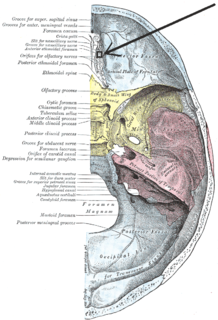
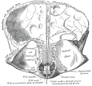
WThe anterior ethmoidal foramen is a small opening in the ethmoid bone in the skull.
 W
WThe carotid canal is the passageway in the temporal bone through which the internal carotid artery enters the middle cranial fossa from the neck. The canal starts on the inferior surface of the temporal bone at the external opening of the carotid canal. The canal ascends at first superiorly, and then, making a bend, runs anteromedially. The canal's internal opening is near the foramen lacerum, above which the internal carotid artery passes on its way anteriorly to the cavernous sinus.
 W
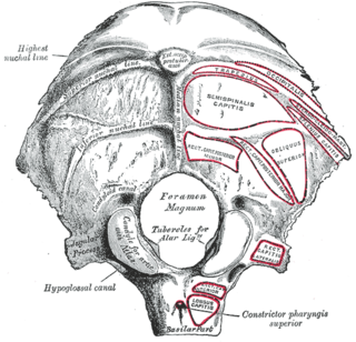
WThe condylar canal is a canal in the condyloid fossa of the lateral parts of occipital bone behind the occipital condyle. Resection of the rectus capitis posterior major and minor muscles reveals the bony recess leading to the condylar canal, which is situated posterior and lateral to the occipital condyle. It is immediately superior to the extradural vertebral artery, which makes a loop above the posterior C1 ring to enter the foramen magnum. The anteriomedial wall of the condylar canal thickens to join the foramen magnum rim and connect to the occipital condyle.
 W
WThe epitympanic recess is a hollow located on the superior/roof aspect of the middle ear.
 W
WThe facial canal, also known as the Fallopian canal, first described by Gabriele Falloppio, is a Z-shaped canal running through the temporal bone from the internal acoustic meatus to the stylomastoid foramen. In humans it is approximately 3 centimeters long, which makes it the longest human osseous canal of a nerve. It is located within the middle ear region, according to its shape it is divided into three main segments: the labyrinthine, the tympanic, and the mastoidal segment. It contains Cranial Nerve VII, also known as the facial nerve.
 W
WThe frontal crest of the frontal bone ends below in a small notch which is converted into a foramen, the foramen cecum, by articulation with the ethmoid.
 W
WThe foramen lacerum is a triangular hole in the base of skull, located between the sphenoid, the apex of the petrous temporal and the basilar part of the occipital.
 W
WThe foramen magnum is a large oval opening (foramen) in the occipital bone of the skull in humans and many other animals. It is one of the several oval or circular openings (foramina) in the base of the skull. The spinal cord, an extension of the medulla oblongata, passes through the foramen magnum as it exits the cranial cavity. Apart from the transmission of the medulla oblongata and its membranes, the foramen magnum transmits the vertebral arteries, the anterior and posterior spinal arteries, the tectorial membranes and alar ligaments. It also transmits the accessory nerve into the skull.
 W
WAt the base of the skull, the foramen ovale is one of the larger of the several holes that transmit nerves through the skull. The foramen ovale is situated in the posterior part of the sphenoid bone, posterolateral to the foramen rotundum.
WThe foramen rotundum is a circular hole in the sphenoid bone that connects the middle cranial fossa and the pterygopalatine fossa.
WThe foramen spinosum is one of two foramina located in the base of the human skull, on the sphenoid bone. It is situated just anterior to the spine of the sphenoid bone, and just lateral to the foramen ovale. The middle meningeal artery, middle meningeal vein, and the meningeal branch of the mandibular nerve pass through the foramen.
 W
WIn the maxilla, occasionally two additional canals are present in the middle line of the palatine process; they are termed the foramina of Scarpa, and when present transmit the nasopalatine nerves, the left passing through the anterior, and the right through the posterior canal.
 W
WThe greater palatine canal is a passage in the skull that transmits the descending palatine artery, vein, and greater and lesser palatine nerves between the pterygopalatine fossa and the oral cavity.
 W
WThe hiatus for the greater petrosal nerve is a small hole in the petrous part of the temporal bone which connects the facial canal to the middle cranial fossa. The greater petrosal nerve travels through it to branch from the facial nerve and reach the middle cranial fossa on its way to the pterygopalatine ganglion.
 W
WThe hiatus for lesser petrosal nerve is a hiatus in the petrous part of the temporal bone which transmits the lesser petrosal nerve. It is located posterior to the groove for the superior petrosal sinus and posterolateral to the jugular foramen.
 W
WThe hypoglossal canal is a foramen in the occipital bone of the skull. It is hidden medially and superiorly to each occipital condyle. The hypoglossal nerve traverses the canal.
WIn the opening of the incisive foramen, the orifices of two lateral canals are visible; they are named the incisive canals or foramina of Stensen.
WIn the human mouth, the incisive foramen, also called anterior palatine foramen, or nasopalatine foramen is a funnel-shaped opening in the bone of the oral hard palate immediately behind the incisor teeth where blood vessels and nerves pass. The incisive foramen is continuous with the incisive canal, this foramen or group of foramina is located behind the central incisor teeth in the incisive fossa of the maxilla.
WThe inferior tympanic canaliculus is a small passage of the tympanic branch of the glossopharyngeal nerve and inferior tympanic artery. In the bony ridge dividing the carotid canal from the jugular fossa is the small inferior tympanic canaliculus. The inferior tympanic canaliculus is near the fossula petrosa which houses inferior ganglion of glossopharyngeal nerve/petrous ganglion from which the tympanic nerve arises.
 W
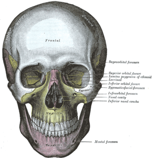
WIn human anatomy, the infraorbital foramen is an opening in the maxillary bone of the skull located below the infraorbital margin of the orbit. It transmits the infraorbital artery and vein, and the infraorbital nerve, a branch of the maxillary nerve. It is typically 6.10 to 10.9 mm from the infraorbital margin.
 W
WThe internal auditory meatus is a canal within the petrous part of the temporal bone of the skull between the posterior cranial fossa and the inner ear.
 W
WThe jugular foramen is a large foramen (opening) in the base of the skull, located behind the carotid canal. It is formed in front by the petrous portion of the temporal bone, and behind by the occipital bone; it is generally larger on the right than on the left side.
 W
WIn human anatomy, the mandibular canal is a canal within the mandible that contains the inferior alveolar nerve, inferior alveolar artery, and inferior alveolar vein. It runs obliquely downward and forward in the ramus, and then horizontally forward in the body, where it is placed under the alveoli and communicates with them by small openings.
 W
WThe mandibular foramen is an opening on the internal surface of the ramus of the mandible for divisions of the mandibular nerve and blood vessels to pass through.
WIn the lateral part of the jugular fossa of the temporal bone is the mastoid canaliculus for the entrance of the auricular branch of the vagus nerve.
WThe mastoid foramen is a hole in the posterior border of the temporal bone. It transmits a Mastoid emissary vein to the sigmoid sinus and a small branch of the occipital artery, the posterior meningeal artery to the dura mater.
 W
WThe mental foramen is one of two foramina (openings) located on the anterior surface of the mandible. It transmits the terminal branches of the inferior alveolar nerve and vessels. The mental foramen descends slightly in toothless individuals.
 W
WThe nasal foramina are foramina which run through the nasal bones.
 W
WThe olfactory foramina, also known as the cribriform foramina, is the grouping of holes located on the cribriform plate. The cribriform plate forms the roof of the nasal cavity, and the olfactory foramina are in the two depressions lateral to the median blade of the cribriform plate called the crista galli. There is a pair of olfactory bulbs of the brain that rest in these two depressions. These holes that make up the olfactory foramina allow passage for about 20 bundles of nerve fibers that make up the olfactory nerve, also known as Cranial Nerve I (CNI), from the nasal cavity to meet with the olfactory bulbs. Therefore, the olfactory foramina are necessary for the human sense of smell. These foramina vary in size and number with age.
 W
WThe optic foramen is the opening to the optic canal. The canal is located in the sphenoid bone; it is bounded medially by the body of the sphenoid and laterally by the lesser wing of the sphenoid.
 W
WThe parietal foramen is an opening for the parietal emissary vein, which drains into the superior sagittal sinus. Occasionally, a small branch of the occipital artery can also pass through it. It is located at the back part of the parietal bone, close to the upper or sagittal border. It is not always present, and its size varies considerably.
 W
WThe petrotympanic fissure is a fissure in the temporal bone that runs from the temporomandibular joint to the tympanic cavity.
 W
WLateral to either olfactory groove are the internal openings of the anterior and posterior ethmoidal foramina.
 W
WThe pterygoid canal is a passage in the sphenoid bone of the skull leading from just anterior to the foramen lacerum in the middle cranial fossa to the pterygopalatine fossa.
WIn the base of the skull, in the great wings of the sphenoid bone, medial to the foramen ovale, a small aperture, the sphenoidal emissary foramen, may occasionally be seen opposite the root of the pterygoid process. When present, it opens below near the scaphoid fossa. Vesalius was the first to describe and illustrate this foramen, and it thus sometimes bears the name of foramen Vesalii. Other names include foramen venosum and canaliculus sphenoidalis.
 W
WThe sphenopalatine foramen is a foramen in the skull that connects the nasal cavity with the pterygopalatine fossa.
 W
WBetween the styloid and mastoid processes of the temporal bone is the stylomastoid foramen
 W
WThe superior orbital fissure is a foramen in the skull, although strictly it is more of a cleft, lying between the lesser and greater wings of the sphenoid bone.
 W
WThe supraorbital foramen, is a bony elongated opening located above the orbit and under the forehead. The supraorbital foramen lies directly under the eyebrow. Sometimes this foramen is incomplete and is then known as the supraorbital notch.
 W
WThe zygomatico-orbital foramina are two canals in the skull, that allow nerves to pass through. The orifices are seen on the orbital process of the zygomatic bone.
WThe zygomaticofacial foramen is a small aperture. It perforates the malar surface of the convex zygomatic bone near its center, for the passage of the zygomaticofacial nerve and vessels. Below this foramen is a slight elevation, which gives origin to the Zygomaticus.