 W
WAdult neurogenesis is the process in which neurons are generated from neural stem cells in the adult. This process differs from prenatal neurogenesis.
 W
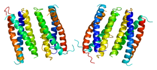
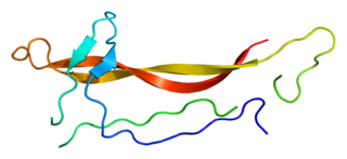
WAgrin is a large proteoglycan whose best-characterised role is in the development of the neuromuscular junction during embryogenesis. Agrin is named based on its involvement in the aggregation of acetylcholine receptors during synaptogenesis. In humans, this protein is encoded by the AGRN gene.
 W
WAbnormal spindle-like microcephaly-associated protein also known as abnormal spindle protein homolog or Asp homolog is a protein that in humans is encoded by the ASPM gene. ASPM is located on chromosome 1, band q31 (1q31). The ASPM gene contains 28 exons and codes for a 3477 amino‐acid‐long protein. The ASPM protein is conserved across species including human, mouse, Drosophila, and C. elegans. Defective forms of the ASPM gene are associated with autosomal recessive primary microcephaly.
 W
WThe autism spectrum encompasses a range of neurodevelopmental conditions, including autism and Asperger syndrome, generally known as autism spectrum disorders (ASD). Individuals on the autistic spectrum experience difficulties with social communication and interaction and also exhibit restricted, repetitive patterns of behavior, interests, or activities. Symptoms are typically recognized between one and two years of age. However, a lot of children are not finally diagnosed until they are older. Final diagnosis could still be given as an adolescent or even as an adult. The term "spectrum" refers to the variation in the type and severity of symptoms. Those in the mild range may function independently, while those with moderate to severe symptoms may require more substantial support in their daily lives. Long-term problems may include difficulties in performing daily tasks, creating and keeping relationships, and maintaining a job.
 W
WBrain-derived neurotrophic factor (BDNF), or abrineurin, is a protein that, in humans, is encoded by the BDNF gene. BDNF is a member of the neurotrophin family of growth factors, which are related to the canonical nerve growth factor. Neurotrophic factors are found in the brain and the periphery. BDNF was first isolated from pig brain in 1982 by Yves-Alain Barde and Hans Thoenen.
 W
WHarold Saxton Burr was E. K. Hunt Professor of Anatomy at Yale University School of Medicine and researcher into bio-electrics.
 W
WA cerebral organoid, or brain organoid, describes an artificially grown, in vitro, miniature organ resembling the brain. Cerebral organoids are created by culturing pluripotent stem cells in a three-dimensional rotational bioreactor, and they develop over a course of months. The brain is an extremely complex system of heterogeneous tissues and consists of a diverse array of neurons. This complexity has made studying the brain and understanding how it works a difficult task in neuroscience, especially when it comes to neurodegenerative diseases. The purpose of creating an in vitro neurological model is to study these diseases in a more simple and variable space. This 3D model is free of many potential in vivo limitations. The varying physiology between human and other mammalian models limits the scope of study in neurological disorders. Cerebral organoids are synthesized tissues that contain several types of nerve cells and have anatomical features that recapitulate regions of the cortex observed in brains. Cerebral organoids are most similar to layers of neurons called the cortex and choroid plexus. In some cases, structures similar to the retina, meninges and hippocampus can form. Stem cells have the potential to grow into many different types of tissues, and their fate is dependent on many factors. Below is an image showing some of the chemical factors that can lead stem cells to differentiate into various neural tissues; a more in-depth table of generating specific organoid identity has been published since. Similar techniques are used on stem cells used to grow cerebral organoids.
 W
WCiliary neurotrophic factor is a protein that in humans is encoded by the CNTF gene.
 W
WThe Disabled-1 (Dab1) gene encodes a key regulator of Reelin signaling. Reelin is a large glycoprotein secreted by neurons of the developing brain, particularly Cajal-Retzius cells. DAB1 functions downstream of Reln in a signaling pathway that controls cell positioning in the developing brain and during adult neurogenesis. It docks to the intracellular part of the Reelin very low density lipoprotein receptor (VLDLR) and apoE receptor type 2 (ApoER2) and becomes tyrosine-phosphorylated following binding of Reelin to cortical neurons. In mice, mutations of Dab1 and Reelin generate identical phenotypes. In humans, Reelin mutations are associated with brain malformations and mental retardation. In mice, Dab1 mutation results in the scrambler mouse phenotype.
 W
WCorticogenesis is the process in which the cerebral cortex of the brain is formed during the development of the nervous system. The cortex is the outer layer of the brain and is composed of up to six layers. Neurons formed in the ventricular zone migrate to their final locations in one of the six layers of the cortex. The process occurs from embryonic day 10 to 17 in mice and between gestational weeks seven to 18 in humans.
 W
WAdele Dorothy Diamond is a professor of neuroscience at the University of British Columbia, where she is currently a Tier 1 Canada Research Chair in Developmental Cognitive Neuroscience. One of the pioneers in the field of developmental cognitive neuroscience, Diamond researches how executive functions are affected by biological and environmental factors, especially in children. Her discoveries have improved treatment for disorders such as phenylketonuria and attention-deficit hyperactivity disorder, and they have impacted early education.
 W
WEnvironmental enrichment is the stimulation of the brain by its physical and social surroundings. Brains in richer, more stimulating environments have higher rates of synaptogenesis and more complex dendrite arbors, leading to increased brain activity. This effect takes place primarily during neurodevelopment, but also during adulthood to a lesser degree. With extra synapses there is also increased synapse activity, leading to an increased size and number of glial energy-support cells. Environmental enrichment also enhances capillary vasculation, providing the neurons and glial cells with extra energy. The neuropil expands, thickening the cortex. Research on rodent brains suggests that environmental enrichment may also lead to an increased rate of neurogenesis.
 W
WThe ependyma is the thin neuroepithelial lining of the ventricular system of the brain and the central canal of the spinal cord. The ependyma is one of the four types of neuroglia in the central nervous system (CNS). It is involved in the production of cerebrospinal fluid (CSF), and is shown to serve as a reservoir for neuroregeneration.
 W
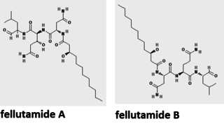
WFellutamide A and B are tripeptide derivatives from Penicillium fellutanum and other fungi. They are potent proteasome inhibitor that stimulates nerve growth factor synthesis in vitro. It strongly inhibits the growth of the tuberculosis-causing bacterium Mycobacterium tuberculosis. Its biosynthetic pathway has been determined in the filamentous fungus Aspergillus nidulans.
 W
WThe floor plate is a structure integral to the developing nervous system of vertebrate organisms. Located on the ventral midline of the embryonic neural tube, the floor plate is a specialized glial structure that spans the anteroposterior axis from the midbrain to the tail regions. It has been shown that the floor plate is conserved among vertebrates, such as zebrafish and mice, with homologous structures in invertebrates such as the fruit fly Drosophila and the nematode C. elegans. Functionally, the structure serves as an organizer to ventralize tissues in the embryo as well as to guide neuronal positioning and differentiation along the dorsoventral axis of the neural tube.
 W
WGanglion mother cells (GMCs) are cells involved in neurogenesis, in non-mammals, that divide only once to give rise to two neurons, or one neuron and one glial cell or two glial cells, and are present only in the central nervous system. They are also responsible for transcription factor expression. While each ganglion mother cell necessarily gives rise to two neurons, a neuroblast can asymmetrically divide multiple times. GMCs are the progeny of type I neuroblasts. Neuroblasts asymmetrically divide during embryogenesis to create GMCs. GMCs are only present in certain species and only during the embryonic and larval stages of life. Recent research has shown that there is an intermediate stage between a GMC and two neurons. The GMC forms two ganglion cells which then develop into neurons or glial cells. Embryonic neurogenesis has been extensively studied in Drosophila melanogaster embryos and larvae.
 W
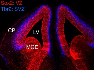
WThe ganglionic eminence (GE) is a transitory structure in the development of the nervous system that guides cell and axon migration. It is present in the embryonic and fetal stages of neural development found between the thalamus and caudate nucleus.
 W
WA growth cone is a large actin-supported extension of a developing or regenerating neurite seeking its synaptic target. Their existence was originally proposed by Spanish histologist Santiago Ramón y Cajal based upon stationary images he observed under the microscope. He first described the growth cone based on fixed cells as "a concentration of protoplasm of conical form, endowed with amoeboid movements". Growth cones are situated on the tips of neurites, either dendrites or axons, of the nerve cell. The sensory, motor, integrative, and adaptive functions of growing axons and dendrites are all contained within this specialized structure.
 W
WIn molecular biology, Human accelerated region 1 is a segment of the human genome found on the long arm of chromosome 20. It is a Human accelerated region. It is located within a pair of overlapping long non-coding RNA genes, HAR1A (HAR1F) and HAR1B (HAR1R).
 W
W W
WInsulin-like growth factor 1 (IGF-1), also called somatomedin C, is a hormone similar in molecular structure to insulin which plays an important role in childhood growth, and has anabolic effects in adults.
 W
WLegius syndrome (LS) is an autosomal dominant condition characterized by cafe au lait spots. It was first described in 2007 and is often mistaken for neurofibromatosis type I (NF-1). It is caused by mutations in the SPRED1 gene. It is also known as neurofibromatosis type 1-like syndrome (NFLS).
 W
WMuSK is a receptor tyrosine kinase required for the formation and maintenance of the neuromuscular junction. It is activated by a nerve-derived proteoglycan called agrin.
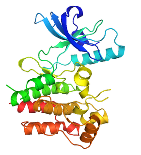
WNerve growth factor (NGF) is a neurotrophic factor and neuropeptide primarily involved in the regulation of growth, maintenance, proliferation, and survival of certain target neurons. It is perhaps the prototypical growth factor, in that it was one of the first to be described. Since it was first isolated by Nobel Laureates Rita Levi-Montalcini and Stanley Cohen in 1956, numerous biological processes involving NGF have been identified, two of them being the survival of pancreatic beta cells and the regulation of the immune system.
 W
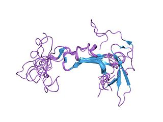
WNetrins are a class of proteins involved in axon guidance. They are named after the Sanskrit word "netr", which means "one who guides". Netrins are genetically conserved across nematode worms, fruit flies, frogs, mice, and humans. Structurally, netrin resembles the extracellular matrix protein laminin.
 W
WNeuregulins or neuroregulins are a family of four structurally related proteins that are part of the EGF family of proteins. These proteins have been shown to have diverse functions in the development of the nervous system and play multiple essential roles in vertebrate embryogenesis including: cardiac development, Schwann cell and oligodendrocyte differentiation, some aspects of neuronal development, as well as the formation of neuromuscular synapses.
 W
WNeurotrophin-3 is a protein that in humans is encoded by the NTF3 gene.
 W
WNeurotrophin-4 (NT-4), also known as neurotrophin-5 (NT-5), is a protein that in humans is encoded by the NTF4 gene. It is a neurotrophic factor that signals predominantly through the TrkB receptor tyrosine kinase.
 W
WParental experience, as well as changing hormone levels during pregnancy and postpartum, cause changes in the parental brain. Displaying maternal sensitivity towards infant cues, processing those cues and being motivated to engage socially with her infant and attend to the infant's needs in any context could be described as mothering behavior and is regulated by many systems in the maternal brain. Research has shown that hormones such as oxytocin, prolactin, estradiol and progesterone are essential for the onset and the maintenance of maternal behavior in rats, and other mammals as well. Mothering behavior has also been classified within the basic drives. Less is known about the paternal brain, but changes in the father's brain occur alongside the mother once the offspring is born.
 W
WPiaget's theory of cognitive development is a comprehensive theory about the nature and development of human intelligence. It was originated by the Swiss developmental psychologist Jean Piaget (1896–1980). The theory deals with the nature of knowledge itself and how humans gradually come to acquire, construct, and use it. Piaget's theory is mainly known as a developmental stage theory. Piaget "was intrigued by the fact that children of different ages made different kinds of mistakes while solving problems". He also believed that children are not like "little adults" who may know less; children just think and speak differently. By Piaget thinking that children have great cognitive abilities, he came up with four different cognitive development stages, which he put out into testing. Within those four stages he managed to group them with different ages. Each stage he realized how children managed to develop their cognitive skills. For example, he believed that children experience the world through actions, representing things with words, thinking logically, and using reasoning.
 W
WRadial glial cells, or radial glial progenitor cells (RGPs), are bipolar-shaped progenitor cells that are responsible for producing all of the neurons in the cerebral cortex. RGPs also produce certain lineages of glia, including astrocytes and oligodendrocytes. Their cell bodies (somata) reside in the embryonic ventricular zone, which lies next to the developing ventricular system.
 W
WThe Radial Unit Hypothesis (RUH) is a conceptual theory of cerebral cortex development, first described by Pasko Rakic. The RUH states that the cerebral cortex develops during embryogenesis as an array of interacting cortical columns, or 'radial units', each of which originates from a transient stem cell layer called the ventricular zone, which contains neural stem cells known as radial glial cells.
 W
WReelin (RELN) is a large secreted extracellular matrix glycoprotein that helps regulate processes of neuronal migration and positioning in the developing brain by controlling cell–cell interactions. Besides this important role in early development, reelin continues to work in the adult brain. It modulates synaptic plasticity by enhancing the induction and maintenance of long-term potentiation. It also stimulates dendrite and dendritic spine development and regulates the continuing migration of neuroblasts generated in adult neurogenesis sites like the subventricular and subgranular zones. It is found not only in the brain but also in the liver, thyroid gland, adrenal gland, Fallopian tube, breast and in comparatively lower levels across a range of anatomical regions.
 W
WThe rhombic lip is a posterior section of the developing metencephalon which can be recognized transiently within the vertebrate embryo. It extends posteriorly from the roof of the fourth ventricle to dorsal neuroepithelial cells. The rhombic lip can be divided into eight structural units based on rhombomeres 1-8 (r1-r8), which can be recognized at early stages of hindbrain development. Producing granule cells and five brainstem nuclei, the rhombic lip plays an important role in developing a complex cerebellar neural system.
 W
WCellular adhesions can be defined as proteins or protein aggregates that form mechanical and chemical linkages between the intracellular and extracellular space. Adhesions serve several critical processes including cell migration, signal transduction, tissue development and repair. Due to this functionality, adhesions and adhesion molecules have been a topic of study within the scientific community. Specifically, it has been found that adhesions are involved in tissue development, plasticity, and memory formation within the central nervous system (CNS), and may prove vital in the generation of CNS-specific therapeutics.
 W
WThe rostral migratory stream (RMS) is a specialized migratory route found in the brain of some animals along which neuronal precursors that originated in the subventricular zone (SVZ) of the brain migrate to reach the main olfactory bulb (OB). The importance of the RMS lies in its ability to refine and even change an animal's sensitivity to smells, which explains its importance and larger size in the rodent brain as compared to the human brain, as our olfactory sense is not as developed. This pathway has been studied in the rodent, rabbit, and both the squirrel monkey and rhesus monkey. When the neurons reach the OB they differentiate into GABAergic interneurons as they are integrated into either the granule cell layer or periglomerular layer.
 W
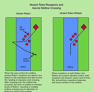
WThe Roundabout (Robo) family of proteins are single-pass transmembrane receptors that are highly conserved across many branches of the animal kingdom, from C. elegans to humans. They were first discovered in Drosophila, through a mutant screen for genes involved in axon guidance. The Drosophila roundabout mutant was named after its phenotype, which resembled the circular traffic junctions. The Robo receptors are most well known for their role in the development of the nervous system, where they have been shown to respond to secreted Slit ligands. One well-studied example is the requirement for Slit-Robo signaling in regulation of axonal midline crossing. Slit-Robo signaling is also critical for many neurodevelopmental processes including formation of the olfactory tract, the optic nerve, and motor axon fasciculation. In addition, Slit-Robo signaling contributes to cell migration and the development of other tissues such as the lung, kidney, liver, muscle and breast. Mutations in Robo genes have been linked to multiple neurodevelopmental disorders in humans.
 W
WHomeobox protein SIX3 is a protein that in humans is encoded by the SIX3 gene.
 W
WThe subgranular zone (SGZ) is a brain region in the hippocampus where adult neurogenesis occurs. The other major site of adult neurogenesis is the subventricular zone (SVZ) in the brain.
WThe subplate, also called the subplate zone, together with the marginal zone and the cortical plate, in the fetus represents the developmental anlage of the mammalian cerebral cortex. It was first described, as a separate transient fetal zone by Ivica Kostović and Mark E. Molliver in 1974.
 W
WThe subventricular zone (SVZ) is a region situated on the outside wall of each lateral ventricle of the vertebrate brain. It is present in both the embryonic and adult brain. In embryonic life, the SVZ refers to a secondary proliferative zone containing neural progenitor cells, which divide to produce neurons in the process of neurogenesis. The primary neural stem cells of the brain and spinal cord, termed radial glial cells, instead reside in the ventricular zone (VZ).
 W
WSynaptic pruning, a phase in the development of the nervous system, is the process of synapse elimination that occurs between early childhood and the onset of puberty in many mammals, including humans. Pruning starts near the time of birth and continues into the mid-20s. During pruning, both the axon and dendrite decay and die off. It was traditionally considered to be complete by the time of sexual maturation, but this was discounted by MRI studies.
 W
WTropomyosin receptor kinase B (TrkB), also known as tyrosine receptor kinase B, or BDNF/NT-3 growth factors receptor or neurotrophic tyrosine kinase, receptor, type 2 is a protein that in humans is encoded by the NTRK2 gene. TrkB is a receptor for brain-derived neurotrophic factor (BDNF).
 W
WThe growth cone is a highly dynamic structure of the developing neuron, changing directionality in response to different secreted and contact-dependent guidance cues; it navigates through the developing nervous system in search of its target. The migration of the growth cone is mediated through the interaction of numerous trophic and tropic factors; netrins, slits, ephrins and semaphorins are four well-studied tropic cues (Fig.1). The growth cone is capable of modifying its sensitivity to these guidance molecules as it migrates to its target; this sensitivity regulation is an important theme seen throughout development.
 W
WTropomyosin receptor kinase C (TrkC), also known as NT-3 growth factor receptor, neurotrophic tyrosine kinase receptor type 3, or TrkC tyrosine kinase is a protein that in humans is encoded by the NTRK3 gene.
 W
WIn vertebrates, the ventricular zone (VZ) is a transient embryonic layer of tissue containing neural stem cells, principally radial glial cells, of the central nervous system (CNS). The VZ is so named because it lines the ventricular system, which contains cerebrospinal fluid (CSF). The embryonic ventricular system contains growth factors and other nutrients needed for the proper function of neural stem cells. Neurogenesis, or the generation of neurons, occurs in the VZ during embryonic and fetal development as a function of the Notch pathway, and the newborn neurons must migrate substantial distances to their final destination in the developing brain or spinal cord where they will establish neural circuits. A secondary proliferative zone, the subventricular zone (SVZ), lies adjacent to the VZ. In the embryonic cerebral cortex, the SVZ contains intermediate neuronal progenitors that continue to divide into post-mitotic neurons. Through the process of neurogenesis, the parent neural stem cell pool is depleted and the VZ disappears. The balance between the rates of stem cell proliferation and neurogenesis changes during development, and species from mouse to human show large differences in the number of cell cycles, cell cycle length, and other parameters, which is thought to give rise to the large diversity in brain size and structure.
 W
WThe zona limitans intrathalamica (ZLI) is a lineage-restriction compartment and primary developmental boundary in the vertebrate forebrain that serves as a signaling center and a restrictive border between the thalamus and the prethalamus.