 W
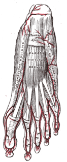
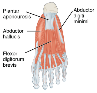
WThe abductor digiti minimi is a muscle which lies along the lateral (outer) border of the foot, and is in relation by its medial margin with the lateral plantar artery, vein and nerves.
 W
WThe abductor hallucis muscle is an intrinsic muscle of the foot. It participates in the abduction and flexion of the great toe.
 W
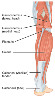
WThe Achilles tendon or heel cord, also known as the calcaneal tendon, is a tendon at the back of the lower leg, and is the thickest in the human body. It serves to attach the plantaris, gastrocnemius (calf) and soleus muscles to the calcaneus (heel) bone. These muscles, acting via the tendon, cause plantar flexion of the foot at the ankle joint, and flexion at the knee.
 W
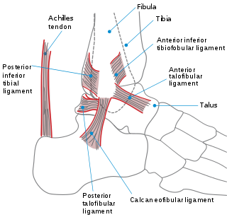
WThe ankle, or the talocrural region, is the region where the foot and the leg meet. The ankle includes three joints: the ankle joint proper or talocrural joint, the subtalar joint, and the inferior tibiofibular joint. The movements produced at this joint are dorsiflexion and plantarflexion of the foot. In common usage, the term ankle refers exclusively to the ankle region. In medical terminology, "ankle" can refer broadly to the region or specifically to the talocrural joint.
 W
WThe anterior compartment of the leg is a fascial compartment of the lower limb. It contains muscles that produce dorsiflexion and participate in inversion and eversion of the foot, as well as vascular and nervous elements including the anterior tibial artery and veins, and the deep fibular nerve.
 W
WThe anterior intermuscular septum of leg or anterior crural intermuscular septum is a band of fascia which separates the lateral from the anterior compartment of leg.
 W
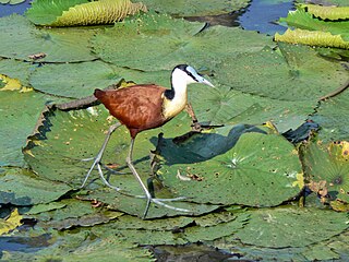
WThe anatomy of bird legs and feet is diverse, encompassing many accommodations to perform a wide variety of functions.
 W
WThe buttocks are two rounded portions of the exterior anatomy of most mammals, located on the posterior of the pelvic region. In humans, the buttocks are located between the lower back and the perineum. They are composed of a layer of exterior skin and underlying subcutaneous fat superimposed on a left and right gluteus maximus and gluteus medius muscles. The two gluteus maximus muscles are the largest muscles in the human body. They are responsible for achieving the upright posture when the body is bent at the waist; maintaining the body in the upright posture by keeping the hip joints extended; and propelling the body forward via further leg (hip) extension when walking or running. In the seated position, the buttocks bear the weight of the upper body and take that weight off the feet.
 W
WThe calf is the back portion of the lower leg in human anatomy. The muscles within the calf correspond to the posterior compartment of the leg. The two largest muscles within this compartment are known together as the calf muscle and attach to the heel via the Achilles tendon. Several other, smaller muscles attach to the knee, the ankle, and via long tendons to the toes.
 W
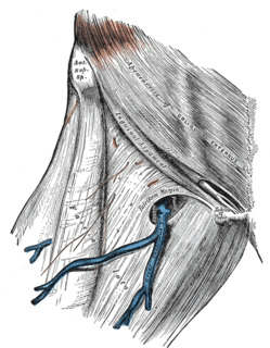
WThe cribriform fascia, fascia cribrosa also Hesselbach's fascia is the portion of fascia covering the saphenous opening in the thigh. It is perforated by the great saphenous vein and by numerous blood and lymphatic vessels..
 W
WThe cuboideonavicular joint is a joint (articulation) in the foot formed between the navicular bone and cuboid bone. The navicular bone is connected with the cuboid bone by the dorsal, plantar, and interosseous cuboideonavicular ligaments.
 W
WThe cuneonavicular joint is a joint (articulation) in the human foot. It is formed between the navicular bone and the three cuneiform bones. The navicular and cuneiform bones are connected by dorsal and plantar ligaments.
WThe deltoid ligament is a strong, flat, triangular band, attached, above, to the apex and anterior and posterior borders of the medial malleolus. The deltoid ligament is composed of: 1. Anterior tibiotalar ligament 2. Tibiocalcaneal ligament 3. Posterior tibiotalar ligament 4. Tibionavicular ligament. It consists of two sets of fibers, superficial and deep.
 W
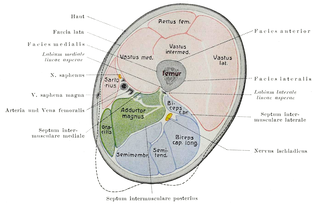
WThe fascia lata is the deep fascia of the thigh. It encloses the thigh muscles and forms the outer limit of the fascial compartments of thigh, which are internally separated by intermuscular septa. The fascia lata is thickened at its lateral side where it forms the iliotibial tract, a structure that runs to the tibia and serves as a site of muscle attachment.
 W
WThe fascial compartments of thigh are the three fascial compartments that divide and contain the thigh muscles. The fascia lata is the strong and deep fascia of the thigh that surrounds the thigh muscles and forms the outer limits of the compartments. Internally the muscle compartments are divided by the lateral and medial intermuscular septa.
 W
WIn human anatomy of the leg, the femoral sheath has three compartments. The lateral compartment contains the femoral artery, the intermediate compartment contains the femoral vein, and the medial and smallest compartment is called the femoral canal. The femoral canal contains efferent lymphatic vessels and a lymph node embedded in a small amount of areolar tissue. It is conical in shape and is about 2 cm long.
 W
WThe femoral ring is the base of the femoral canal. It is directed upward and is oval in form, its long diameter being directed transversely and measuring about 1.25 cm. Part of the intestine can sometimes pass through the femoral ring into the femoral canal causing a femoral hernia.
WThe femoral sheath is formed by a prolongation downward, behind the inguinal ligament, of the abdominal fascia, the transverse fascia being continued down in front of the femoral vessels and the iliac fascia behind them. The femoral sheath is contained within the femoral triangle.
 W
WThe femoral triangle is an anatomical region of the upper third of the thigh. It is a subfascial space which appears as a triangular depression below the inguinal ligament when the thigh is flexed, abducted and laterally rotated.
 W
WThe gluteal sulcus is an area of the body of humans and anthropoid apes, described by a horizontal crease formed by the inferior aspect of the buttocks and the posterior upper thigh. The gluteal sulcus is formed by the posterior horizontal skin crease of the hip joint and overlying fat, and is not formed by the lower border of the gluteus maximus muscle, which crosses the fold obliquely. It is one of the major defining features of the buttocks. Children with developmental dysplasia of the hips are born with uneven gluteal folds and can be diagnosed with physical examination and sonogram.
 W
WThe human leg, in the general word sense, is the entire lower limb of the human body, including the foot, thigh and even the hip or gluteal region. However, the definition in human anatomy refers only to the section of the lower limb extending from the knee to the ankle, also known as the crus. Legs are used for standing, and all forms of locomotion including recreational such as dancing, and constitute a significant portion of a person's mass. Female legs generally have greater hip anteversion and tibiofemoral angles, but shorter femur and tibial lengths than those in males.
 W
WThe iliopectineal bursa or the iliopsoas bursa is a large synovial bursa that separates the external surface of the hip joint capsule from the normally just the tendon of the iliopsoas muscle.
 W
WThe iliotibial tract or iliotibial band is a longitudinal fibrous reinforcement of the fascia lata. The action of the muscles associated with the ITB flex, extend, abduct, and laterally and medially rotate the hip. The ITB contributes to lateral knee stabilization. During knee extension the ITB moves anterior to the lateral condyle of the femur, while ~30 degrees knee flexion, the ITB moves posterior to the lateral condyle. However, it has been suggested that this is only an illusion due to the changing tension in the anterior and posterior fibers during movement. It originates at the anterolateral iliac tubercle portion of the external lip of the iliac crest and inserts at the lateral condyle of the tibia at Gerdy's tubercle. The figure shows only the proximal part of the iliotibial tract.
 W
WThe inferior extensor retinaculum of the foot is a Y-shaped band placed in front of the ankle-joint, the stem of the Y being attached laterally to the upper surface of the calcaneus, in front of the depression for the interosseous talocalcaneal ligament; it is directed medialward as a double layer, one lamina passing in front of, and the other behind, the tendons of the peroneus tertius and extensor digitorum longus.
 W
WThe intercuneiform joints are the joints the cuneiform bones.
WThe interosseous membrane of the leg extends between the interosseous crests of the tibia and fibula, helps stabilize the Tib-Fib relationship and separates the muscles on the front from those on the back of the leg.
WThe intertarsal joint are the joints of the tarsal bones in the foot. There are seven specific inter tarsal joints (articulations) in the human foot:Subtalar joint Talocalcaneonavicular joint Calcaneocuboid joint Cuneonavicular joint Cuboideonavicular joint Intercuneiform joints
 W
WThe knee bursae are the fluid-filled sacs and synovial pockets that surround and sometimes communicate with the knee joint cavity. The bursae are thin-walled, and filled with synovial fluid. They represent the weak point of the joint, but also provide enlargements to the joint space. They can be grouped into either communicating and non-communicating bursae or, after their location – frontal, lateral, or medial.
 W
WA lap is a surface created between the knee and hips of a biped when it is in a seated or lying down position. The lap of a parent or loved one is seen as a physically and psychologically comfortable place for a child to sit. In countries where Christmas is celebrated, it has been a tradition for children to sit on the lap of a person dressed as Santa Claus to tell Santa what they want for Christmas, and have their picture taken, but this practice has since been questioned in some of these countries, where this sort of contact between children and unfamiliar adults raises concerns.
 W
WThe lateral intermuscular septum of thigh is a fold of deep fascia in the thigh.
 W
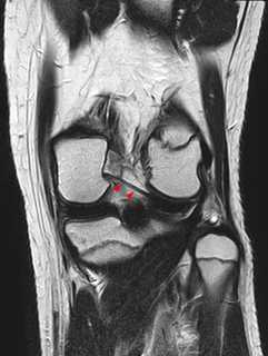
WThe lateral meniscus is a fibrocartilaginous band that spans the lateral side of the interior of the knee joint. It is one of two menisci of the knee, the other being the medial meniscus. It is nearly circular and covers a larger portion of the articular surface than the medial. It can occasionally be injured or torn by twisting the knee or applying direct force, as seen in contact sports.
 W
WA leg is a weight-bearing and locomotive anatomical structure, usually having a columnar shape. During locomotion, legs function as "extensible struts". The combination of movements at all joints can be modeled as a single, linear element capable of changing length and rotating about an omnidirectional "hip" joint.
 W
WLeg hair is hair that grows on the legs of humans, generally appearing after the onset of puberty. For hygienic or aesthetic reasons and for some sports, people shave, wax, epilate, or use hair removal creams to remove the hair from their legs: see leg shaving.
 W
WThe medial intermuscular septum of thigh is a fold of deep fascia in the thigh.
WThe mucous sheaths of the tendons around the ankle protect tendons in the ankle. All the tendons crossing the ankle-joint are enclosed for part of their length in mucous sheaths which have an almost uniform length of about 8 cm. each.
 W
WThe peroneal retinacula are fibrous retaining bands which bind down the tendons of the peroneus longus and brevis as they run across the side of the ankle..
 W
WThe pes is the zoological term for the distal portion of the hind limb of tetrapod animals. It is the part of the pentadactyl limb that includes the metatarsals and digits (phalanges). During evolution, it has taken many forms and served a variety of functions. It can be represented by the foot of primates, the lower hind limb of hoofed animals, the lower portion of the leg of dinosaurs including birds or the rear paw. It is also represented in the rear 'paddle' of extinct marine reptiles, such as plesiosaurs. The oldest types of tetrapods had seven or eight digits.
 W
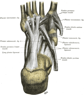
WThe plantar fascia is the thick connective tissue (aponeurosis) which supports the arch on the bottom of the foot. It runs from the tuberosity of the calcaneus forward to the heads of the metatarsal bones.
 W
WThe popliteal fossa is a shallow depression located at the back of the knee joint. The bones of the popliteal fossa are the femur and the tibia. Like other flexion surfaces of large joints, it is an area where blood vessels and nerves pass relatively superficially, and with an increased number of lymph nodes.
 W
WThe Posterior meniscofemoral ligament is a small fibrous band of the knee joint. It attaches to the posterior area of the lateral meniscus and crosses superiorly and medially behind the posterior cruciate ligament to attach to the medial condyle of the femur.
 W
WThe prepatellar bursa is a frontal bursa of the knee joint. It is a superficial bursa with a thin synovial lining located between the skin and the patella.
 W
WA slight ridge is sometimes seen commencing about the middle of the intertrochanteric crest, and reaching vertically downward for about 5 cm. along the back part of the body: it is called the linea quadrata, and gives attachment to the Quadratus femoris and a few fibers of the Adductor magnus.
WIn anatomy, the saphenous opening is an oval opening in the upper mid part of the fascia lata of the thigh. It lies 3–4 cm below and lateral to the pubic tubercle and is about 3 cm long and 1.5 cm wide.
 W
WSteatopygia is the state of having substantial levels of tissue on the buttocks and thighs. This build is not confined to the gluteal regions, but extends to the outside and front of the thighs, and tapers to the knee producing a curvaceous figure. The term is from the Greek stéar (στέαρ), meaning "tallow", and pugḗ (πυγή), meaning "rump".
WThe superior extensor retinaculum of the foot is the upper part of the extensor retinaculum of foot which extends from the ankle to the heelbone.
 W
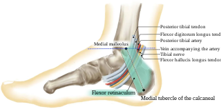
WThe tarsal tunnel is found along the inner leg posterior to the medial malleolus.
WIn human anatomy, the thigh is the area between the hip (pelvis) and the knee. Anatomically, it is part of the lower limb.