 W
WAcantholysis is the loss of intercellular connections, such as desmosomes, resulting in loss of cohesion between keratinocytes, seen in diseases such as pemphigus vulgaris. It is absent in bullous pemphigoid, making it useful for differential diagnosis.
 W
WAlbinism is a congenital disorder characterized in humans by the complete or partial absence of pigment in the skin, hair and eyes. Albinism is associated with a number of vision defects, such as photophobia, nystagmus, and amblyopia. Lack of skin pigmentation makes for more susceptibility to sunburn and skin cancers. In rare cases such as Chédiak–Higashi syndrome, albinism may be associated with deficiencies in the transportation of melanin granules. This also affects essential granules present in immune cells leading to increased susceptibility to infection.
 W
WAmelanism is a pigmentation abnormality characterized by the lack of pigments called melanins, commonly associated with a genetic loss of tyrosinase function. Amelanism can affect fish, amphibians, reptiles, birds, and mammals including humans. The appearance of an amelanistic animal depends on the remaining non-melanin pigments. The opposite of amelanism is melanism, a higher percentage of melanin.
 W
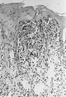
WAtypia is a histopathologic term for a structural abnormality in a cell, i.e. it is used to describe atypical cells.
 W
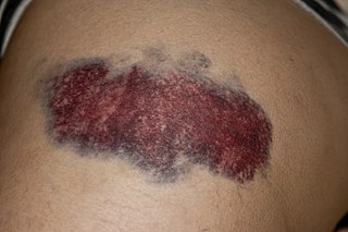
WA bruise, also known as a contusion, is a type of hematoma of tissue, the most common cause being capillaries damaged by trauma, causing localized bleeding that extravasates into the surrounding interstitial tissues. Most bruises are not very deep under the skin so that the bleeding causes a visible discoloration. The bruise then remains visible until the blood is either absorbed by tissues or cleared by immune system action. Bruises, which do not blanch under pressure, can involve capillaries at the level of skin, subcutaneous tissue, muscle, or bone. Bruises are not to be confused with other similar-looking lesions. These lesions include petechia, purpura, and ecchymosis.
 W
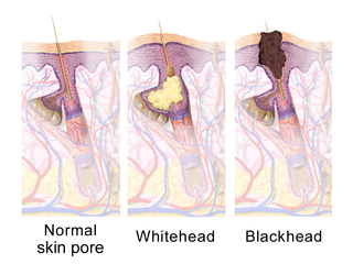
WA comedo is a clogged hair follicle (pore) in the skin. Keratin combines with oil to block the follicle. A comedo can be open (blackhead) or closed by skin (whitehead) and occur with or without acne. The word comedo comes from the Latin comedere, meaning 'to eat up', and was historically used to describe parasitic worms; in modern medical terminology, it is used to suggest the worm-like appearance of the expressed material.
 W
WA dermatomycosis is a skin disease caused by a fungus. This excludes dermatophytosis.
 W
WDermatoscopy is the examination of skin lesions with a dermatoscope.
 W
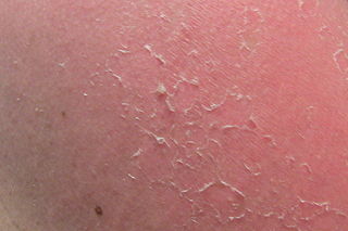
WDesquamation, commonly called skin peeling, is the shedding of the outermost membrane or layer of a tissue, such as the skin. The term is from Latin desquamare 'to scrape the scales off a fish'.
 W
WErythema is redness of the skin or mucous membranes, caused by hyperemia in superficial capillaries. It occurs with any skin injury, infection, or inflammation. Examples of erythema not associated with pathology include nervous blushes.
 W
WErythrism or erythrochroism refers to an unusual reddish pigmentation of an animal's hair, skin, feathers, or eggshells.
 W
WAn exanthem is a widespread rash occurring on the outside of the body and usually occurring in children. An exanthem can be caused by toxins, drugs, or microorganisms, or can result from autoimmune disease.
 W
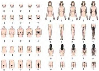
WThe Ferriman–Gallwey score is a method of evaluating and quantifying hirsutism in women. The method was originally published in 1961 by D. Ferriman and J.D. Gallwey in the Journal of Clinical Endocrinology.
 W
WThe Fitzpatrick scale is a numerical classification schema for human skin color. It was developed in 1975 by Thomas B. Fitzpatrick as a way to estimate the response of different types of skin to ultraviolet (UV) light. It was initially developed on the basis of skin color to measure the correct dose of UVA for PUVA therapy, and when the initial testing based only on hair and eye colour resulted in too high UVA doses for some, it was altered to be based on the patient's reports of how their skin responds to the sun; it was also extended to a wider range of skin types. The Fitzpatrick scale remains a recognized tool for dermatological research into human skin pigmentation.
 W
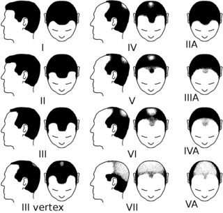
WThe Hamilton–Norwood scale is used to classify the stages of male pattern baldness. The stages are described with a number from 1 to 7.
 W
WHyperkeratosis is thickening of the stratum corneum, often associated with the presence of an abnormal quantity of keratin, and also usually accompanied by an increase in the granular layer. As the corneum layer normally varies greatly in thickness in different sites, some experience is needed to assess minor degrees of hyperkeratosis.
 W
WHyperpigmentation is the darkening of an area of skin or nails caused by increased melanin.
 W
WHypopigmentation is characterized specifically as an area of skin becoming lighter than the baseline skin color, but not completely devoid of pigment. This is not to be confused with depigmentation, which is characterized as the absence of all pigment. It is caused by melanocyte or melanin depletion, or a decrease in the amino acid tyrosine, which is used by melanocytes to make melanin. Some common genetic causes include mutations in the tyrosinase gene or OCA2 gene. As melanin pigments tend to be in the skin, eye, and hair, these are the commonly affected areas in those with hypopigmentation.
 W
WIntertrigo refers to a type of inflammatory rash (dermatitis) of the superficial skin that occurs within a person's body folds. These areas are more susceptible to irritation and subsequent infection due to factors that promote skin breakdown such as moisture, friction, and exposure to bodily secretions such as sweat, urine or feces. Areas of the body which are more likely to be affected by intertrigo include the inframammary fold, intergluteal cleft, armpits, and spaces between the fingers or toes. Skin affected by intertrigo is more prone to infection than intact skin.
 W
WKeratosis is a growth of keratin on the skin or on mucous membranes stemming from keratinocytes, the prominent cell type in the epidermis. More specifically, it can refer to:actinic keratosis chronic scar keratosis hydrocarbon keratosis keratosis pilaris seborrheic keratosis
 W
WLeucism is a wide variety of conditions which result in the partial loss of pigmentation in an animal—which causes white, pale, or patchy coloration of the skin, hair, feathers, scales or cuticles, but not the eyes. It is occasionally spelled leukism. Some genetic conditions that result in a "leucistic" appearance include piebaldism, Waardenburg syndrome, vitiligo, Chédiak–Higashi syndrome, and Melanophilin mutations. Pale patches of skin, feathers, or fur can also result from injury.
 W
WThe Ludwig scale is a method of classifying female pattern baldness, and ranges from stages I to III.
 W
WA malar rash, also called butterfly rash, is a medical sign consisting of a characteristic form of facial rash. It is often seen in lupus erythematosus but is not pathognomonic - it is also seen in other diseases such as pellagra, dermatomyositis, and Bloom syndrome. While some literature has described the slapped-cheek rash of fifth disease as a malar rash, it differs slightly in that the nose is typically spared.
 W
WThe Malpighian layer of the skin is generally defined as both the stratum basale and stratum spinosum as a unit, although it is occasionally defined as the stratum basale specifically,or the stratum spinosum specifically.
 W
WThe term melanism refers to black pigment and is derived from the Greek: μελανός. Melanism is the increased development of the dark-colored pigment melanin in the skin or hair.
 W
WNevus is a nonspecific medical term for a visible, circumscribed, chronic lesion of the skin or mucosa. The term originates from nævus, which is Latin for "birthmark"; however, a nevus can be either congenital or acquired. Common terms, including mole, birthmark, and beauty mark, are used to describe nevi, but these terms do not distinguish specific types of nevi from one another.
 W
WPagetoid is a term used in dermatology to refer to "upward spreading" of abnormal cells in the epidermis. It is uncommon and a possible indication of a precancerous or cancerous condition. Cells display pagetoid growth when they invade the upper epidermis from below. Squamous cell carcinoma, melanoma in situ, Pagetoid Bowen's disease, ocular sebaceous carcinoma and other carcinomas can all display pagetoid growth.
 W
WPallor is a pale color of the skin that can be caused by illness, emotional shock or stress, stimulant use, or anemia, and is the result of a reduced amount of oxyhaemoglobin and may also be visible as pallor of the conjunctivae of the eyes on physical examination.
 W
WA papule is a circumscribed, solid elevation of skin with no visible fluid, varying in area from a pinhead to 1 cm. Papules can be brown, purple, pink or red in color, and can cluster into a papular rash. They may open when scratched and become infected and crusty. Larger non-blisterform elevated lesions may be termed nodules.
 W
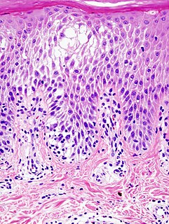
WParakeratosis is a mode of keratinization characterized by the retention of nuclei in the stratum corneum. In mucous membranes, parakeratosis is normal. In the skin, this process leads to the abnormal replacement of annular squames with nucleated cells. Parakeratosis is associated with the thinning or loss of the granular layer and is usually seen in diseases of increased cell turnover, whether inflammatory or neoplastic. Parakeratosis is seen in the plaques of psoriasis and in dandruff.
 W
WPhysical urticaria is a distinct subgroup of the urticaria that are induced by an exogenous physical stimulus rather than occurring spontaneously. There are seven subcategories that are recognized as independent diseases. Physical urticaria is known to be painful, itchy and physically unappealing; it can recur for months to years of a person's life.
 W
WA pimple is a kind of comedo that results from excess sebum and dead skin cells getting trapped in the pores of the skin. In its aggravated state, it may evolve into a pustule or papules. Pimples can be treated by acne medications, antibiotics, and anti-inflammatories prescribed by a physician, or various over the counter remedies purchased at a pharmacy.
 W
WPoliosis, also called poliosis circumscripta, is the decrease or absence of melanin in head hair, eyebrows, eyelashes or any other hairy area. It is popularly known as white forelock when it affects hair directly above the forehead.
 W
WPurpura is a condition of red or purple discolored spots on the skin that do not blanch on applying pressure. The spots are caused by bleeding underneath the skin secondary to platelet disorders, vascular disorders, coagulation disorders, or other causes. They measure 3–10 mm, whereas petechiae measure less than 3 mm, and ecchymoses greater than 1 cm.
 W
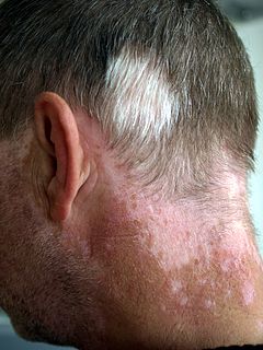
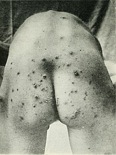
WPyoderma means any skin disease that is pyogenic. These include superficial bacterial infections such as impetigo, impetigo contagiosa, ecthyma, folliculitis, Bockhart's impetigo, furuncle, carbuncle, tropical ulcer, etc. Autoimmune conditions include pyoderma gangrenosum. Pyoderma affects more than 111 million children worldwide, making it one of the three most common skin disorders in children along with scabies and tinea.
 W
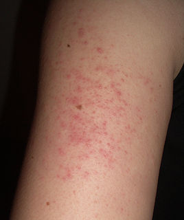
WA rash is a change of the human skin which affects its color, appearance, or texture.
 W
WA sebaceous filament is a tiny collection of sebum and dead skin cells around a hair follicle, which usually takes the form of a small, yellow to off-white hair-like strand when expressed from the skin..
 W
WA skin fissure is a cutaneous condition in which there is a linear-like cleavage of skin, sometimes defined as extending into the dermis. It is smaller than a skin laceration.
 W
WSpongiosis is mainly intercellular edema in the epidermis, and is characteristic of eczematous dermatitis, manifested clinically by intraepidermal vesicles, "juicy" papules, and/or lichenification. It is a severe case of eczema that affects the epidermis, dermis and/or subcutaneous skin tissues. The three types of spongiotic are acute, subacute and chronic. A dermatologist can diagnose acute spongiotic by examining the skin during an office visit but a biopsy is needed for an accurate diagnosis of the type.
 W
WIn medicine, a targetoid object is a structure or lesion that has the appearance of a target or is target-like.
 W
WTelangiectasias, also known as spider veins, are small dilated blood vessels that can occur near the surface of the skin or mucous membranes, measuring between 0.5 and 1 millimeter in diameter. These dilated blood vessels can develop anywhere on the body but are commonly seen on the face around the nose, cheeks and chin. Dilated blood vessels can also develop on the legs, although when they occur on the legs, they often have underlying venous reflux or "hidden varicose veins". When found on the legs, they are found specifically on the upper thigh, below the knee joint and around the ankles.
 W
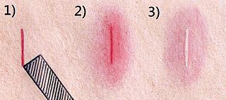
WThe triple response of Lewis is a cutaneous response that occurs from firm stroking of the skin, which produces an initial red line, followed by a flare around that line, and then finally a wheal.
 W
WAn ulcer is a sore on the skin or a mucous membrane, accompanied by the disintegration of tissue. Ulcers can result in complete loss of the epidermis and often portions of the dermis and even subcutaneous fat. Ulcers are most common on the skin of the lower extremities and in the gastrointestinal tract. An ulcer that appears on the skin is often visible as an inflamed tissue with an area of reddened skin. A skin ulcer is often visible in the event of exposure to heat or cold, irritation, or a problem with blood circulation.
 W
WVacuolar interface dermatitis is a dermatitis with vacuolization at the dermoepidermal junction, with lymphocytic inflammation at the epidermis and dermis.
 W
WVacuolization is the formation of vacuoles or vacuole-like structures, within or adjacent to cells. Perinuclear vacuolization of epidermal keratinocytes is most likely inconsequential when not observed in combination with other pathologic findings. In dermatopathology "vacuolization" often refers specifically to vacuoles in the basal cell-basement membrane zone area, where it is an unspecific sign of disease. It may be a sign of for example vacuolar interface dermatitis, which in turn has many causes.
 W
WThe vermilion border, also called margin or zone, is the normally sharp demarcation between the lip and the adjacent normal skin. It is where lipstick is sometimes applied. It represents the change in the epidermis from highly keratinized external skin to less keratinized internal skin. It has no sebaceous glands, sweat glands, or facial hair.
 W
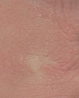
WXeroderma, xerosis or xerosis cutis, or simply dry skin, is a skin condition characterized by excessively dry skin. The medical term xeroderma is derived from the Greek words meaning dry skin.
 W
WXerostomia, also known as dry mouth, is dryness in the mouth, which may be associated with a change in the composition of saliva, or reduced salivary flow, or have no identifiable cause.