 W
WAn acceleromyograph is a piezoelectric myograph, used to measure the force produced by a muscle after it has undergone nerve stimulation. Acceleromyographs may be used, during anaesthesia when muscle relaxants are administered, to measure the depth of neuromuscular blockade and to assess adequacy of recovery from these agents at the end of surgery. Acceleromyography is classified as quantitative neuromuscular monitoring.
 W
WMuscles are described using unique anatomical terminology according to their actions and structure.
 W
WArchitectural gear ratio, also called anatomical gear ratio (AGR) is a feature of pennate muscle defined by the ratio between the longitudinal strain of the muscle and muscle fiber strain. It is sometimes also defined as the ratio between muscle-shortening velocity and fiber-shortening velocity.
 W
WThe axillary spaces are anatomic spaces. through which axillary contents leave the axilla. They consist of the quadrangular space, triangular space, and triangular interval. It is bounded by teres major, teres minor, medial border of the humerus, and long head of triceps brachii.
 W
WCardiac muscle is one of three types of vertebrate muscles, with the other two being skeletal and smooth muscles. It is an involuntary, striated muscle that constitutes the main tissue of the walls of the heart. The myocardium forms a thick middle layer between the outer layer of the heart wall and the inner layer, with blood supplied via the coronary circulation. It is composed of individual heart muscle cells (cardiomyocytes) joined together by intercalated discs, encased by collagen fibers and other substances that form the extracellular matrix.
 W
WThe central governor is a proposed process in the brain that regulates exercise in regard to a neurally calculated safe exertion by the body. In particular, physical activity is controlled so that its intensity cannot threaten the body’s homeostasis by causing anoxic damage to the heart muscle. The central governor limits exercise by reducing the neural recruitment of muscle fibers. This reduced recruitment causes the sensation of fatigue. The existence of a central governor was suggested to explain fatigue after prolonged strenuous exercise in long-distance running and other endurance sports, but its ideas could also apply to other causes of exertion-induced fatigue.
 W
WThe conjoint tendon is a structure formed from the lower part of the common aponeurosis of the internal oblique muscle and the transversus abdominis as it inserts into the crest of the pubis and pectineal line immediately behind the superficial inguinal ring. It is usually conjoint with the tendon of the internal oblique muscle, but they may be separate as well. It forms the medial part of the posterior wall of the inguinal canal.
 W
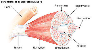
WThe endomysium, meaning within the muscle, is a wispy layer of areolar connective tissue that ensheaths each individual myocyte. It also contains capillaries and nerves. It overlies the muscle fiber's cell membrane: the sarcolemma. Endomysium is the deepest and smallest component of muscle connective tissue. This thin layer helps provide an appropriate chemical environment for the exchange of calcium, sodium, and potassium, which is essential for the excitation and subsequent contraction of a muscle fiber.
 W
WIn adult animals, trunk muscles can be broadly divided into hypaxial muscles, which lie ventral to the horizontal septum of the vertebrae and epaxial muscles, which lie dorsal to the septum. Hypaxial muscles include some vertebral muscles, the diaphragm, the abdominal muscles, and all limb muscles. The serratus posterior inferior and serratus posterior superior are innervated by the ventral primary ramus and are hypaxial muscles. Epaxial muscles include other (dorsal) muscles associated with the vertebrae, ribs, and base of the skull. In humans, the erector spinae, the transversospinal muscles, the splenius and suboccipital muscles are the only epaxial muscles.
WEpimysium is the fibrous tissue envelope that surrounds skeletal muscle. It is a layer of dense irregular connective tissue which ensheaths the entire muscle and protects muscles from friction against other muscles and bones. It is continuous with fascia and other connective tissue wrappings of muscle including the endomysium and perimysium. It is also continuous with tendons, where it becomes thicker and collagenous.
 W
WAn extensor expansion is the special connective attachments by which the extensor tendons insert into the phalanges.
 W
WGalvanism is a term invented by the late 18th C. physicist and chemist, Alessandro Volta, to refer to the generation of electrical current by chemical action. The term also came to refer to the discoveries of its namesake, Luigi Galvani, specifically the generation of electrical current within biological organisms and the contraction/convulsion of biological muscle tissue upon contact with electrical current. While Volta theorized and later demonstrated the phenomenon of his "Galvanism" to be replicable with otherwise inert materials, Galvani thought his discovery to be a confirmation of the existence of "animal electricity," a vital force which gave life to organic matter.
 W
WHypertrophy is the increase in the volume of an organ or tissue due to the enlargement of its component cells. It is distinguished from hyperplasia, in which the cells remain approximately the same size but increase in number. Although hypertrophy and hyperplasia are two distinct processes, they frequently occur together, such as in the case of the hormonally-induced proliferation and enlargement of the cells of the uterus during pregnancy.
 W
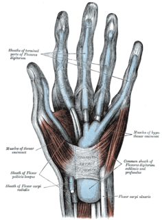
WThe hypothenar muscles are a group of three muscles of the palm that control the motion of the little finger.
WLateral to the inguinal aponeurotic falx there is a ligamentous band originating from the lower margin of the transversalis fascia and extending down in front of the inferior epigastric artery to the superior ramus of the pubis; it is termed the interfoveolar ligament of Hesselbach and sometimes contains a few muscular fibers.
 W
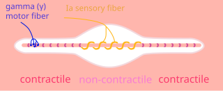
WIntrafusal muscle fibers are skeletal muscle fibers that serve as specialized sensory organs (proprioceptors) that detect the amount and rate of change in length of a muscle. They constitute the muscle spindle and are innervated by both sensory (afferent) and motor (efferent) fibers. Gamma efferents from small multipolar cells from anterior gray column innervate it. These form a part of neuromuscular spindles. Intrafusal muscle fibers are walled off from the rest of the muscle by an outer connective tissue sheath consisting of flattened fibroblasts and collagen. This sheath has a spindle or "fusiform" shape, hence the name "intrafusal".
 W
WIntramuscular injection, often abbreviated IM, is the injection of a substance into a muscle. In medicine, it is one of several methods for parenteral administration of medications. Intramuscular injection may be preferred because muscles have larger and more numerous blood vessels than subcutaneous tissue, leading to faster absorption than subcutaneous or intradermal injections. Medication administered via intramuscular injection also is not subject to the first-pass metabolism effect which affects oral medications.
 W
WThe iris dilator muscle, is a smooth muscle of the eye, running radially in the iris and therefore fit as a dilator. The pupillary dilator consists of a spokelike arrangement of modified contractile cells called myoepithelial cells. These cells are stimulated by the sympathetic nervous system. When stimulated, the cells contract, widening the pupil and allowing more light to enter the eye.
WThe iris sphincter muscle is a muscle in the part of the eye called the iris. It encircles the pupil of the iris, appropriate to its function as a constrictor of the pupil.
WA key component in lateral force transmission in skeletal muscle is the extracellular matrix (ECM). Skeletal muscle is a complex biological material that is composed of muscle fibers and an ECM consisting of the epimysium, perimysium, and endomysium. It can be described as a collagen fiber-reinforced composite. The ECM has at least three functions: (1) to provide a framework binding muscle fibers together and ensure their proper alignment, (2) to transmit the forces, either from active muscle contraction or ones passively imposed on it, and (3) providing lubricated surfaces between muscle fibers and bundles enabling the muscle to change shape. The mechanical properties of skeletal muscle depend on both the properties of muscle fibers and the ECM, and the interaction between the two. Contractile forces are transmitted laterally within intramuscular connective tissue to the epimysium and then to the tendon. Due to the nature of skeletal muscle, direct measurements are not possible, but many indirect studies and analyses have shown that the ECM is an important part of force transmission during muscle contraction.
 W
WThis is a table of skeletal muscles of the human anatomy.
 W
WIn facial anatomy, the modiolus is a chiasma of facial muscles held together by fibrous tissue, located lateral and slightly superior to each angle of the mouth. It is important in moving the mouth, facial expression and in dentistry. It is extremely important in relation to stability of lower denture, because of the strength and variability of movement of the area. It derives its motor nerve supply from the facial nerve, and its blood supply from labial branches of the facial artery.
 W
WMuscle is a soft tissue found in most animals. Muscle cells contain protein filaments of actin and myosin that slide past one another, producing a contraction that changes both the length and the shape of the cell. Muscles function to produce force and motion. They are primarily responsible for maintaining and changing posture, locomotion, as well as movement of internal organs, such as the contraction of the heart and the movement of food through the digestive system via peristalsis.
 W
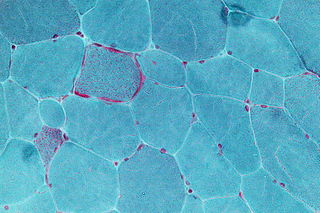
WIn medicine, a muscle biopsy is a procedure in which a piece of muscle tissue is removed from an organism and examined microscopically. A muscle biopsy can lead to the discovery of problems with the nervous system, connective tissue, vascular system, or musculoskeletal system.
 W
WMuscle coactivation occurs when agonist and antagonist muscles surrounding a joint contract simultaneously to provide joint stability. It is also known as muscle cocontraction, since two muscle groups are contracting at the same time. It is able to be measured using electromyography (EMG) from the contractions that occur. The general mechanism of it is still widely unknown. It is believed to be important in joint stabilization, as well as general motor control.
WMuscle contraction is the activation of tension-generating sites within muscle fibers. In physiology, muscle contraction does not necessarily mean muscle shortening because muscle tension can be produced without changes in muscle length, such as when holding a heavy book or a dumbbell at the same position. The termination of muscle contraction is followed by muscle relaxation, which is a return of the muscle fibers to their low tension-generating state.
 W
WMuscle contractures can occur for many reasons, such as paralysis, muscular atrophy, and forms of muscular dystrophy. Fundamentally, the muscle and its tendons shorten, resulting in reduced flexibility. For example, in the case of partial paralysis the loss of strength and muscle control tend to be greater in some muscles than in others, leading to an imbalance between the various muscle groups around specific joints. Case in point: when the muscles which dorsiflex are less functional than the muscles which plantarflex a contracture occurs, giving the foot a progressively downward angle and loss of flexibility. Various interventions can slow, stop, or even reverse muscle contractures, ranging from physical therapy to surgery. A common cause for having the ankle lose its flexibility in this manner is from having sheets tucked in at the foot of the bed when sleeping. The weight of the sheets keep the feet plantarflexed all night. Correcting this by not tucking the sheets in at the foot of the bed, or by sleeping with the feet hanging off the bed when in the prone position, is part of correcting this imbalance.
WA muscle fascicle is a bundle of skeletal muscle fibers surrounded by perimysium, a type of connective tissue.
 W
WMuscle hypertrophy involves an increase in size of skeletal muscle through a growth in size of its component cells. Two factors contribute to hypertrophy: sarcoplasmic hypertrophy, which focuses more on increased muscle glycogen storage; and myofibrillar hypertrophy, which focuses more on increased myofibril size.
 W
WMuscle tissue is a soft tissue that composes muscles in animal bodies, and gives rise to muscles' ability to contract. This is opposed to other components or tissues in muscle such as tendons or perimysium. It is formed during embryonic development through a process known as myogenesis. Muscle tissue consists of elongated cells also called as muscle fibers. This tissue is responsible for movements in our body. Muscles contain special proteins called contractile protein which contract and relax to cause movement.
 W
WIn human anatomy, the muscles of the hip joint are those muscles that cause movement in the hip. Most modern anatomists define 17 of these muscles, although some additional muscles may sometimes be considered. These are often divided into four groups according to their orientation around the hip joint: the gluteal group; the lateral rotator group; the adductor group; and the iliopsoas group.
 W
WMyogenesis is the formation of muscular tissue, particularly during embryonic development.
 W
WMyotoxins are small, basic peptides found in snake venoms,, and lizard venoms. This involves a non-enzymatic mechanism that leads to severe muscle necrosis. These peptides act very quickly, causing instantaneous paralysis to prevent prey from escaping and eventually death due to diaphragmatic paralysis.
WA nuclear chain fiber is a specialized sensory organ contained within a muscle. Nuclear chain fibers are intrafusal fibers that, along with nuclear bag fibers, make up the muscle spindle responsible for the detection of changes in muscle length.
 W
WOrthotics is a medical specialty that focuses on the design and application of orthoses. An orthosis is "an externally applied device used to modify the structural and functional characteristics of the neuromuscular and skeletal system". An orthotist is the primary medical clinician responsible for the prescription, manufacture and management of orthoses. An orthosis may be used to:Control, guide, limit and/or immobilize an extremity, joint or body segment for a particular reason Restrict movement in a given direction Assist movement generally Reduce weight bearing forces for a particular purpose Aid rehabilitation from fractures after the removal of a cast Otherwise correct the shape and/or function of the body, to provide easier movement capability or reduce pain
WPerimysium is a sheath of connective tissue that groups muscle fibers into bundles or fascicles.
 W
WIn muscle physiology, physiological cross-sectional area (PCSA) is the area of the cross section of a muscle perpendicular to its fibers, generally at its largest point. It is typically used to describe the contraction properties of pennate muscles. It is not the same as the anatomical cross-sectional area (ACSA), which is the area of the crossection of a muscle perpendicular to its longitudinal axis. In a non-pennate muscle the fibers are parallel to the longitudinal axis, and therefore PCSA and ACSA coincide.
 W
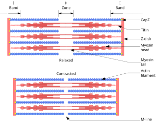
WA sarcomere is the complicated unit of striated muscle tissue. It is the repeating unit between two Z lines. Skeletal muscles are composed of tubular muscle cells which are formed in a process known as myogenesis. Muscle fibers contain numerous tubular myofibrils. Myofibrils are composed of repeating sections of sarcomeres, which appear under the microscope as alternating dark and light bands. Sarcomeres are composed of long, fibrous proteins as filaments that slide past each other when a muscle contracts or relaxes. The costamere is a different component that connects the sarcomere to the sarcolemma.
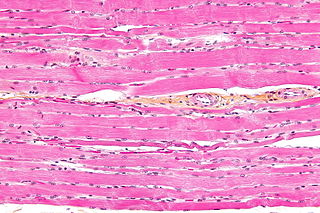
WSkeletal muscle is one of three major muscle types, the others being cardiac muscle and smooth muscle. It is a form of striated muscle tissue which is under the voluntary control of the somatic nervous system. Most skeletal muscles are attached to bones by bundles of collagen fibers known as tendons.
 W
WThe skeletal-muscle pump is a collection of skeletal muscles that aid the heart in the circulation of blood. It is especially important in increasing venous return to the heart, but may also play a role in arterial blood flow.
 W
WThe sliding filament theory explains the mechanism of muscle contraction based on muscle proteins that slide past each other to generate movement. According to the sliding filament theory, the myosin (thick) filaments of muscle fibers slide past the actin (thin) filaments during muscle contraction, while the two groups of filaments remain at relatively constant length.
 W
WSmooth muscle is an involuntary non-striated muscle. It is divided into two subgroups; the single-unit (unitary) and multiunit smooth muscle. Within single-unit cells, the whole bundle or sheet contracts as a syncytium.
 W
WStriated muscle tissue is a muscle tissue that features repeating functional units called sarcomeres. The presence of sarcomeres manifests as a series of bands visible along the muscle fibers, which is responsible for the striated appearance observed in microscopic images of this tissue. There are two types of striated muscles:Cardiac muscle Skeletal muscle
 W
WT-tubules are extensions of the cell membrane that penetrate into the centre of skeletal and cardiac muscle cells. With membranes that contain large concentrations of ion channels, transporters, and pumps, T-tubules permit rapid transmission of the action potential into the cell, and also play an important role in regulating cellular calcium concentration. Through these mechanisms, T-tubules allow heart muscle cells to contract more forcefully by synchronising calcium release throughout the cell. T-tubule structure may be affected by disease, potentially contributing to heart failure and arrhythmias. Although these structures were first seen in 1897, research into T-tubule biology is ongoing.
 W
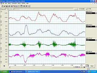
WTensiomyography (TMG) is a measuring method for detection of skeletal muscles’ contractile properties. Tensiomyography assesses muscle mechanical response based on radial muscle belly displacement induced by a single electrical stimulus. It is performed using the TMG S2 system. A tensiomyography measurement instrument includes an electrical stimulator and data acquisition subunit (1), a digital sensor (2), a tripod with manipulating hand (3), and muscle electrodes (4) that work with an essential software interface installed on a PC.
 W
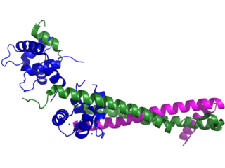
WTitin, also known as connectin, is a protein that in humans is encoded by the TTN gene. Titin is a giant protein, greater than 1 µm in length, that functions as a molecular spring which is responsible for the passive elasticity of muscle. It comprises 244 individually folded protein domains connected by unstructured peptide sequences. These domains unfold when the protein is stretched and refold when the tension is removed.
WIn the histology of skeletal muscle, a triad is the structure formed by a T tubule with a sarcoplasmic reticulum (SR) known as the terminal cisterna on either side. Each skeletal muscle fiber has many thousands of triads, visible in muscle fibers that have been sectioned longitudinally. In mammals, triads are typically located at the A-I junction; that is, the junction between the A and I bands of the sarcomere, which is the smallest unit of a muscle fiber.
 W
WTroponin, or the troponin complex, is a complex of three regulatory proteins that are integral to muscle contraction in skeletal muscle and cardiac muscle, but not smooth muscle. Measurements of cardiac-specific troponins I and T are extensively used as diagnostic and prognostic indicators in the management of myocardial infarction and acute coronary syndrome. Blood troponin levels may be used as a diagnostic marker for stroke, although the sensitivity of this measurement is low.