 W
WThe anterior interventricular sulcus is one of two grooves that separates the ventricles of the heart, the other being the posterior interventricular sulcus.
 W
WThe aorta is the main and largest artery in the human body, originating from the left ventricle of the heart and extending down to the abdomen, where it splits into two smaller arteries. The aorta distributes oxygenated blood to all parts of the body through the systemic circulation.
 W
WThe aortic sac or aortic bulb is a dilated structure in mammalian embryos, lined by endothelial cells located just above the truncus arteriosus. It is the primordial vascular channel from which the aortic arches arise and is homologous to the ventral aorta of gill-bearing vertebrates.
 W
WThe aortic valve is a valve in the human heart between the left ventricle and the aorta. It is one of the two semilunar valves of the heart, the other being the pulmonary valve. The heart has four valves; the other two are the mitral and the tricuspid valves. The aortic valve normally has three cusps or leaflets, although in 1–2% of the population it is found to congenitally have two leaflets. The aortic valve is the last structure in the heart the blood travels through before stopping the flow through the systemic circulation.
WThe aorticopulmonary septum is developmentally formed from neural crest, specifically the cardiac neural crest, and actively separates the aorta and pulmonary arteries and fuses with the interventricular septum within the heart during heart development.
 W
WThe atrioventricular node or AV node is a part of the electrical conduction system of the heart that coordinates the top of the heart. It electrically connects the atria and ventricles. The AV node lies at the lower back section of the interatrial septum near the opening of the coronary sinus, and conducts the normal electrical impulse from the atria to the ventricles. The AV node is quite compact.
 W
WThe atrioventricular septum is a septum of the heart between the right atrium (RA) and the left ventricle (LV).
WThe atrium is the upper chamber through which blood enters the ventricles of the heart. There are two atria in the human heart – the left atrium receives blood from the pulmonary (lung) circulation, and the right atrium receives blood from the venae cavae. The atria receive blood while relaxed (diastole), then contract (systole) to move blood to the ventricles. All animals with a closed circulatory system have at least one atrium. Humans have two atria.
 W
WIn the heart's conduction system, Bachmann's bundle is a branch of the anterior internodal tract that resides on the inner wall of the left atrium. It is a broad band of cardiac muscle that passes from the right atrium, between the superior vena cava and the ascending aorta. Bachmann's bundle is, during normal sinus rhythm, the preferential path for electrical activation of the left atrium. It is therefore considered to be part of the "atrial conduction system" of the heart.
 W
WThe Björk–Shiley valve is a mechanical artificial heart valve. The valve was co-invented by American engineer Donald Shiley and Swedish heart surgeon Viking Björk.
 W
WThe bundle branches, or Tawara branches, are offshoots of the bundle of His in the heart's ventricle. They play an integral role in the electrical conduction system of the heart by transmitting cardiac action potentials from the bundle of His to the Purkinje fibers.
WDiscovered in 1893 by Swiss-born cardiologist and anatomist Wilhelm His Jr., the bundle of His (BH) or His bundle (HB) ( "hiss") is a collection of heart muscle cells specialized for electrical conduction. As part of the electrical conduction system of the heart, it transmits the electrical impulses from the AV node to the point of the apex of the fascicular branches via the bundle branches. The fascicular branches then lead to the Purkinje fibers, which provide electrical conduction to the ventricles, causing the cardiac muscle of the ventricles to contract at a paced interval.
 W
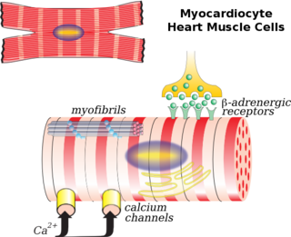
WCardiac muscle is one of three types of vertebrate muscles, with the other two being skeletal and smooth muscles. It is an involuntary, striated muscle that constitutes the main tissue of the walls of the heart. The myocardium forms a thick middle layer between the outer layer of the heart wall and the inner layer, with blood supplied via the coronary circulation. It is composed of individual heart muscle cells (cardiomyocytes) joined together by intercalated discs, encased by collagen fibers and other substances that form the extracellular matrix.
 W
WCardiac muscle cells or cardiomyocytes are the muscle cells (myocytes) that make up the cardiac muscle. Each myocardial cell contains myofibrils, which are specialized organelles consisting of long chains of sarcomeres, the fundamental contractile units of muscle cells.
WThe contraction of cardiac muscle in all animals is initiated by electrical impulses known as action potentials. The rate at which these impulses fire, controls the rate of cardiac contraction, that is, the heart rate. The cells that create these rhythmic impulses, setting the pace for blood pumping, are called pacemaker cells, and they directly control the heart rate. They make up the cardiac pacemaker, that is, the natural pacemaker of the heart. In most humans, the concentration of pacemaker cells in the sinoatrial (SA) node is the natural pacemaker, and the resultant rhythm is a sinus rhythm.
 W
WThe term cardium comes from the Greek word for 'heart'. It is a frequently encountered term in physiology where it is incorporated into scientific nomenclature.
 W
WThe chordae tendineae , colloquially known as the heart strings, are tendon-resembling fibrous cords of connective tissue that connect the papillary muscles to the tricuspid valve and the mitral valve in the heart.
 W
WThe coronary arteries are the arterial blood vessels of coronary circulation, which transport oxygenated blood to the heart muscle. The heart requires a continuous supply of oxygen to function and survive, much like any other tissue or organ of the body.
 W
WCoronary circulation is the circulation of blood in the blood vessels that supply the heart muscle (myocardium). Coronary arteries supply oxygenated blood to the heart muscle, and cardiac veins drain away the blood once it has been deoxygenated. Because the rest of the body, and most especially the brain, needs a steady supply of oxygenated blood that is free of all but the slightest interruptions, the heart is required to function continuously. Therefore its circulation is of major importance not only to its own tissues but to the entire body and even the level of consciousness of the brain from moment to moment. Interruptions of coronary circulation quickly cause heart attacks, in which the heart muscle is damaged by oxygen starvation. Such interruptions are usually caused by ischemic heart disease and sometimes by embolism from other causes like obstruction in blood flow through vessels.
WThe atria of the heart are separated from the ventricles by the coronary sulcus. The structure contains the trunks of the nutrient vessels of the heart, and is deficient in front, where it is crossed by the root of the pulmonary trunk. On the posterior surface of the heart, the coronary sulcus contains the coronary sinus
 W
WThe crux cordis or crux of the heart is the area on the lower back side of the heart where the coronary sulcus and the posterior interventricular sulcus meet. It is important surgically because the atrioventricular nodal artery, a small but vital vessel, passes in proximity to the crux of the heart. It is the anastomotic point of right and left coronary artery.
WThe interventricular septum is the stout wall separating the ventricles, the lower chambers of the heart, from one another.
 W
WThe endocardium is the innermost layer of tissue that lines the chambers of the heart. Its cells are embryologically and biologically similar to the endothelial cells that line blood vessels. The endocardium also provides protection to the valves and heart chambers.
 W
WThe fold of the left vena cava, ligament of the left vena cava, or vestigial fold of Marshall, is a triangular fold of the serous pericardium that lies between the left pulmonary artery and subjacent pulmonary vein.
 W
WBuilt in 1953, the Giant Heart exhibit, originally called the "Engine of Life" exhibit, is one of the most popular and notable exhibits at the Franklin Institute. The exhibit is roughly two stories tall and 35-feet in diameter. A walk-through exhibit, visitors can explore the different areas of the heart.
 W
WA heart valve is a one-way valve that normally allows blood to flow in only one direction through the heart. The four valves are commonly represented in a mammalian heart that determines the pathway of blood flow through the heart. A heart valve opens or closes incumbent on differential blood pressure on each side.
 W
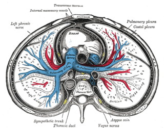
WThe heart is a muscular organ in most animals, which pumps blood through the blood vessels of the circulatory system. The pumped blood carries oxygen and nutrients to the body, while carrying metabolic waste such as carbon dioxide to the lungs. In humans, the heart is approximately the size of a closed fist and is located between the lungs, in the middle compartment of the chest.
WThe infundibulum is a conical pouch formed from the upper and left angle of the right ventricle in the chordate heart, from which the pulmonary trunk arises. It develops from the bulbus cordis. Typically, the infundibulum refers to the corresponding internal structure, whereas the conus arteriosus refers to the external structure. Defects in infundibulum development can result in a heart condition known as tetrology of Fallot.
WIntercalated discs are microscopic identifying features of cardiac muscle. Cardiac muscle consists of individual heart muscle cells (cardiomyocytes) connected by intercalated discs to work as a single functional organ or syncytium. By contrast, skeletal muscle consists of multinucleated muscle fibers and exhibits no intercalated discs. Intercalated discs support synchronized contraction of cardiac tissue. They occur at the Z line of the sarcomere and can be visualized easily when observing a longitudinal section of the tissue.
WThe left border of heart is shorter than the right border, full, and rounded: it is formed mainly by the left ventricle, but to a slight extent, above, by the left atrium.
WThe mitral valve, also known as the bicuspid valve or left atrioventricular valve, is a valve with two flaps in the heart that lies between the left atrium and the left ventricle. The mitral valve and the tricuspid valve are known collectively as the atrioventricular valves because they lie between the atria and the ventricles of the heart.
 W
WThe papillary muscles are muscles located in the ventricles of the heart. They attach to the cusps of the atrioventricular valves via the chordae tendineae and contract to prevent inversion or prolapse of these valves on systole. The papillary muscles constitute about 10% of the total heart mass.
WThe pectinate muscles are parallel ridges in the walls of the atria of the heart. They are so-called because of their resemblance to the teeth of a comb as in pecten.
 W
WPericardial fluid is the serous fluid secreted by the serous layer of the pericardium into the pericardial cavity. The pericardium consists of two layers, an outer fibrous layer and the inner serous layer. This serous layer has two membranes which enclose the pericardial cavity into which is secreted the pericardial fluid. The fluid is similar to the cerebrospinal fluid of the brain which also serves to cushion and allow some movement of the organ.
 W
WThere are two pericardial sinuses: transverse and oblique.The sinus, enclosed between the limbs of the inverted U of the venous mesocardium lies behind the left atrium and in between left and right pulmonary veins. This is known as the oblique sinus. The passage between the venous and arterial mesocardia—i.e., behind the aorta and pulmonary trunk and anterior to the superior vena cava —is termed the transverse sinus. Also, the sinus that forms in the pericardial cavity where the dorso-mesentary pericardium reside. Note: This sinus is clinically important because passing one end of clamp through the sinus, and the other end anterior to the aorta/pulmonary trunk will allow complete blockage of blood output. This is done during some heart surgeries.
WThe pericardium, also called pericardial sac, is a double-walled sac containing the heart and the roots of the great vessels. The pericardial sac has two layers, a serous layer and a fibrous layer. It encloses the pericardial cavity which contains pericardial fluid.
WThe posterior interventricular sulcus or posterior longitudinal sulcus is one of the two grooves that separates the ventricles of the heart and is on the diaphragmatic surface of the heart near the right margin. The other groove is the anterior interventricular sulcus, situated on the sternocostal surface of the heart, close to its left margin.
WThe pulmonary valve is the semilunar valve of the heart that lies between the right ventricle and the pulmonary artery and has three cusps. Similar to the aortic valve, the pulmonary valve opens in ventricular systole, when the pressure in the right ventricle rises above the pressure in the pulmonary artery. At the end of ventricular systole, when the pressure in the right ventricle falls rapidly, the pressure in the pulmonary artery will close the pulmonary valve.
WThe Purkinje fibers are located in the inner ventricular walls of the heart, just beneath the endocardium in a space called the subendocardium. The Purkinje fibers are specialised conducting fibers composed of electrically excitable cells. They are larger than cardiomyocytes with fewer myofibrils and many mitochondria. They conduct cardiac action potentials more quickly and efficiently than any other cells in the heart. Purkinje fibers allow the heart's conduction system to create synchronized contractions of its ventricles, and are essential for maintaining a consistent heart rhythm.
WThe right atrioventricular orifice is the large oval aperture of communication between the right atrium and ventricle.
WThe right border of the heart is a long border on the surface of the heart, and is formed by the right atrium.The atrial portion is rounded and almost vertical; it is situated behind the third, fourth, and fifth right costal cartilages about 1.25 cm. from the margin of the sternum. The ventricular portion, thin and sharp, is named the acute margin; it is nearly horizontal, and extends from the sternal end of the sixth right coastal cartilage to the apex of the heart.
WThe sinoatrial node is a group of cells located in the wall of the right atrium of the heart. These cells have the ability to spontaneously produce an electrical impulse, that travels through the heart via the electrical conduction system causing it to contract. In a healthy heart, the SA node continuously produces action potential, setting the rhythm of the heart and so is known as the heart's natural pacemaker. The rate of action potential production is influenced by nerves that supply it.
WThe smallest cardiac veins are minute valveless veins in the walls of all four heart chambers. The veins are sometimes accurately referred to as vessels, but they are frequently confused with a distinct set of artery connections eponymously referred to as the "vessels of Wearn". In his 1928 publication, Wearn himself referred to the arterio-cameral connections as thebesian. However, Wearn's 1933 and 1941 publications emphatically described the thebesian veins as distinct from arterio-cameral vessels. Thus, there is effort to add the term vessels of Wearn and/or arteriae cardiacae minimae to the TA and FLA databases, but international collaboration appears necessary to accomplish this task.
WThe fibrous pericardium is attached to the posterior surface of the sternum by the superior and inferior sternopericardiac ligaments ; the upper passing to the manubrium, and the lower to the xiphoid process.
WThe chordae tendineae , colloquially known as the heart strings, are tendon-resembling fibrous cords of connective tissue that connect the papillary muscles to the tricuspid valve and the mitral valve in the heart.
WThe tricuspid valve, or right atrioventricular valve, is on the right dorsal side of the mammalian heart, at the superior portion of the right ventricle. The function of the valve is to prevent back flow (regurgitation) of blood from the right ventricle into the right atrium during right ventricular contraction: systole.
WThe valve of the coronary sinus is a semicircular fold of the lining membrane of the right atrium, at the orifice of the coronary sinus. It is situated at the base of the inferior vena cava.
WThe valve of the inferior vena cava is a venous valve that lies at the junction of the inferior vena cava and right atrium.
WA ventricle is one of two large chambers toward the bottom of the heart that collect and expel blood received from an atrium towards the peripheral beds within the body and lungs. The atrium primes the pump.