 W
WAngiomyxoma is a myxoid tumor involving the blood vessels.
 W
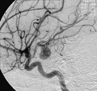
WAn aneurysm is an outward bulging, likened to a bubble or balloon, caused by a localized, abnormal, weak spot on a blood vessel wall. Aneurysms may be a result of a hereditary condition or an acquired disease. Aneurysms can also be a nidus for clot formation (thrombosis) and embolization. The word is from Greek: ἀνεύρυσμα, aneurysma, "dilation", from ἀνευρύνειν, aneurynein, "to dilate". As an aneurysm increases in size, the risk of rupture increases, leading to uncontrolled bleeding. Although they may occur in any blood vessel, particularly lethal examples include aneurysms of the Circle of Willis in the brain, aortic aneurysms affecting the thoracic aorta, and abdominal aortic aneurysms. Aneurysms can arise in the heart itself following a heart attack, including both ventricular and atrial septal aneurysms. There are congenital atrial septal aneurysms, a rare heart defect.
 W
WIn medicine (gastroenterology), angiodysplasia is a small vascular malformation of the gut. It is a common cause of otherwise unexplained gastrointestinal bleeding and anemia. Lesions are often multiple, and frequently involve the cecum or ascending colon, although they can occur at other places. Treatment may be with colonoscopic interventions, angiography and embolization, medication, or occasionally surgery.
 W
WIn medicine, aortoiliac occlusive disease, is a form of central artery disease involving the blockage of the abdominal aorta as it transitions into the common iliac arteries.
 W
WArteriolosclerosis is a form of cardiovascular disease involving hardening and loss of elasticity of arterioles or small arteries and is most often associated with hypertension and diabetes mellitus. Types include hyaline arteriolosclerosis and hyperplastic arteriolosclerosis, both involved with vessel wall thickening and luminal narrowing that may cause downstream ischemic injury. The following two terms whilst similar, are distinct in both spelling and meaning and may easily be confused with arteriolosclerosis.Arteriosclerosis is any hardening of medium or large arteries Atherosclerosis is a hardening of an artery specifically due to an atheromatous plaque. The term atherogenic is used for substances or processes that cause atherosclerosis.
 W
WArteriosclerosis is the thickening, hardening, and loss of elasticity of the walls of arteries. This process gradually restricts the blood flow to one's organs and tissues and can lead to severe health risks brought on by atherosclerosis, which is a specific form of arteriosclerosis caused by the buildup of fatty plaques, cholesterol, and some other substances in and on the artery walls. It can be brought on by smoking, a bad diet, or many genetic factors. Atherosclerosis is the primary cause of coronary artery disease (CAD) and stroke, with multiple genetic and environmental contributions. Genetic-epidemiologic studies have identified a surprisingly long list of genetic and non-genetic risk factors for CAD. However, such studies indicate that family history is the most significant independent risk factor.
 W
WAn atheroma, or atheromatous plaque ("plaque"), is an abnormal accumulation of material in the inner layer of the wall of an artery.
 W
WAtherosclerosis is a disease in which the wall of the artery develops abnormalities, called lesions. These lesions may lead to narrowing due to the buildup of atheromatous plaque. Initially, there are generally no symptoms. When severe, it can result in coronary artery disease, stroke, peripheral artery disease, or kidney problems, depending on which arteries are affected. Symptoms, if they occur, generally do not begin until middle age.
 W
WBrain ischemia is a condition in which there is insufficient blood flow to the brain to meet metabolic demand. This leads to poor oxygen supply or cerebral hypoxia and thus leads to the death of brain tissue or cerebral infarction / ischemic stroke. It is a sub-type of stroke along with subarachnoid hemorrhage and intracerebral hemorrhage.
 W
WCapillaritis is where the capillaries, usually of the legs or lungs, are inflamed.
 W
WCardiac allograft vasculopathy (CAV) is a common long-term complication of heart transplantation affecting up to half of people within 10 years. It arises when the blood vessels supplying the transplanted heart gradually narrow and restrict its blood flow, subsequently leading to impairment of the heart muscle or sudden death. The other major causes of death following heart transplantation include graft failure, organ rejection and infection.
 W
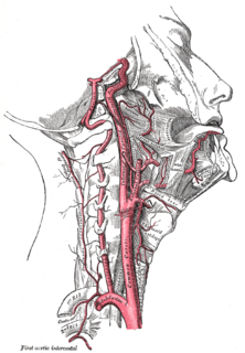
WCarotid artery dissection is a separation of the layers of the artery wall supplying oxygen-bearing blood to the head and brain and is the most common cause of stroke in young adults.
 W
WCarotid artery stenosis is a narrowing or constriction of any part of the carotid arteries, usually caused by atherosclerosis.
 W
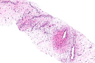
WCerebral amyloid angiopathy (CAA), is a form of angiopathy in which amyloid beta peptide deposits in the walls of small to medium blood vessels of the central nervous system and meninges. The term congophilic is sometimes used because the presence of the abnormal aggregations of amyloid can be demonstrated by microscopic examination of brain tissue after staining with Congo red. The amyloid material is only found in the brain and as such the disease is not related to other forms of amyloidosis.
 W
WCholesterol embolism occurs when cholesterol is released, usually from an atherosclerotic plaque, and travels as an embolus in the bloodstream to lodge causing an obstruction in blood vessels further away. Most commonly this causes skin symptoms, gangrene of the extremities and sometimes kidney failure; problems with other organs may arise, depending on the site at which the cholesterol crystals enter the bloodstream. When the kidneys are involved, the disease is referred to as atheroembolic renal disease. The diagnosis usually involves biopsy from an affected organ. Cholesterol embolism is treated by removing the cause and giving supportive therapy; statin drugs have been found to improve the prognosis.
 W
WChronic cerebrospinal venous insufficiency is a term invented by Italian researcher Paolo Zamboni in 2008 to describe compromised flow of blood in the veins draining the central nervous system. Zamboni hypothesized that it might play a role in the cause or development of multiple sclerosis (MS). Zamboni also devised a surgical procedure which the media nicknamed a liberation procedure or liberation therapy, involving venoplasty or stenting of certain veins. Zamboni's ideas about CCSVI are very controversial, with significantly more detractors than supporters, and any treatments based on his ideas are considered experimental.
 W
WChronic venous insufficiency (CVI) is a medical condition in which blood pools in the veins, straining the walls of the vein. The most common cause of CVI is superficial venous reflux which is a treatable condition. As functional venous valves are required to provide for efficient blood return from the lower extremities, this condition typically affects the legs. If the impaired vein function causes significant symptoms, such as swelling and ulcer formation, it is referred to as chronic venous disease. It is sometimes called chronic peripheral venous insufficiency and should not be confused with post-thrombotic syndrome in which the deep veins have been damaged by previous deep vein thrombosis.
 W
WCLOVES syndrome is an extremely rare overgrowth syndrome with complex vascular anomalies. CLOVES syndrome affects people with various symptoms, ranging from mild fatty soft-tissue tumors to vascular malformations encompassing the spine or internal organs. CLOVES syndrome is closely linked to other overgrowth disorders like proteus syndrome, Klippel–Trénaunay syndrome, Sturge–Weber syndrome, and hemihypertrophy, to name a few. It can also cause fat to move to the back, and many cases undescended testicles.
 W
WDiabetic nephropathy, also known as diabetic kidney disease, is the chronic loss of kidney function occurring in those with diabetes mellitus. Diabetic nephropathy is one of the leading causes of chronic kidney disease (CKD) and end-stage renal disease (ESRD) globally. Protein loss in the urine due to damage to the glomeruli may become massive, and cause a low serum albumin with resulting generalized body swelling (edema) and result in the nephrotic syndrome. Likewise, the estimated glomerular filtration rate (eGFR) may progressively fall from a normal of over 90 ml/min/1.73m2 to less than 15, at which point the patient is said to have end-stage renal disease. It usually is slowly progressive over years.
 W
WA dural arteriovenous fistula (DAVF) or Malformation, is an abnormal direct connection (fistula) between a meningeal artery and a meningeal vein or dural venous sinus.
 W
WFamilial aortic dissection or FAD refers to the splitting of the wall of the aorta in either the arch, ascending or descending portions. FAD is thought to be passed down as an autosomal dominant disease and once inherited will result in dissection of the aorta, and dissecting aneurysm of the aorta, or rarely aortic or arterial dilation at a young age. Dissection refers to the actual tearing open of the aorta. However, the exact gene(s) involved has not yet been identified. It can occur in the absence of clinical features of Marfan syndrome and of systemic hypertension. Over time this weakness, along with systolic pressure, results in a tear in the aortic intima layer thus allowing blood to enter between the layers of tissue and cause further tearing. Eventually complete rupture of the aorta occurs and the pleural cavity fills with blood. Warning signs include chest pain, ischemia, and hemorrhaging in the chest cavity. This condition, unless found and treated early, usually results in death. Immediate surgery is the best treatment in most cases. FAD is not to be confused with PAU and IMH, both of which present in ways similar to that of familial aortic dissection.
 W
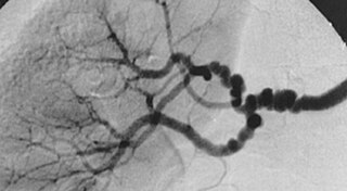
WFibromuscular dysplasia (FMD) is a non-atherosclerotic, non-inflammatory disease of the blood vessels that causes abnormal growth within the wall of an artery. FMD has been found in nearly every arterial bed in the body although the most common arteries affected are the renal and carotid arteries.
 W
WA flow diverter is an endovascular prosthesis used to treat intracranial aneurysms. It is placed in the aneurysm's parent artery, covering the neck, in order to divert blood flow and determine a progressive thrombosis of the sac. Flow diverting stents consist of structural Cobalt-chrome or Nitinol alloy wires and often a set of radiopaque wires woven together in a flexible braid.
 W
WHemorrhagic infarcts are infarcts commonly caused by occlusion of veins, with red blood cells entering the area of the infarct, or an artery occlusion of an organ with collaterals or dual circulation. This is commonly seen in brain, lungs, and the GI tract, areas referred to as having "loose tissue," or dual circulation. Loose-textured tissue allows red blood cells released from damaged vessels to diffuse through the necrotic tissue. White infarcts can become hemorrhagic with reperfusion. Compare to Anemic infarct. Hemorrhagic infarction is also associated with testicular torsion.
 W
WHereditary cystatin C amyloid angiopathy (HCCAA) is a rare, fatal amyloid disease in young people in Iceland caused by a mutation in cystatin C. Most of the families with the defect gene can be traced to a region in the northwest of Iceland, around Breiðafjörður.
 W
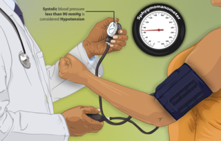
WHypotension is low blood pressure. Blood pressure is the force of blood pushing against the walls of the arteries as the heart pumps out blood. Blood pressure is indicated by two numbers, the systolic blood pressure and the diastolic blood pressure, which are the maximum and minimum blood pressures, respectively. A systolic blood pressure of less than 90 millimeters of mercury or diastolic of less than 60 mm Hg is generally considered to be hypotension. Different numbers apply to children. However, in practice, blood pressure is considered too low only if noticeable symptoms are present.
 W
WInferior vena cava syndrome (IVCS) is a constellation of symptoms resulting from obstruction of the inferior vena cava. It can be caused by physical invasion or compression by a pathological process or by thrombosis within the vein itself. It can also occur during pregnancy. Pregnancy leads to high venous pressure in the lower limbs, decreased blood return to the heart, decreased cardiac output due to obstruction of the inferior vena cava, sudden rise in venous pressure which can lead to placental separation, and a decrease in kidney function. All of these issues can arise from lying in the supine position during late pregnancy which can cause compression of the inferior vena cava by the uterus. Symptoms of late pregnancy inferior vena cava syndrome consist of intense pain in the right hand side, muscle twitching, hypotension, and fluid retention.
 W
WInflammatory aortic aneurysm (IAA), also known as Inflammatory abdominal aortic aneurysm (IAAA), is a type of abdominal aortic aneurysm (AAA) where the walls of the aneurysm become thick and inflamed. Similar to AAA, IAA occurs in the abdominal region. IAA is closely associated and believed to be a response to and extensive peri-anuerysmal fibrosis, which is the formation of excess fibrous connective tissue in an organ or tissue in a reparative or reactive process IAA accounts for 5-10% of aortic aneurysms. IAA occurs mainly in a population that is on average younger by 10 years than most AAA patients. Some common symptoms of IAA may include back pain, abdominal tenderness, fevers, weight loss or elevated Erythrocyte sedimentation rate (ESR) levels. Corticosteroids and other immunosuppressive drugs have been found to decrease symptoms and the degree of peri-aortic inflammation and fibrosis
 W
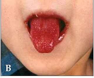
WKawasaki disease is a syndrome of unknown cause that results in a fever and mainly affects children under 5 years of age. It is a form of vasculitis, where blood vessels become inflamed throughout the body. The fever typically lasts for more than five days and is not affected by usual medications. Other common symptoms include large lymph nodes in the neck, a rash in the genital area, and red eyes, lips, palms, or soles of the feet. Within three weeks of the onset, the skin from the hands and feet may peel, after which recovery typically occurs. In some children, coronary artery aneurysms form in the heart.
 W
WLoeys–Dietz syndrome (LDS) is an autosomal dominant genetic connective tissue disorder. It has features similar to Marfan syndrome and Ehlers–Danlos syndrome. The disorder is marked by aneurysms in the aorta, often in children, and the aorta may also undergo sudden dissection in the weakened layers of the wall of the aorta. Aneurysms and dissections also can occur in arteries other than the aorta. Because aneurysms in children tend to rupture early, children are at greater risk for dying if the syndrome is not identified. Surgery to repair aortic aneurysms is essential for treatment.
 W
WMönckeberg's arteriosclerosis, or Mönckeberg's sclerosis, is a form of arteriosclerosis or vessel hardening, where calcium deposits are found in the muscular middle layer of the walls of arteries. It is an example of dystrophic calcification. This condition occurs as an age-related degenerative process. However, it can occur in pseudoxanthoma elasticum and idiopathic arterial calcification of infancy as a pathological condition, as well. Its clinical significance and cause are not well understood and its relationship to atherosclerosis and other forms of vascular calcification are the subject of disagreement. Mönckeberg's arteriosclerosis is named after Johann Georg Mönckeberg, who first described it in 1903.
 W
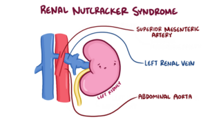
WThe nutcracker syndrome (NCS) results most commonly from the compression of the left renal vein (LRV) between the abdominal aorta (AA) and superior mesenteric artery (SMA), although other variants exist. The name derives from the fact that, in the sagittal plane and/or transverse plane, the SMA and AA appear to be a nutcracker crushing a nut. Furthermore, the venous return from the left gonadal vein returning to the left renal vein is blocked, thus causing testicular pain . There is a wide spectrum of clinical presentations and diagnostic criteria are not well defined, which frequently results in delayed or incorrect diagnosis. This condition is not to be confused with superior mesenteric artery syndrome, which is the compression of the third portion of the duodenum by the SMA and the AA.
 W
WObliterating endarteritis is severe proliferating endarteritis that results in an occlusion of the lumen of the artery. Obliterating endarteritis can occur due to a variety of medical conditions such as a complication of radiation poisoning, tuberculosis meningitis or a syphilis infection.
 W
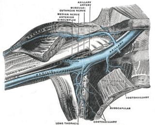
WPaget–Schroetter disease, is a form of upper extremity deep vein thrombosis (DVT), a medical condition in which blood clots form in the deep veins of the arms. These DVTs typically occur in the axillary and/or subclavian veins.
 W
WPhlegmasia cerulea dolens is an uncommon severe form of deep venous thrombosis which results from extensive thrombotic occlusion of the major and the collateral veins of an extremity. It is characterized by sudden severe pain, swelling, cyanosis and edema of the affected limb. There is a high risk of massive pulmonary embolism, even under anticoagulation. Foot gangrene may also occur. An underlying malignancy is found in 50% of cases. Usually, it occurs in those afflicted by a life-threatening illness.
 W
WPortal vein thrombosis (PVT) is a vascular disease of the liver that occurs when a blood clot occurs in the hepatic portal vein, which can lead to increased pressure in the portal vein system and reduced blood supply to the liver. The mortality rate is approximately 1 in 10.
 W
WPostural orthostatic tachycardia syndrome (POTS) is a condition in which a change from lying to standing causes an abnormally large increase in heart rate. This occurs with symptoms that may include lightheadedness, trouble thinking, blurred vision or weakness. Other commonly associated conditions include Ehlers–Danlos syndrome, mast cell activation syndrome, irritable bowel syndrome, insomnia, chronic headaches, chronic fatigue syndrome and fibromyalgia.
 W
WPulmonary insufficiency is a condition in which the pulmonary valve is incompetent and allows backflow from the pulmonary artery to the right ventricle of the heart during diastole. While a small amount of backflow may occur ordinarily, it is usually only shown on an echocardiogram and is harmless. More pronounced regurgitation that is noticed through a routine physical examination is a medical sign of disease and warrants further investigation. If it is secondary to pulmonary hypertension it is referred to as a Graham Steell murmur.
 W
WPylephlebitis is an uncommon thrombophlebitis of the portal vein or any of its branches that is caused by infection. It is usually a complication of intra-abdominal sepsis, most often following diverticulitis, perforated appendicitis, or peritonitis. Considered uniformly lethal in the pre-antibiotic era, it still carries a mortality of 10-30%.
WSack–Barabas syndrome is an older name for the medical condition Ehlers-Danlos syndrome, vascular type. It affects the body's blood vessels and organs, making them prone to rupture.
 W
WSuperficial vein thrombosis (SVT) is a type blood clot in a vein, which forms in a superficial vein near the surface of the body. Usually there is thrombophlebitis, which is an inflammatory reaction around a thrombosed vein, presenting as a painful induration with redness. SVT itself has limited significance when compared to a deep vein thrombosis (DVT), which occurs deeper in the body at the deep venous system level. However, SVT can lead to serious complications, and is therefore no longer regarded as a benign condition. If the blood clot is too near the saphenofemoral junction there is a higher risk of pulmonary embolism, a potentially life-threatening complication.
 W
WSusac's syndrome is a very rare form of microangiopathy characterized by encephalopathy, branch retinal artery occlusions and hearing loss. The cause is unknown but the current thinking is that antibodies are produced against endothelial cells in tiny arteries which leads to damage and the symptoms related to the illness. Despite this being an extremely rare disease, there are 4 registries collecting data on the illness; two are the United States, one in Germany, and one in Portugal.
 W
WTakayasu's arteritis is a form of large vessel granulomatous vasculitis with massive intimal fibrosis and vascular narrowing, most commonly affecting young or middle-age women of Asian descent, though anyone can be affected. It mainly affects the aorta and its branches, as well as the pulmonary arteries. Females are about 8–9 times more likely to be affected than males.
 W
WA thoracic aortic aneurysm is an aortic aneurysm that presents primarily in the thorax.
WThrombophlebitis is a phlebitis related to a thrombus. When it occurs repeatedly in different locations, it is known as thrombophlebitis migrans.
 W
WA varicocele is an abnormal enlargement of the pampiniform venous plexus in the scrotum. This plexus of veins drains blood from the testicles back to the heart. The vessels originate in the abdomen and course down through the inguinal canal as part of the spermatic cord on their way to the testis. Varicoceles occur in around 15% to 20% of all men. The incidence of varicocele increase with age.
 W
WVascular disease is a class of diseases of the blood vessels – the arteries and veins of the circulatory system of the body. It is a subgroup of cardiovascular disease. Disorders in this vast network of blood vessels, can cause a range of health problems which can be severe or prove fatal.
 W
WDysautonomia or autonomic dysfunction is a condition in which the autonomic nervous system (ANS) does not work properly. This may affect the functioning of the heart, bladder, intestines, sweat glands, pupils, and blood vessels. Dysautonomia has many causes, not all of which may be classified as neuropathic. A number of conditions can feature dysautonomia, such as Parkinson's disease, HIV/AIDS, multiple system atrophy, autonomic failure, postural orthostatic tachycardia syndrome, Ehlers-Danlos syndrome, autoimmune autonomic ganglionopathy, and autonomic neuropathy.
 W
WVenous ulcers are wounds that are thought to occur due to improper functioning of venous valves, usually of the legs. They are the major occurrence of chronic wounds, occurring in 70% to 90% of leg ulcer cases. Venous ulcers develop mostly along the medial distal leg, and can be painful with negative effects on quality of life.
 W
WVertebral artery dissection (VAD) is a flap-like tear of the inner lining of the vertebral artery, which is located in the neck and supplies blood to the brain. After the tear, blood enters the arterial wall and forms a blood clot, thickening the artery wall and often impeding blood flow. The symptoms of vertebral artery dissection include head and neck pain and intermittent or permanent stroke symptoms such as difficulty speaking, impaired coordination and visual loss. It is usually diagnosed with a contrast-enhanced CT or MRI scan.