 W
WStaining is a technique used to enhance contrast in samples, generally at the microscopic level. Stains and dyes are frequently used in histology and in the medical fields of histopathology, hematology, and cytopathology that focus on the study and diagnoses of disease at a microscopic level. Stains may be used to define biological tissues, cell populations, or organelles within individual cells.
 W
WAcid-fastness is a physical property of certain bacterial and eukaryotic cells, as well as some sub-cellular structures, specifically their resistance to decolorization by acids during laboratory staining procedures. Once stained as part of a sample, these organisms can resist the acid and/or ethanol-based decolorization procedures common in many staining protocols, hence the name acid-fast.
 W
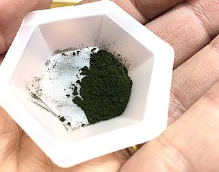
WAlcian blue is any member of a family of polyvalent basic dyes, of which the Alcian blue 8G has been historically the most common and the most reliable member. It is used to stain acidic polysaccharides such as glycosaminoglycans in cartilages and other body structures, some types of mucopolysaccharides, sialylated glycocalyx of cells etc. For many of these targets it is one of the most widely used cationic dyes for both light and electron microscopy. Use of alcian blue has historically been a popular staining method in histology especially for light microscopy in paraffin embedded sections and in semithin resin sections. The tissue parts that specifically stain by this dye become blue to bluish-green after staining and are called "Alcianophilic". Alcian blue staining can be combined with H&E staining, PAS staining and van Gieson staining methods. Alcian blue can be used to quantitate acidic glycans both in microspectrophotometric quantitation in solution or for staining glycoproteins in polyacrylamide gels or on western blots. Biochemists had used it to assay acid polysaccharides in urine since the 1960s for diagnosis of diseases like mucopolysaccharidosis but from 1970's, partly due to lack of availability of Alcian and partly due to length and tediousness of the procedure, alternative methods had to be developed e.g. Dimethyl methylene blue method.
 W
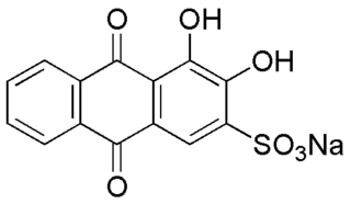
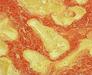
WAlizarin Red S (also known as C.I. Mordant Red 3, Alizarin Carmine, and C.I 58005. It is a water-soluble sodium salt of Alizarin sulfonic acid with a chemical formula of C14H7NaO7S. Alizarin Red S was discovered by Graebe and Libermann in 1871. In the field of histology alizarin Red S is been used to stain calcium deposits in tissues, and in geology to stain and differentiate carbonate minerals.
 W
WThe auramine–rhodamine stain (AR), also known as the Truant auramine–rhodamine stain, is a histological technique used to visualize acid-fast bacilli using fluorescence microscopy, notably species in the Mycobacterium genus. Acid-fast organisms display a reddish-yellow fluorescence. Although the auramine–rhodamine stain is not as specific for acid-fast organisms as the Ziehl–Neelsen stain, it is more affordable and more sensitive, therefore it is often utilized as a screening tool.
 W
WBiebrich scarlet is a molecule used in Lillie's trichrome.
 W
WThe Bielschowsky technique is a silver staining method used in histochemistry for the visualization of nerve fibers, including multipolar interneurons in the cerebellum.
 W
WCarbol fuchsin, carbol-fuchsin, or carbolfuchsin, is a mixture of phenol and basic fuchsin, used in bacterial staining procedures. It is commonly used in the staining of mycobacteria as it has an affinity for the mycolic acids found in their cell membranes.
 W
WA counterstain is a stain with colour contrasting to the principal stain, making the stained structure easily visible using a microscope.
 W
WCresyl violet is an organic compound with the chemical formula C19H18ClN3O. It is a basic dye and is used as a common stain in histology.
 W
WThe Dieterle stain is a way of marking tissue for microscopic examination. The key reagent of Dieterle stain is silver nitrate. It can stain microbes like Treponema pallidum in grey or black and background in yellow.
 W
WDiff-Quik is a commercial Romanowsky stain variant used to rapidly stain and differentiate a variety of pathology specimens. It is most frequently used for blood films and cytopathological smears, including fine needle aspirates. The Diff-Quik procedure is based on a modification of the Wright-Giemsa stain pioneered by Harleco in the 1970s, and has advantages over the routine Wright-Giemsa staining technique in that it reduces the 4-minute process into a much shorter operation and allows for selective increased eosinophilic or basophilic staining depending upon the time the smear is left in the staining solutions.
 W
WEosin methylene blue is a selective stain for Gram-negative bacteria. EMB contains dyes that are toxic to Gram-positive bacteria. EMB is the selective and differential medium for coliforms. It is a blend of two stains, eosin and methylene blue in the ratio of 6:1. EMB is a differential microbiological medium, which slightly inhibits the growth of Gram-positive bacteria and provides a color indicator distinguishing between organisms that ferment lactose and those that do not. Organisms that ferment lactose display "nucleated colonies"—colonies with dark centers.
 W
WEosin Y also called C.I. 45380 or C.I. Acid Red 87, is the form of eosin most commonly used in histology, most notably in the H&E stain. Eosin Y is also widely used in the Papanicolaou stain and the Romanowsky type cytologic stains. It is also used as a photosensitizer in organic synthesis.
 W
WFeulgen stain is a staining technique discovered by Robert Feulgen and used in histology to identify chromosomal material or DNA in cell specimens. It is darkly stained. It depends on acid hydrolysis of DNA, therefore fixating agents using strong acids should be avoided.
 W
WField stain is a histological method for staining of blood smears. It is used for staining thick blood films in order to discover malarial parasites. Field's stain is a version of a Romanowsky stain, used for rapid processing of the specimens.
 W
WG-banding, G banding or Giemsa banding is a technique used in cytogenetics to produce a visible karyotype by staining condensed chromosomes. It is useful for identifying genetic diseases through the photographic representation of the entire chromosome complement. The metaphase chromosomes are treated with trypsin and stained with Giemsa stain. Heterochromatic regions, which tend to be rich with adenine and thymine (AT-rich) DNA and relatively gene-poor, stain more darkly in G-banding. In contrast, less condensed chromatin (Euchromatin)—which tends to be rich with guanine and cytosine (GC-rich) and more transcriptionally active—incorporates less Giemsa stain, and these regions appear as light bands in G-banding. The pattern of bands are numbered on each arm of the chromosome from the centromere to the telomere. This numbering system allows any band on the chromosome to be identified and described precisely. The reverse of G‑bands is obtained in R‑banding. Banding can be used to identify chromosomal abnormalities, such as translocations, because there is a unique pattern of light and dark bands for each chromosome.
 W
WThe gallocyanin stain, also known as the gallocyanin-chromalum stain, is a stain of the oxazine group for total nucleic acids. It is prepared from gallocyanin and is an ideal method for numerous slides that need to be stained serially, equivalently, and reproducible.
 W
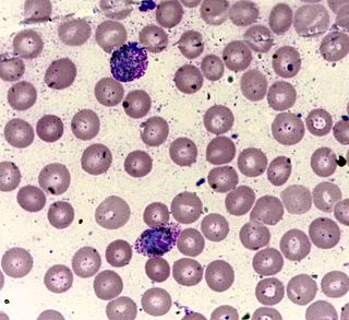
WGiemsa stain, named after German chemist and bacteriologist Gustav Giemsa, is a nucleic acid stain used in cytogenetics and for the histopathological diagnosis of malaria and other parasites.
 W
WGolgi's method is a silver staining technique that is used to visualize nervous tissue under light microscopy. The method was discovered by Camillo Golgi, an Italian physician and scientist, who published the first picture made with the technique in 1873. It was initially named the black reaction by Golgi, but it became better known as the Golgi stain or later, Golgi method.
 W
WGömöri trichrome stain is a histological stain used on muscle tissue.
 W
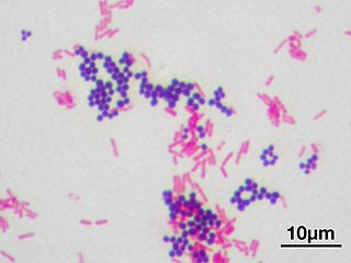
WGram stain or Gram staining, also called Gram's method, is a method of staining used to distinguish and classify bacterial species into two large groups: gram-positive bacteria and gram-negative bacteria. The name comes from the Danish bacteriologist Hans Christian Gram, who developed the technique.
 W
WGram-negative bacteria are bacteria that do not retain the crystal violet stain used in the gram-staining method of bacterial differentiation. They are characterized by their cell envelopes, which are composed of a thin peptidoglycan cell wall sandwiched between an inner cytoplasmic cell membrane and a bacterial outer membrane.
WIn bacteriology, gram-positive bacteria are bacteria that give a positive result in the Gram stain test, which is traditionally used to quickly classify bacteria into two broad categories according to their cell wall.
 W
WIn pathology, the Grocott-Gomori's methenamine silver stain, abbreviated GMS, is a popular staining method in histology. The stain was originally named after György Gömöri, the Hungarian physician who developed the stain.
 W
WHematoxylin and eosin stain or haematoxylin and eosin stain is one of the principal tissue stains used in histology. It is the most widely used stain in medical diagnosis and is often the gold standard. For example, when a pathologist looks at a biopsy of a suspected cancer, the histological section is likely to be stained with H&E.
 W
WHistology, also known as microscopic anatomy or microanatomy, is the branch of biology which studies the microscopic anatomy of biological tissues. Histology is the microscopic counterpart to gross anatomy, which looks at larger structures visible without a microscope. Although one may divide microscopic anatomy into organology, the study of organs, histology, the study of tissues, and cytology, the study of cells, modern usage places these topics under the field of histology. In medicine, histopathology is the branch of histology that includes the microscopic identification and study of diseased tissue. In the field of paleontology, the term paleohistology refers to the histology of fossil organisms.
 W
WImmunocytochemistry (ICC) is a common laboratory technique that is used to anatomically visualize the localization of a specific protein or antigen in cells by use of a specific primary antibody that binds to it. The primary antibody allows visualization of the protein under a fluorescence microscope when it is bound by a secondary antibody that has a conjugated fluorophore. ICC allows researchers to evaluate whether or not cells in a particular sample express the antigen in question. In cases where an immunopositive signal is found, ICC also allows researchers to determine which sub-cellular compartments are expressing the antigen.
 W
WImmunohistochemistry (IHC) is the most common application of immunostaining. It involves the process of selectively identifying antigens (proteins) in cells of a tissue section by exploiting the principle of antibodies binding specifically to antigens in biological tissues. IHC takes its name from the roots "immuno", in reference to antibodies used in the procedure, and "histo", meaning tissue. Albert Coons conceptualized and first implemented the procedure in 1941.
 W
WJones' stain, also Jones stain, is a methenamine silver-Periodic acid-Schiff stain used in pathology. It is also referred to as methenamine PAS which is commonly abbreviated MPAS.
 W
WLeishman stain, also known as Leishman's stain, is used in microscopy for staining blood smears. It is generally used to differentiate between and identify white blood cells, malaria parasites, and trypanosomas. It is based on a methanolic mixture of "polychromed" methylene blue and eosin. The methanolic stock solution is stable and also serves the purpose of directly fixing the smear eliminating a prefixing step. If a working solution is made by dilution with an aqueous buffer, the resulting mixture is very unstable and cannot be used for long. Leishman stain is named after its inventor, the Scottish pathologist William Boog Leishman. It is a version of the Romanowsky stain, and is thus similar to and partially replaceable by Giemsa stain, Jenner's stain, and Wright's stain.
 W
WLuxol fast blue stain, abbreviated LFB stain or simply LFB, is a commonly used stain to observe myelin under light microscopy, created by Heinrich Klüver and Elizabeth Barrera in 1953. LFB is commonly used to detect demyelination in the central nervous system (CNS), but cannot discern myelination in the peripheral nervous system.
 W
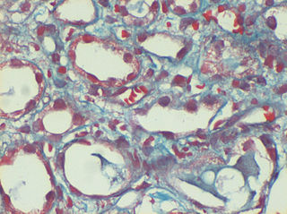
WMasson's trichrome is a three-colour staining protocol used in histology. The recipes evolved from Claude L. Pierre Masson's (1880–1959) original formulation have different specific applications, but all are suited for distinguishing cells from surrounding connective tissue.
 W
WPapanicolaou stain is a multichromatic (multicolored) cytological staining technique developed by George Papanicolaou in 1942. The Papanicolaou stain is one of the most widely used stains in cytology, where it is used to aid pathologists in making a diagnosis. Although most notable for its use in the detection of cervical cancer in the Pap test or Pap smear, it is also used to stain non-gynecological specimen preparations from a variety of bodily secretions and from small needle biopsies of organs and tissues. Papanicolaou published three formulations of this stain in 1942, 1954, and 1960.
 W
WPeriodic acid–Schiff–diastase stain is a periodic acid–Schiff (PAS) stain used in combination with diastase, an enzyme that breaks down glycogen. PAS-D is a stain often used by pathologists as an ancillary study in making a histologic diagnosis on paraffin-embedded tissue specimens. PAS stain typically gives a magenta color in the presence of glycogen. When PAS and diastase are used together, a light pink color replaces the deep magenta. Differences in the intensities of the two stains can be attributed to different glycogen concentrations and can be used to semiquantify glycogen in samples. In practice, the tissue is deparaffinized, the diastase incubates, and then the PAS stain is applied.
 W
WPeriodic acid–Schiff (PAS) is a staining method used to detect polysaccharides such as glycogen, and mucosubstances such as glycoproteins, glycolipids and mucins in tissues. The reaction of periodic acid oxidizes the vicinal diols in these sugars, usually breaking up the bond between two adjacent carbons not involved in the glycosidic linkage or ring closure in the ring of the monosaccharide units that are parts of the long polysaccharides, and creating a pair of aldehydes at the two free tips of each broken monosaccharide ring. The oxidation condition has to be sufficiently regulated so as to not oxidize the aldehydes further. These aldehydes then react with the Schiff reagent to give a purple-magenta color. A suitable basic stain is often used as a counterstain.
 W
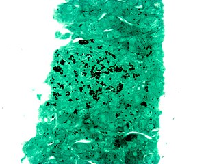
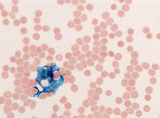
WPerls Prussian blue is a commonly used method in histology, histopathology, and clinical pathology to detect the presence of iron in tissue or cell samples. Perls Prussian Blue derives its name from the German pathologist Max Perls (1843–1881), who described the technique in 1867. The method does not involve the application of a dye, but rather causes the pigment Prussian blue to form directly within the tissue. The method stains mostly iron in the ferric state which includes ferritin and hemosiderin, rather than iron in the ferrous state.
 W
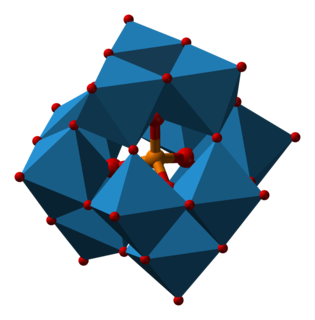
WPhosphotungstic acid (PTA), tungstophosphoric acid (TPA), is a heteropoly acid with the chemical formula H3PW12O40. It is normally present as a hydrate. EPTA is the name of ethanolic phosphotungstic acid, its alcohol solution used in biology. It has the appearance of small, colorless-grayish or slightly yellow-green crystals, with melting point 89 °C (24 H2O hydrate). It is odorless and soluble in water (200 g/100 ml). It is not especially toxic, but is a mild acidic irritant. The compound is known by a variety of different names and acronyms (see 'other names' section of infobox).
 W
WPhosphotungstic acid haematoxylin (PTAH) is a mix of haematoxylin with phosphotungstic acid, used in histology for staining.
 W
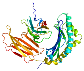
WProlactin-inducible protein also known as gross cystic disease fluid protein 15 (GCDFP-15), extra-parotid glycoprotein (EP-GP), gp17 seminal actin-binding protein (SABP) or BRST2 is a protein that in humans is encoded by the PIP gene. It is upregulated by prolactin and androgens and downregulated by estrogen.
 W
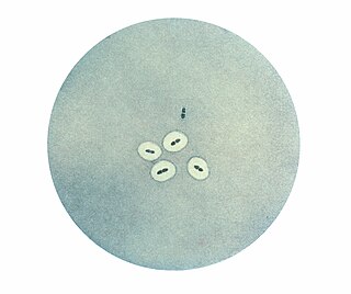
WThe quellung reaction, also called the Neufeld reaction, is a biochemical reaction in which antibodies bind to the bacterial capsule of Streptococcus pneumoniae, Klebsiella pneumoniae, Neisseria meningitidis, Bacillus anthracis, Haemophilus influenzae, Escherichia coli, and Salmonella. The antibody reaction allows these species to be visualized under a microscope. If the reaction is positive, the capsule becomes opaque and appears to enlarge.
 W
WIn pathology, the reticulin stain, is a popular staining method in histology. It is used to visualize reticular fiber and used extensively in liver histopathology.
 W
WRhodamine B is a chemical compound and a dye. It is often used as a tracer dye within water to determine the rate and direction of flow and transport. Rhodamine dyes fluoresce and can thus be detected easily and inexpensively with fluorometers.
 W
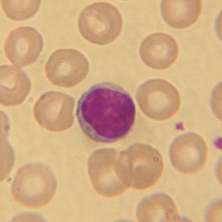
WRomanowsky staining, also known as Romanowsky–Giemsa staining, is a prototypical staining technique that was the forerunner of several distinct but similar stains widely used in hematology and cytopathology. Romanowsky-type stains are used to differentiate cells for microscopic examination in pathological specimens, especially blood and bone marrow films, and to detect parasites such as malaria within the blood. Stains that are related to or derived from the Romanowsky-type stains include Giemsa, Jenner, Wright, Field, May–Grünwald and Leishman stains. The staining technique is named after the Russian physician Dmitri Leonidovich Romanowsky (1861–1921), who was one of the first to recognize its potential for use as a blood stain.
 W
WThe Schaeffer–Fulton stain is a technique designed to isolate endospores by staining any present endospores green, and any other bacterial bodies red. The primary stain is malachite green, and the counterstain is safranin, which dyes any other bacterial bodies red.
 W
WSupravital staining is a method of staining used in microscopy to examine living cells that have been removed from an organism. It differs from intravital staining, which is done by injecting or otherwise introducing the stain into the body. Thus a supravital stain may have a greater toxicity, as only a few cells need to survive it a short while. The term "vital stain" is used by some authors to refer specifically to an intravital stain, and by others interchangeably with a supravital stain, the core concept being that the cell being examined is still alive. As the cells are alive and unfixed, outside the body, supravital stains are temporary in nature.
 W
WThionine, also known as Lauth's violet, is the salt of a heterocyclic compound. It was firstly synthesised by Charles Lauth. A variety of salts are known including the chloride and acetate, called respectively thionine chloride and thionine acetate. The dye is structurally related to methylene blue, which also features a phenothiazine core. The dye's name is frequently misspelled, with omission of the e. The -ine ending indicates that the compound is an amine..
 W
WToluidine blue is a basic thiazine metachromatic dye with high affinity for acidic tissue components.
 W
WVan Gieson's stain is a mixture of picric acid and acid fuchsin. It is the simplest method of differential staining of collagen and other connective tissue. It was introduced to histology by American neuropsychiatrist and pathologist Ira Van Gieson.
 W
WThe Warthin–Starry stain (WS) is a silver nitrate-based staining method used in histology. It was first introduced in 1920 by American pathologists Aldred Scott Warthin (1866-1931) and Allen Chronister Starry (1890-1973), for the detection of spirochetes. It has been considered a standard stain for the detection of spirochetes, and is also used to stain Helicobacter pylori, Lawsonia intracellularis, Microsporidia, and particulates. It is also important for confirmation of Bartonella henselae, a causative organism in cat-scratch disease.
 W
WWright's stain is a hematologic stain that facilitates the differentiation of blood cell types. It is classically a mixture of eosin (red) and methylene blue dyes. It is used primarily to stain peripheral blood smears, urine samples, and bone marrow aspirates, which are examined under a light microscope. In cytogenetics, it is used to stain chromosomes to facilitate diagnosis of syndromes and diseases.
 W
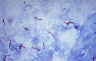
WZiehl-Neelsen staining is a type of acid-fast stain, first introduced by Paul Ehrlich. Ziehl–Neelsen staining is a bacteriological stain used to identify acid-fast organisms, mainly Mycobacteria. It is named for two German doctors who modified the stain: the bacteriologist Franz Ziehl (1859–1926) and the pathologist Friedrich Neelsen (1854–1898).