 W
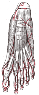
WThe abductor digiti minimi is a muscle which lies along the lateral (outer) border of the foot, and is in relation by its medial margin with the lateral plantar artery, vein and nerves.
 W
WThe abductor hallucis muscle is an intrinsic muscle of the foot. It participates in the abduction and flexion of the great toe.
 W
WThe Adductor hallucis arises by two heads—oblique and transverse and is responsible for adducting the big toe. It has two heads, both are innervated by the lateral plantar nerve.
 W
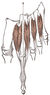
WIn human anatomy, the dorsal interossei of the foot are four muscles situated between the metatarsal bones.
 W
WThe extensor digitorum brevis muscle is a muscle on the upper surface of the foot that helps extend digits 2 through 4.
 W
WThe extensor digitorum longus is a pennate muscle, situated at the lateral part of the front of the leg.
 W
WThe extensor hallucis brevis is a muscle on the top of the foot that helps to extend the big toe.
WThe extensor hallucis longus is a thin muscle, situated between the tibialis anterior and the extensor digitorum longus, that functions to extend the big toe and dorsiflects the foot, and assists with foot eversion and inversion.
 W
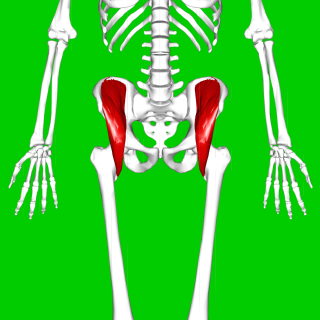
WThe external obturator muscle, obturator externus muscle is a flat, triangular muscle, which covers the outer surface of the anterior wall of the pelvis.
 W
WThe fascial compartments of the leg are the four fascial compartments that separate and contain the muscles of the lower leg. The compartments are divided by septa formed from the fascia. The compartments usually have nerve and blood supplies separate from their neighbours. All of the muscles within a compartment will generally be supplied by the same nerve.
 W
WThe Flexor digiti minimi brevis lies under the metatarsal bone on the little toe, and resembles one of the Interossei.
WThe flexor digitorum brevis lies in the middle of the sole of the foot, immediately above the central part of the plantar aponeurosis, with which it is firmly united.
 W
WThe flexor digitorum longus is situated on the tibial side of the leg. At its origin it is thin and pointed, but it gradually increases in size as it descends. This muscle serves to curl the second, third, fourth, and fifth toes.
 W
WThe Flexor hallucis brevis is a muscle of the foot that flexes the big toe.
 W
WThe flexor hallucis longus muscle (FHL) is one of the three deep muscles of the posterior compartment of the leg that attaches to the plantar surface of the distal phalanx of the great toe. The other deep muscles are the flexor digitorum longus and tibialis posterior; the tibialis posterior is the most powerful of these deep muscles. All three muscles are innervated by the tibial nerve which comprises half of the sciatic nerve.
 W
WThe gastrocnemius muscle is a superficial two-headed muscle that is in the back part of the lower leg of humans. It runs from its two heads just above the knee to the heel, a three joint muscle. The muscle is named via Latin, from Greek γαστήρ (gaster) 'belly' or 'stomach' and κνήμη (knḗmē) 'leg', meaning 'stomach of leg'.
WThe gemelli muscles are the inferior gemellus muscle and the superior gemellus muscle, two small accessory fasciculi to the tendon of the internal obturator muscle. The gemelli muscles belong to the lateral rotator group of six muscles of the hip that rotate the femur in the hip joint.
 W
WThe gluteal muscles are a group of three muscles which make up the buttocks: the gluteus maximus, gluteus medius and gluteus minimus. The three muscles originate from the ilium and sacrum and insert on the femur. The functions of the muscles include extension, abduction, external rotation, and internal rotation of the hip joint.
 W
WThe gluteus maximus is the main extensor muscle of the hip. It is the largest and outermost of the three gluteal muscles and makes up a large part of the shape and appearance of each side of the hips. Its thick fleshy mass, in a quadrilateral shape, forms the prominence of the buttocks. The other gluteal muscles are the medius and minimus, and sometimes informally these are collectively referred to as the "glutes".
 W
WThe gluteus medius, one of the three gluteal muscles, is a broad, thick, radiating muscle. It is situated on the outer surface of the pelvis.
 W
WThe gluteus minimus, or glutæus minimus, the smallest of the three gluteal muscles, is situated immediately beneath the gluteus medius.
 W
WThe iliacus is a flat, triangular muscle which fills the iliac fossa. It forms the lateral portion of iliopsoas, providing flexion of the thigh and lower limb at the acetabulofemoral joint.
 W
WThe iliopsoas refers to the joined psoas and the iliacus muscles. The two muscles are separate in the abdomen, but usually merge in the thigh. They are usually given the common name iliopsoas. The iliopsoas muscle joins to the femur at the lesser trochanter. It acts as the strongest flexor of the hip.
WThe internal obturator muscle or obturator internus muscle originates on the medial surface of the obturator membrane, the ischium near the membrane, and the rim of the pubis.
 WW
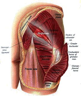
WWThe lateral rotator group is a group of six small muscles of the hip which all externally (laterally) rotate the femur in the hip joint. It consists of the following muscles: Piriformis, gemellus superior, obturator internus, gemellus inferior, quadratus femoris and the obturator externus.
 W
WThe lumbricals are four small skeletal muscles, accessory to the tendons of the flexor digitorum longus and numbered from the medial side of the foot; they arise from these tendons, as far back as their angles of division, each springing from two tendons, except the first. So the first lumbrical is unipennate and the second, third and fourth are bipennate.
WThe peroneus brevis muscle lies under cover of the peroneus longus, and is the shorter and smaller of the peroneus muscles.
 W
WIn human anatomy, the peroneus longus is a superficial muscle in the lateral compartment of the leg, and acts to evert and plantarflex the ankle.
 W
WThe peroneus muscles are a group of muscles in the leg. While the muscle group exists in many variations, it is normally composed of three muscles: peroneus longus, brevis and tertius. The peroneus muscles originates from lower two-thirds of the lateral surface of the shaft of the fibula and the anterior and posterior inter-muscular septa of the leg. It inserts onto the metatarsals.
 W
WThe peroneus tertius is a muscle of the human body located in the lower limb.
 W
WThe piriformis is a muscle in the gluteal region of the lower limbs. It is one of the six muscles in the lateral rotator group.
 W
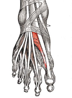
WIn human anatomy, plantar interossei muscles are three muscles located between the metatarsal bones in the foot.
 W
WThe plantaris is one of the superficial muscles of the superficial posterior compartment of the leg, one of the fascial compartments of the leg.
 W
WThe popliteus muscle in the leg is used for unlocking the knees when walking, by laterally rotating the femur on the tibia during the closed chain portion of the gait cycle. In open chain movements, the popliteus muscle medially rotates the tibia on the femur. It is also used when sitting down and standing up. It is the only muscle in the posterior (back) compartment of the lower leg that acts just on the knee and not on the ankle. The gastrocnemius muscle acts on both joints.
WThe posterior compartment of the leg is one of the fascial compartments of the leg and is divided further into deep and superficial compartments.
 W
WThe posterior intermuscular septum of leg, or posterior crural intermuscular septum is a band of fascia which separates the lateral compartment of leg.
 W
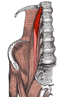
WThe psoas major is a long fusiform muscle located in the lateral lumbar region between the vertebral column and the brim of the lesser pelvis. It joins the iliacus muscle to form the iliopsoas. In animals, this muscle is equivalent to the tenderloin.
 W
WThe psoas minor is a long, slender skeletal muscle which, when present, is located anterior to the psoas major muscle.
WThe quadratus femoris is a flat, quadrilateral skeletal muscle. Located on the posterior side of the hip joint, it is a strong external rotator and adductor of the thigh, but also acts to stabilize the femoral head in the acetabulum. Quadratus femoris use in the Meyer's muscle pedicle grafting to prevent avascular necrosis of femur head.
 W
WThe quadratus plantae is separated from the muscles of the first layer by the lateral plantar vessels and nerve. It acts to aid in flexing the 2nd to 5th toes and is one of the few muscles in the foot with no homolog in the hand.
 W
WThe quadriceps femoris is a large muscle group that includes the four prevailing muscles on the front of the thigh.
 W
WThe sartorius muscle is the longest muscle in the human body. It is a long, thin, superficial muscle that runs down the length of the thigh in the anterior compartment.
 W
WThe semitendinosus is a long superficial muscle in the back of the thigh. It is so named because it has a very long tendon of insertion. It lies posteromedially in the thigh, superficial to the semimembranosus.
 W
WIn humans and some other mammals, the soleus is a powerful muscle in the back part of the lower leg. It runs from just below the knee to the heel, and is involved in standing and walking. It is closely connected to the gastrocnemius muscle and some anatomists consider them to be a single muscle, the triceps surae. Its name is derived from the Latin word "solea", meaning "sandal".
 W
WThe tensor fasciae latae is a muscle of the thigh. Together with the gluteus maximus, it acts on the iliotibial band and is continuous with the iliotibial tract, which attaches to the tibia. The muscle assists in keeping the balance of the pelvis while standing, walking, or running.
 W
WTensor vastus intermedius is a muscle in the anterior compartment of thigh. It lies between the vastus intermedius and the vastus lateralis. The structure has been previously reported but has not been described nor illustrated in any textbook. The term tensor vastus intermedius was given by Grob et al. in 2016.
 W
WThe tibialis anterior is a muscle in humans that originates along the upper two-thirds of the lateral (outside) surface of the tibia and inserts into the medial cuneiform and first metatarsal bones of the foot. It acts to dorsiflex and invert the foot. This muscle is mostly located near the shin.
 W
WThe tibialis posterior is the most central of all the leg muscles, and is located in the deep posterior compartment of the leg.
 W
WThe triceps surae is a pair of muscles located at the calf – the two- headed gastrocnemius and the soleus. These muscles both insert into the calcaneus, the bone of the heel of the human foot, and form the major part of the muscle of the posterior leg, commonly known as the calf muscle.
WThe vastus intermedius (Cruraeus) arises from the front and lateral surfaces of the body of the femur in its upper two-thirds, sitting under the rectus femoris muscle and from the lower part of the lateral intermuscular septum. Its fibers end in a superficial aponeurosis, which forms the deep part of the quadriceps femoris tendon.
WThe vastus lateralis, also called the ''vastus externus'' is the largest and most powerful part of the quadriceps femoris, a muscle in the thigh. Together with other muscles of the quadriceps group, it serves to extend the knee joint, moving the lower leg forward. It arises from a series of flat, broad tendons attached to the femur, and attaches to the outer border of the patella. It ultimately joins with the other muscles that make up the quadriceps in the quadriceps tendon, which travels over the knee to connect to the tibia. The vastus lateralis is the recommended site for intramuscular injection in infants less than 7 months old and those unable to walk, with loss of muscular tone.
WThe vastus medialis is an extensor muscle located medially in the thigh that extends the knee. The vastus medialis is part of the quadriceps muscle group.
WThe vastus muscles are three of the four muscles that make up the quadriceps femoris muscle of the thigh. The three muscles are the vastus intermedius, the vastus lateralis, and the vastus medialis located in the middle, on the outside, and inside of the thigh, respectively. The fourth muscle is the rectus femoris muscle a large fleshy muscle which covers the front and sides of the femur.