 W
WCardiology is a branch of medicine that deals with the disorders of the heart as well as some parts of the circulatory system. The field includes medical diagnosis and treatment of congenital heart defects, coronary artery disease, heart failure, valvular heart disease and electrophysiology. Physicians who specialize in this field of medicine are called cardiologists, a specialty of internal medicine. Pediatric cardiologists are pediatricians who specialize in cardiology. Physicians who specialize in cardiac surgery are called cardiothoracic surgeons or cardiac surgeons, a specialty of general surgery.
 W
WAccelerated idioventricular rhythm is a ventricular rhythm with a rate of between 40 and 120 beats per minute. Idioventricular means “relating to or affecting the cardiac ventricle alone” and refers to any ectopic ventricular arrhythmia. Accelerated idioventricular arrhythmias are distinguished from ventricular rhythms with rates less than 40 and those faster than 120. Though some other references limit to between 60 and 100 beats per minute. It is also referred to as AIVR and "slow ventricular tachycardia."
 W
WAdvanced cardiac life support, or advanced cardiovascular life support, often referred to by its acronym, "ACLS", refers to a set of clinical algorithms for the urgent treatment of cardiac arrest, stroke, myocardial infarction, and other life-threatening cardiovascular emergencies. Outside North America, Advanced Life Support (ALS) is used.
 W
WThe Antihypertensive and Lipid Lowering Treatment to Prevent Heart Attack Trial, also known as ALLHAT, was a randomized, double-blind, active-controlled study comparing at the same time, four different classes of antihypertensive drugs with the rate of coronary heart disease (CHD) events in ‘high-risk’ people with hypertension. Participants were initially randomised to chlorthalidone (diuretic) versus doxazosin, lisinopril (ACE-inhibitor), and amlodipine.
 W
WAn artificial heart is a device that replaces the heart. Artificial hearts are typically used to bridge the time to heart transplantation, or to permanently replace the heart in the case that a heart transplant is impossible. Although other similar inventions preceded it from the late 1940s, the first artificial heart to be successfully implanted in a human was the Jarvik-7 in 1982, designed by a team including Willem Johan Kolff and Robert Jarvik.
 W
WAn artificial heart valve is a one-way valve implanted into a person's heart to replace a valve that is not functioning properly. Artificial heart valves can be separated into three broad classes: mechanical heart valves, bioprosthetic tissue valves and engineered tissue valves.
 W
WAtrial flutter (AFL) is a common abnormal heart rhythm that starts in the atrial chambers of the heart. When it first occurs, it is usually associated with a fast heart rate and is classified as a type of supraventricular tachycardia. Atrial flutter is characterized by a sudden-onset (usually) regular abnormal heart rhythm on an electrocardiogram (ECG) in which the heart rate is fast. Symptoms may include a feeling of the heart beating too fast, too hard, or skipping beats, chest discomfort, difficulty breathing, a feeling as if one's stomach has dropped, a feeling of being light-headed, or loss of consciousness.
 W
WBendopnea is a newly described symptom of heart failure, meaning shortness of breath when leaning forward. It was introduced by Thibodeau et al. in 2014. Patients with heart failure often experience this when bending over to tie a shoe or putting socks on. It has been defined as occurring within 30 seconds of bending over, but could occur in as few as 8 seconds. When a patient is in heart failure, it often means the ventricular filling pressures are high at baseline. When said person bends forward, it causes a further increase in ventricular filling pressures, especially in patients with lower cardiac indices. The term "bendopnea" was coined to be easily identifiable among patients and physicians.
 W
WIn medicine, a stent is any device which is inserted into a blood vessel or other internal duct to expand it to prevent or alleviate a blockage. Traditionally, such devices are fabricated from metal mesh and remain in the body permanently or until removed through further surgical intervention. A bioresorbable stent serves the same purpose, but is manufactured from a material that may dissolve or be absorbed in the body.
 W
WBranchial hearts are myogenic accessory pumps found in coleoid cephalopods that supplement the action of the main, systemic heart. Each consists of a single chamber and they are always paired, being located at the base of the gills. They pump blood through the gills via the afferent branchial veins. Since they only circulate venous blood, branchial hearts function under predominantly anaerobic conditions. Branchial hearts also appear to be involved in hemocyanin synthesis.
 W
WThe central governor is a proposed process in the brain that regulates exercise in regard to a neurally calculated safe exertion by the body. In particular, physical activity is controlled so that its intensity cannot threaten the body’s homeostasis by causing anoxic damage to the heart muscle. The central governor limits exercise by reducing the neural recruitment of muscle fibers. This reduced recruitment causes the sensation of fatigue. The existence of a central governor was suggested to explain fatigue after prolonged strenuous exercise in long-distance running and other endurance sports, but its ideas could also apply to other causes of exertion-induced fatigue.
 W
WCerebral circulation is the movement of blood through a network of cerebral arteries and veins supplying the brain. The rate of cerebral blood flow in an adult human is typically 750 milliliters per minute, or about 15% of cardiac output. Arteries deliver oxygenated blood, glucose and other nutrients to the brain. Veins carry "used or spent" blood back to the heart, to remove carbon dioxide, lactic acid, and other metabolic products.
 W
WContrast-enhanced ultrasound (CEUS) is the application of ultrasound contrast medium to traditional medical sonography. Ultrasound contrast agents rely on the different ways in which sound waves are reflected from interfaces between substances. This may be the surface of a small air bubble or a more complex structure. Commercially available contrast media are gas-filled microbubbles that are administered intravenously to the systemic circulation. Microbubbles have a high degree of echogenicity. There is a great difference in echogenicity between the gas in the microbubbles and the soft tissue surroundings of the body. Thus, ultrasonic imaging using microbubble contrast agents enhances the ultrasound backscatter, (reflection) of the ultrasound waves, to produce a sonogram with increased contrast due to the high echogenicity difference. Contrast-enhanced ultrasound can be used to image blood perfusion in organs, measure blood flow rate in the heart and other organs, and for other applications.
 W
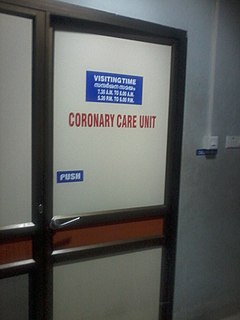
WA coronary care unit (CCU) or cardiac intensive care unit (CICU) is a hospital ward specialized in the care of patients with heart attacks, unstable angina, cardiac dysrhythmia and various other cardiac conditions that require continuous monitoring and treatment.
 W
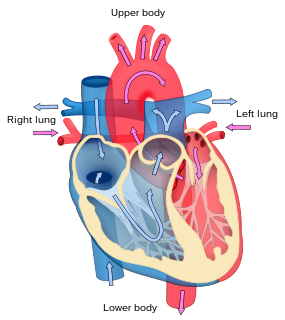
WCoronary circulation is the circulation of blood in the blood vessels that supply the heart muscle (myocardium). Coronary arteries supply oxygenated blood to the heart muscle, and cardiac veins drain away the blood once it has been deoxygenated. Because the rest of the body, and most especially the brain, needs a steady supply of oxygenated blood that is free of all but the slightest interruptions, the heart is required to function continuously. Therefore its circulation is of major importance not only to its own tissues but to the entire body and even the level of consciousness of the brain from moment to moment. Interruptions of coronary circulation quickly cause heart attacks, in which the heart muscle is damaged by oxygen starvation. Such interruptions are usually caused by ischemic heart disease and sometimes by embolism from other causes like obstruction in blood flow through vessels.
 W
WCoxsackie B is a group of six serotypes of Coxsackievirus, a pathogenic enterovirus, that trigger illness ranging from gastrointestinal distress to full-fledged pericarditis and myocarditis.
 W
WThe CPK-MB test is a cardiac marker used to assist diagnoses of an acute myocardial infarction. It measures the blood level of CK-MB, the bound combination of two variants of the enzyme phosphocreatine kinase.
 W
WAn echocardiography, echocardiogram, cardiac echo or simply an echo, is an ultrasound of the heart.
 W
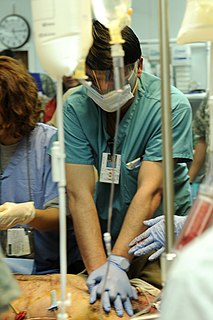
WEndomyocardial biopsy (EMB) is an invasive procedure used routinely to obtain small samples of heart muscle, primarily for detecting rejection of a donor heart following heart transplantation. It is also used as a diagnostic tool in some heart diseases.
 W
WIn the fetal heart, the foramen ovale, also foramen Botalli, or the ostium secundum of Born, allows blood to enter the left atrium from the right atrium. It is one of two fetal cardiac shunts, the other being the ductus arteriosus. Another similar adaptation in the fetus is the ductus venosus. In most individuals, the foramen ovale closes at birth. It later forms the fossa ovalis.
 W
WThe fossa ovalis is a depression in the right atrium of the heart, at the level of the interatrial septum, the wall between right and left atrium. The fossa ovalis is the remnant of a thin fibrous sheet that covered the foramen ovale during fetal development.
 W
WThe fourth heart sound or S4 is an extra heart sound that occurs during late diastole, immediately before the normal two "lub-dub" heart sounds (S1 and S2). It occurs just after atrial contraction and immediately before the systolic S1 and is caused by the atria contracting forcefully in an effort to overcome an abnormally stiff or hypertrophic ventricle.
 W
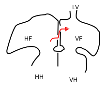
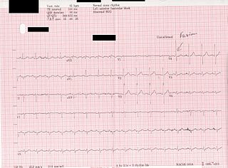
WA fusion beat occurs when electrical impulses from different sources act upon the same region of the heart at the same time. If it acts upon the ventricular chambers it is called a ventricular fusion beat, whereas colliding currents in the atrial chambers produce atrial fusion beats.
 W
WThe Heart Protection Study was a randomized controlled trial run by the Clinical Trial Service Unit, and funded by the Medical Research Council (MRC) and the British Heart Foundation (BHF) in the United Kingdom. It studied the use of the cholesterol lowering drug, simvastatin 40 mg and vitamin supplementation in people who were at risk of cardiovascular disease. It was led by Jane Armitage, an epidemiologist at the Clinical Trial Service Unit.
 W
WHeart rate variability (HRV) is the physiological phenomenon of variation in the time interval between heartbeats. It is measured by the variation in the beat-to-beat interval.
 W
WHeart sounds are the noises generated by the beating heart and the resultant flow of blood through it. Specifically, the sounds reflect the turbulence created when the heart valves snap shut. In cardiac auscultation, an examiner may use a stethoscope to listen for these unique and distinct sounds that provide important auditory data regarding the condition of the heart.
 W
WHeart-type fatty acid binding protein (hFABP) also known as mammary-derived growth inhibitor is a protein that in humans is encoded by the FABP3 gene.
 W
WThe hexaxial reference system, better known as the Cabrera system, is a convention to present the extremity leads of the 12 lead electrocardiogram, that provides an illustrative logical sequence that helps interpretation of the ECG, especially to determine the heart's electrical axis in the frontal plane. The most practical way of using this is by arranging extremity leads according to the Cabrera system, reversing polarity of lead aVR and presenting ECG complexes in the order. Then determine the direction the maximal ECG vector is "pointing", i.e. in which lead there are most positive amplitude - this direction is the electrical axis - see diagram. Example: If lead I has the highest amplitude, the axis is approximately 0°. Conversely, if lead III has the most negative amplitude it means the vector is pointing away from this lead, i.e. towards -60°.
 W
WIntima–media thickness (IMT), also called intimal medial thickness, is a measurement of the thickness of tunica intima and tunica media, the innermost two layers of the wall of an artery. The measurement is usually made by external ultrasound and occasionally by internal, invasive ultrasound catheters. Measurements of the total wall thickness of blood vessels can also be done using other imaging modalities.
 W
WAn isochrone map in geography and urban planning is a map that depicts the area accessible from a point within a certain time threshold. An isochrone is defined as "a line drawn on a map connecting points at which something occurs or arrives at the same time". In hydrology and transportation planning isochrone maps are commonly used to depict areas of equal travel time. The term is also used in cardiology as a tool to visually detect abnormalities using body surface distribution.
 W
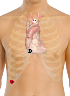
WA Lewis Lead is a modified ECG lead used to detect atrial flutter waves when atrial flutter is suspected clinically, based on signs and symptoms, but is not definitely demonstrated on the standard 12 lead ECG. In order to create the Lewis Lead, the right arm electrode is moved to the manubrium adjacent to the sternum. Then the left arm electrode is moved to the right, fifth intercostal space adjacent to the sternum. The left leg electrode is placed on the right lower costal margin. The Lewis Lead is then read as Lead I on the ECG and, since in most patients it will be roughly perpendicular to the wave of ventricular depolarization, atrial flutter waves may be more apparent.
 W
WA lipoprotein is a biochemical assembly whose primary purpose is to transport hydrophobic lipid molecules in water, as in blood plasma or other extracellular fluids. They consist of a Triglyceride and Cholesterol center, surrounded by a phospholipid outer shell, with the hydrophilic portions oriented outward toward the surrounding water and lipophilic portions oriented inward toward the lipid center. A special kind of protein, called apolipoprotein, is embedded in the outer shell, both stabilising the complex and giving it a functional identity that determines its fate.
 W
WLipoprotein(a) is a low-density lipoprotein variant containing a protein called apolipoprotein(a). Genetic and epidemiological studies have identified lipoprotein(a) as a risk factor for atherosclerosis and related diseases, such as coronary heart disease and stroke.
 W
WLow-density lipoprotein (LDL) is one of the five major groups of lipoprotein which transport all fat molecules around the body in the extracellular water. These groups, from least dense to most dense, are chylomicrons, very low-density lipoprotein (VLDL), intermediate-density lipoprotein (IDL), low-density lipoprotein and high-density lipoprotein (HDL). LDL delivers fat molecules to cells. LDL is involved in atherosclerosis, a process in which it is oxidized within the walls of arteries.
 W
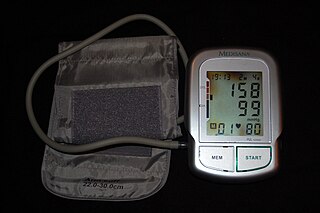
WPathophysiology is a branch of medicine which explains the function of the body as it relates to diseases and conditions. The pathophysiology of hypertension is an area which attempts to explain mechanistically the causes of hypertension, which is a chronic disease characterized by elevation of blood pressure. Hypertension can be classified by cause as either essential or secondary. About 90–95% of hypertension is essential hypertension. Some authorities define essential hypertension as that which has no known explanation, while others define its cause as being due to overconsumption of sodium and underconsumption of potassium. Secondary hypertension indicates that the hypertension is a result of a specific underlying condition with a well-known mechanism, such as chronic kidney disease, narrowing of the aorta or kidney arteries, or endocrine disorders such as excess aldosterone, cortisol, or catecholamines. Persistent hypertension is a major risk factor for hypertensive heart disease, coronary artery disease, stroke, aortic aneurysm, peripheral artery disease, and chronic kidney disease.
 W
WPulmonary angiography is medical fluoroscopic procedure used to visualize the pulmonary arteries and much less frequently, the pulmonary veins.
 W
WPulse oximetry is a noninvasive method for monitoring a person's oxygen saturation. Though its reading of peripheral oxygen saturation (SpO2) is not always identical to the more desirable reading of arterial oxygen saturation (SaO2) from arterial blood gas analysis, the two are correlated well enough that the safe, convenient, noninvasive, inexpensive pulse oximetry method is valuable for measuring oxygen saturation in clinical use.
 W
WReflex syncope is a brief loss of consciousness due to a neurologically induced drop in blood pressure. Before an affected person passes out, there may be sweating, a decreased ability to see, or ringing in the ears. Occasionally, the person may twitch while unconscious. Complications of reflex syncope include injury due to a fall.
 W
WRestenosis is the recurrence of stenosis, a narrowing of a blood vessel, leading to restricted blood flow. Restenosis usually pertains to an artery or other large blood vessel that has become narrowed, received treatment to clear the blockage and subsequently become renarrowed. This is usually restenosis of an artery, or other blood vessel, or possibly a vessel within an organ.
 W
WRhythm interpretation is an important part of healthcare in Emergency Medical Services (EMS). Trained medical personnel can determine different treatment options based on the cardiac rhythm of a patient. There are many common heart rhythms that are part of a few different categories, sinus arrhythmia, atrial arrhythmia, ventricular arrhythmia. Rhythms can be evaluated by measuring a few key components of a rhythm strip, the PQRST sequence, which represents one cardiac cycle, the ventricular rate, which is the rate at which the ventricles contract, and the atrial rate, which is the rate at which the atria contract.
 W
WSATRO-EKG - a computer program analysing electro-cardiology signals. It is based on the SFHAM model. It facilitates the evaluation of electrical activity of myocardium, and therefore, early detection of ischemic changes in the heart.
WThe Scandinavian Simvastatin Survival Study, was a multicentre, randomized, double-blind, placebo-controlled clinical trial, which provided the initial data that supported the use of the cholesterol-lowering drug, simvastatin, in people with a moderately raised cholesterol and coronary heart disease (CHD); that is people who had previously had a heart attack or angina. The study was sponsored by the pharmaceutical company Merck and enrolled 4,444 people from 94 centres in Scandinavia.
WA sinus rhythm is any cardiac rhythm in which depolarisation of the cardiac muscle begins at the sinus node. It is characterised by the presence of correctly oriented P waves on the electrocardiogram (ECG). Sinus rhythm is necessary, but not sufficient, for normal electrical activity within the heart.
 W
WA sonographer is a healthcare professional who specialises in the use of ultrasonic imaging devices to produce diagnostic images, scans, videos or three-dimensional volumes of anatomy and diagnostic data, frequently a radiographer, but may be any healthcare professional with the appropriate training. The requirements for clinical practice vary greatly by country. Sonography requires specialised education and skills to acquire, view, analyze, and optimize information in the image. Due to the high levels of decisional latitude and diagnostic input, sonographers have a high degree of responsibility in the diagnostic process. Many countries require medical sonographers to have professional certification. Sonographers have core knowledge in ultrasound physics, cross-sectional anatomy, physiology, and pathology.
 W
WA split S2 is a finding upon auscultation of the S2 heart sound.
 W
WSports cardiology is an emerging subspecialty field of Cardiology. It may also be considered a subspecialty field of Sports medicine, or alternatively a hybrid subspecialty that spans cardiology and sports medicine. Emergency medicine is another medical specialty that has some overlap with Sports Cardiology. Sports cardiology is now considered to be a distinct subspecialty in Europe and the USA, with a core curriculum developed in both regions. In Europe it has traditionally been grouped under Preventive Cardiology, but the subspecialty of Sports Cardiology is now considered a distinct field. In the USA, it has developed from being a special interest area to a distinct subspecialty as well.
 W
WIn the late 1800s, telegraphy was developing as a way for distant communication. Messages were converted to dots and dashes that were sent as electric pulses and could be converted to sound or visual signals at the distant site. That conversion was done by a coil in a galvanometer, which had a limited frequency. Clément Adair, a French engineer, replaced the coil with a much faster wire or "string" producing the first string galvanometer. Augustus Waller had discovered electrical activity from the heart and produced the first electrocardiogram in 1887. But his equipment was slow. Physiologists worked to find a better instrument. In 1901, Willem Einthoven described the science background and potential utility of a string galvanometer, stating "Mr. Adair as already built an instrument with a wires stretched between poles of a magnet. It was a telegraph receiver." Although Einthoven is sometimes credited with inventing it, he did not invent the string galvanometer. He was a leader in applying the string galvanometer to physiology and medicine, leading to today's electrocardiography. Einthoven was awarded the 1924 Nobel prize in Physiology or Medicine for his work.
 W
WSynCardia Systems, LLC, headquartered in Tucson, Arizona, was founded in 2001 and is the sole manufacturer and provider of the world's only clinically proven and commercially approved Total Artificial Heart.
WThe third heart sound or S3 is a rare extra heart sound that occurs soon after the normal two "lub-dub" heart sounds (S1 and S2). S3 is associated with heart failure.
 W
WTwiddler's syndrome is a malfunction of a pacemaker due to manipulation of the device and the subsequent dislodging of the leads from their intended location. As the leads move, they stop pacing the heart and can cause strange symptoms such as phrenic nerve stimulation resulting in abdominal pulsing or brachial plexus stimulation resulting in rhythmic arm twitching.
 W
WVA conduction, also named Ventriculoatrial conduction and sometimes referred to as Retrograde conduction, is the conduction backward phenomena in the heart, where the conduction comes from the ventricles or from the AV node into and through the atria.
 W
WVectorcardiography (VCG) is a method of recording the magnitude and direction of the electrical forces that are generated by the heart by means of a continuous series of vectors that form curving lines around a central point.
 W
WA ventricular assist device (VAD) is an electromechanical device for assisting cardiac circulation, which is used either to partially or to completely replace the function of a failing heart. The function of VADs is different from that of artificial cardiac pacemakers; some are for short-term use, typically for patients recovering from myocardial infarction and for patients recovering from cardiac surgery; some are for long-term use, typically for patients suffering from advanced heart failure.
 W
WVentricular natriuretic peptide or brain natriuretic peptide (BNP), also known as B-type natriuretic peptide, is a hormone secreted by cardiomyocytes in the heart ventricles in response to stretching caused by increased ventricular blood volume.
 W
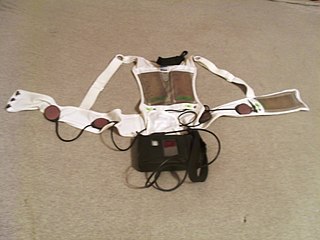
WA wearable cardioverter defibrillator (WCD) is a device worn by patients who are at risk for sudden cardiac arrest (SCA). A WCD allows physicians time to assess for their patient's arrhythmic risk and make appropriate plans.
 W
WWindkessel effect is a term used in medicine to account for the shape of the arterial blood pressure waveform in terms of the interaction between the stroke volume and the compliance of the aorta and large elastic arteries and the resistance of the smaller arteries and arterioles. Windkessel when loosely translated from German to English means 'air chamber', but is generally taken to imply an elastic reservoir. The walls of large elastic arteries contain elastic fibers, formed of elastin. These arteries distend when the blood pressure rises during systole and recoil when the blood pressure falls during diastole. Since the rate of blood entering these elastic arteries exceeds that leaving them via the peripheral resistance, there is a net storage of blood in the aorta and large arteries during systole, which discharges during diastole. The compliance of the aorta and large elastic arteries is therefore analogous to a capacitor.