 W
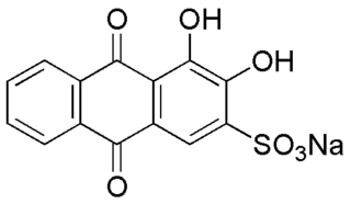
WAlizarin Red S (also known as C.I. Mordant Red 3, Alizarin Carmine, and C.I 58005. It is a water-soluble sodium salt of Alizarin sulfonic acid with a chemical formula of C14H7NaO7S. Alizarin Red S was discovered by Graebe and Libermann in 1871. In the field of histology alizarin Red S is been used to stain calcium deposits in tissues, and in geology to stain and differentiate carbonate minerals.
 W
WThe Bielschowsky technique is a silver staining method used in histochemistry for the visualization of nerve fibers, including multipolar interneurons in the cerebellum.
 W
WHistochemistry and Cell Biology is a peer-reviewed scientific journal in the field of molecular histology and cell biology, publishing original articles dealing with the localization and identification of molecular components, metabolic activities, and cell biological aspects of cells and tissues. The journal covers the development, application, and evaluation of methods and probes that can be used in the entire area of histochemistry and cell biology. The journal is published by Springer Science+Business Media and the official journal of the Society for Histochemistry. Earlier names of the journal are Histochemie and Histochemistry. The editors-in-chief are Jürgen Roth, Takehiko Koji, Michael Schrader and Douglas J. Taatjes.
 W
WHistology, also known as microscopic anatomy or microanatomy, is the branch of biology which studies the microscopic anatomy of biological tissues. Histology is the microscopic counterpart to gross anatomy, which looks at larger structures visible without a microscope. Although one may divide microscopic anatomy into organology, the study of organs, histology, the study of tissues, and cytology, the study of cells, modern usage places these topics under the field of histology. In medicine, histopathology is the branch of histology that includes the microscopic identification and study of diseased tissue. In the field of paleontology, the term paleohistology refers to the histology of fossil organisms.
 W
WThe Journal of Histochemistry and Cytochemistry is a peer-reviewed scientific journal of cell biology established in 1953. It covers research in the structure and function of cells, tissues, and organs as well as components of development, differentiation, and disease, as well as microscopy and imaging techniques. The journal is the official publication of The Histochemical Society and is published by SAGE Publications. The editor-in-chief as of January 1, 2016 is Stephen M. Hewitt of the National Cancer Institute, National Institutes of Health, in Bethesda Maryland, USA. The immediate past editor-in-chief is John R. Couchman. The journal is published online and in print monthly. Journal content is available for free after twelve months.
 W
WLuxol fast blue stain, abbreviated LFB stain or simply LFB, is a commonly used stain to observe myelin under light microscopy, created by Heinrich Klüver and Elizabeth Barrera in 1953. LFB is commonly used to detect demyelination in the central nervous system (CNS), but cannot discern myelination in the peripheral nervous system.
 W
WThe major histocompatibility complex in sexual selection concerns how major histocompatibility complex (MHC) molecules allow for immune system surveillance of the population of protein molecules in a host's cells. In 1976, Yamazaki et al. demonstrated a sexual selection mate choice by male mice for females of a different MHC.
 W
WPalaeoimmunology or paleo-immunology is the analysis using histochemical techniques to look at the matrix proteins in historic and pre-historic materials. Modern immunological assays are used to detect the presence of specific antigens in the sample material. Specimens subject to immunoassays have usually been preserved in a way that has prevented biomolecular targets from degrading. This has either been achieved through natural preservative circumstances, such as accelerated fossilization, or through artificial mummification. Regardless of the path taken to achieve this state, preservation has occurred before the denaturing of antigenic targets. The purpose of applying immunological assays to archaeological materials is to better understand the biochemical makeup and composition of these pre-historic samples. Antigenic elements within these materials may reveal information regarding the "life" and "death" of the sample being studied.